R E S E A R C H A R T I C L E
Open Access
The role of segmental nodes in the pathological
staging of non-small cell lung cancer
Zhen-xuan Li
1,2†, Hong Yang
1†, Ke-lin She
1,3, Ming-xing Zhang
1, Han-qing Xie
1, Peng Lin
1, Lan-jun Zhang
1and Xiao-dong Li
1*Abstract
Background:Segmental nodes are not examined routinely in current clinical practice for lung cancer, the role of segmental nodes in pathological staging of non-small cell lung cancer after radical resection was investigated.
Methods:A total of 113 consecutive non-small cell lung cancer patients who underwent radical resection between
June 2009 and December 2011 were retrospectively reviewed. All the operations were performed by the same group of surgeons. N2 nodes, hilar nodes, interlobar nodes and some lobar nodes were collected during surgery. The removed lung lobes were dissected routinely along lobar and segmental bronchi to collect lobar nodes and segmental nodes. The collected lymph nodes were separately labeled for histological examination.
Results:The detection rates of hilar nodes, interlobar nodes, lobar nodes and segmental nodes were 61.1%, 85.0%, 75.2% and 80.5%, respectively. The metastasis rates of hilar nodes, interlobar nodes, lobar nodes and segmental nodes were 5.3%, 10.5%, 16.8% and 14.2%, respectively. There were 68 cases of N0 disease, 16 cases of N1 disease and 29 cases of N2 disease. If an analysis of segmental lymph nodes had been omitted, six patients (37.5% of N1 disease) would have been down-staged to N0, and two cases of multiple-zone N1 disease would have been misdiagnosed as single-zone N1 disease, one patient would have been misdiagnosed as N2 disease with skip metastases.
Conclusion:Segmental nodes play an important role in the accurate staging of non-small cell lung cancer, and routinely dissecting the segmental bronchi to collect the lymph nodes is feasible and may be necessary.
Keywords:Non-small cell lung cancer, Lymph node, Segmental node, Pathological staging, Radical resection
Background
Accurate pathological staging of lymph node involvement has been recognized as a key factor in the management of lung cancer, which includes the postsurgical treatment se-lection and prognosis prediction. The 5-year survival rates of N0, single-zone N1, multiple-zone N1 and N2 disease for surgically managed patients with non-small cell lung cancer (NSCLC) are 56%, 48%, 35% and 22%, respectively [1]. Based on the current staging, adjuvant therapy can be used to improve survival [2,3]. Referring to the Inter-national Association for the Study of Lung Cancer (IASLC) lymph node map [4], levels 10–12 lymph nodes (hilar nodes, interlobar nodes and lobar nodes) are
examined routinely in current clinical practice, but level 13 lymph nodes (segmental nodes) are not. The role of segmental nodes in the pathological staging of lung cancer has rarely been reported. Accordingly, we retro-spectively analyzed the influence of segmental nodes for patients with non-small cell lung cancer undergoing radical resections, which were performed by the same group of surgeons.
Methods
This study was approved by the Sun Yat-Sen University Cancer Center Institutional Review Board of Clinical Research. The need for informed consent from patients was waived due to its retrospective design.
* Correspondence:[email protected]
†Equal contributors
1
State Key Laboratory of Oncology in South China, Sun Yat-sen University Cancer Center, 651 Dongfeng Rd, East, Guangzhou, PR China
Full list of author information is available at the end of the article
Patient eligibility
Between June 2009 and December 2011, 127 consecutive patients with lung cancer underwent surgical resection in our treatment group at the Sun Yat-Sen University Cancer Center. The inclusion criteria are listed here: (1) Patients with non-small cell lung cancer who underwent pul-monary resection plus complete mediastinal lymph node dissection (MLND), (2) patients who underwent R0 (microscopically complete) resection and (3) patients who did not have distant metastasis. One hundred and thirteen patients were enrolled. All patients underwent computed tomography (CT) scans of the chest and upper abdomen with intravenous contrast, bronchoscopy, and brain magnetic resonance imaging prior to surgery. Some patients underwent positron emission computed tomography (PET), PET-CT, bone scanning or mediasti-noscopy for exact clinical staging. The 7th edition of the AJCC and UICC TNM classification for lung cancer was used for staging [5].
Surgical technique
Pulmonary resection plus complete MLND by thoracot-omy or video-assisted thoracoscopic surgery (VATS) was performed, and extended resection was performed in some cases to achieve R0 resection. The technique
“lymph node dissection” was defined as the en bloc
removal of all tissue that may contain cancer cells, including the lymph nodes and surrounding fatty tissue within anatomic landmarks, such as the trachea, bron-chus, superior vena cava, aorta and its branches, pul-monary vessels, and pericardium [6]. The details of the procedure were described in our previous report [7]. Complete MLND means that levels 2–4 and 7–9 lymph nodes were en bloc removed for right lung cancer patients, and levels 4–9 lymph nodes were removed for
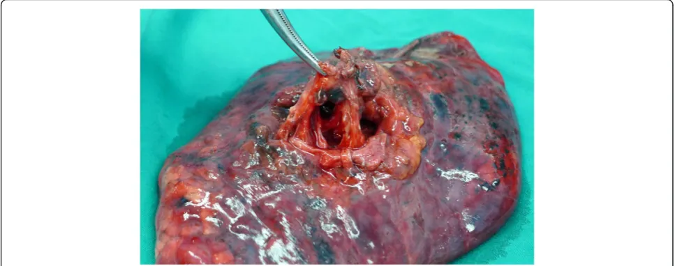
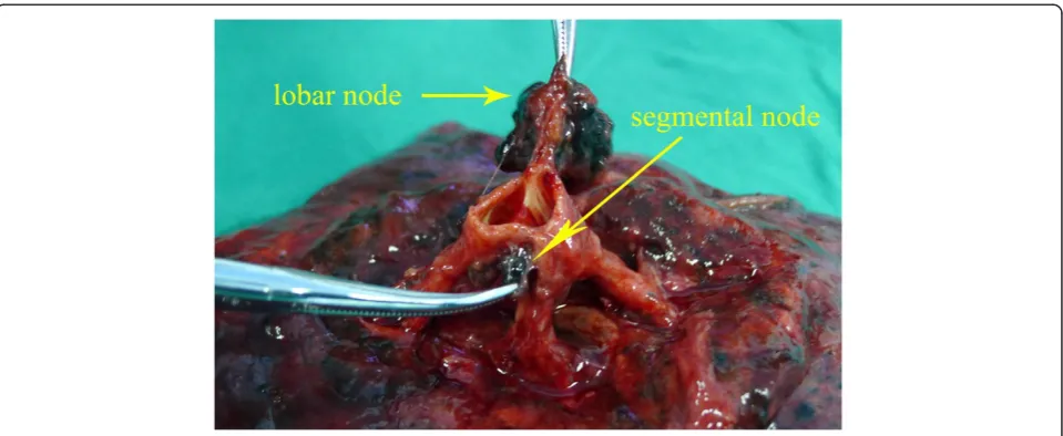
left lung cancer patients [7]. Levels 10–12 lymph nodes were collected during the surgeries. After surgery, the pulmonary lobes that had been removed were routinely dissected by the surgeon to collect levels 12–13 lymph nodes. Segmental nodes are defined as the lymph nodes adjacent to the segmental bronchi [4], whose upper border is the origin of the segmental bronchi, and whose lower border is the origin of the subsegmental bronchi. We usually cut lung tissue with scissors along the bronchial wall and the segmental bronchial wall to gather the lobar and segmental nodes (Figures 1 and 2). The collected lymph nodes were separately labeled for histological examination.
Statistical methodology
Numerical data were expressed as mean ± standard deviation or median, categorical data were expressed as frequencies or ratios. The chi-square or Fisher exact tests were used to analyze differences among the categorical data. All statistical tests reported in the manuscript were two-sided. Values ofp< 0.05 were considered to be statis-tically significant. All statistical analyses were performed using the SPSS 13.0 for Windows (SPSS Inc, Chicago, IL).
Results
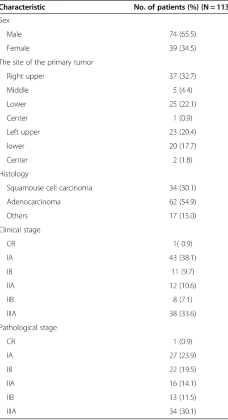
Patient characteristics were shown in Table 1. The median age was 60.0 years (range, 33–76 years). The median tumor diameter was 3.0 cm (range, 0.5-11 cm). Thora-cotomy was performed for 57 patients, while VATS was performed for 56 patients. Patients underwent lobec-tomy or bilobeclobec-tomy (n = 102), lobeclobec-tomy with a bron-chial sleeve resection (n = 4) and pneumonectomy (n = 7). Four patients underwent neoadjuvant chemotherapy, of which one case achieved complete response (CR), and three cases achieved partial response (PR). One patient
undergoing neoadjuvant chemotherapy had N1 disease with only segmental lymph node involvement. All patients underwent complete MLND, and no perioperative deaths occurred.
The mean numbers of N2 nodes, hilar nodes, interlobar nodes, lobar nodes and segmental nodes were 23.4 ± 10.5, 2.0 ± 2.3, 3.1 ± 2.4, 2.9 ± 2.7, and 2.2 ± 1.8, respectively. The detection rates for hilar nodes, interlobar nodes, lobar nodes and segmental nodes were 61.1%, 85.0%, 75.2% and 80.5%, respectively. The metastasis rates for hilar nodes, interlobar nodes, lobar nodes and segmental nodes were 5.3%, 10.6%, 16.8% and 14.2%, respectively (Table 2). There were 29 cases of N2 disease, 16 cases of N1 disease and 68 cases of N0 disease. One patient with a left upper-lobe tumor had metastatic lymph nodes at levels 5, 6 and 13. Eleven patients presented with skip metas-tases (N2 disease in the absence of N1 metastasis). Lymph node stations were grouped together into six zones by IASLC [1]: peripheral (levels 12–14) or hilar (levels 10 and 11) for N1 nodes and upper or lower me-diastinal, aortopulmonary, and subcarinal for N2 nodes. We identified 3 patients with multiple-zone N1 disease and 13 patients with single-zone N1 disease (Table 3). Forty-five patients had metastatic lymph nodes, of which 35.6% (16/45) had segmental node metastasis. Among patients with N1 disease, the cases with meta-static segmental nodes accounted for 56.3% (9/16), which is a greater percentage than other N1 stations (level 10, 6.3%; level 11, 31.3%; level 12, 31.3%). If an analysis of segmental nodes had been omitted, six pa-tients (37.5% of N1 disease) would have been down-staged to N0. Furthermore, two cases of multiple-zone N1 disease would have been misdiagnosed as single-zone N1 disease. In addition, one patient who had a left upper-lobe tumor with metastatic lymph nodes at levels
5, 6 and 13 would have been misdiagnosed with AP zone disease in the absence of N1 metastases.
In the subgroup of patients who had tumor diameters >3 cm, the detection rate for segmental nodes was higher than for patients who had tumor diameters≤3 cm (50/55 vs. 41/58, P = 0.007). Patients with history of smoking had higher detection rates of segmental nodes (53/60 vs. 38/53, P = 0.026). Detection rates for segmen-tal nodes were not significantly different, irrespective of location, histology, differentiation, and clinical staging. The metastasis rates of segmental nodes were not statis-tically different, regardless of tumor size, history of smoking, location, histology, differentiation and clinical staging.
Discussion
Accurate pathological staging of lymph node involvement plays an important role in the prediction of prognosis and the selection of treatment strategies for lung cancer. Pulmonary resection plus complete MLND has proven effective in obtaining accurate N staging [8,9]. Among the previous complete MLND randomized controlled trials [8-12], Wu Y [8] and Izbicki J R [10] did not mention the details of N1 disease in their study. Passlick B [11] and Sugi K [12] did not explicitly report whether they dis-sected segmental nodes, and no metastatic segmental nodes in N1 disease were reported. The American College of Surgeons Oncology Group Z0030 trial [13] mentioned segmental nodes. In the Z0030 study, segmental nodes were harvested from only 11.5% (60/524) of the patients who underwent MLND. However, in our study, segmental nodes were harvested from 80.5% (91/113) of the patients. Overall, the detection of segmental nodes has generally been overlooked in current clinical practice.
In our study, level 13 had a high detection rate. It has been suggested that lymph nodes are mainly distributed at the intersection of vessels [14]. Lymphatic drainage of the lungs occurs via a superficial subpleural lymph plexus and a deep plexus of lymph vessels accompanying the bronchi. Both groups drain through the hilar or bronchopulmonary nodes to the tracheobronchial nodes
and thence to the mediastinal lymph trunks [14]. Based on this, the segmental nodes could often be involved during lung cancer metastasis. In our study, level 13 has the highest metastasis rate in N1 disease, which is cor-respond with the lymphatic drainage accompanying the bronchi.
Moreover, level 13 could be the single station of meta-static lymph nodes in lung cancer patients, as has been demonstrated in some reports [15-18] with rates from 3.2% to 29.7%. Maeshima A M [19] reported patients with pN1 status who had only level 13/14 lymph node metastasis had an intermediate 5-year disease-free sur-vival rate between that of patients with pN0 status and other patients with pN1 status. They supported the con-clusion that routine examination of level 13/14 lymph nodes is important for accurate pathologic staging and for the prediction of clinical outcome of patients with NSCLC. In our study, 6 patients had single station of metastatic segmental nodes. If an analysis of segmental nodes had been omitted, these patients would have been down-staged to N0. We will further our study to invest-gate whether segmental nodes metastasis impact survival or recurrence.
N1 disease has a heterogeneous prognosis in relation to node descriptors [1,16,20-23]. The prognosis of single-zone N1 disease has proven better than multiple-zone N1 disease [1,20], and single-station N1 disease has a better prognosis than multiple-station N1 disease [20-23]. N2 disease also has a heterogeneous prognosis with regard to node descriptors. In the absence of N1 metastasis, AP zone disease was associated with better survival rates in patients with left upper-lobe tumors [1]. N2 disease with skip metastases had a more favorable prognosis than N2 disease that lacked skip metastases [24-27]. If the segmental nodes had not been examined in our study, two patients with multiple-zone N1 disease would have been misdiagnosed as single-zone N1; three cases with multiple-station N1 disease would be mis-judged as single-station N1; and one patient who had a left upper-lobe tumor with metastases at levels 5, 6 and 13
Table 3 Metastatic type of N1 disease
Lymph node station No. of patients
Multiple-zone N1 3
Levels 13 + 11 1
Levels 13 + 10 1
Levels 12 + 11 1
Single-zone N1 13
Levels 13 6
Level 12 3
Level 11 3
Level 13 +12 1
Table 2 The detection and metastasis of N1 nodes
ND DR NM MR
Level 10 69 61.1% 6 5.3%
Level 11 96 85.0% 12 10.6%
Level 12 85 75.2% 19 16.8%
Level 13 91 80.5% 16 14.2%
ND, number of cases that have lymph nodes were detected;DR, detection rate; NM, number of cases that have metastatic lymph nodes;MR, metastasis rate.
Table 1 Patient characteristics
Characteristic No. of patients (%) (N = 113)
Sex
Male 74 (65.5)
Female 39 (34.5)
The site of the primary tumor
Right upper 37 (32.7)
Middle 5 (4.4)
Lower 25 (22.1)
Center 1 (0.9)
Left upper 23 (20.4)
lower 20 (17.7)
Center 2 (1.8)
Histology
Squamouse cell carcinoma 34 (30.1)
Adenocarcinoma 62 (54.9)
Others 17 (15.0)
Clinical stage
CR 1( 0.9)
IA 43 (38.1)
IB 11 (9.7)
IIA 12 (10.6)
IIB 8 (7.1)
IIIA 38 (33.6)
Pathological stage
CR 1 (0.9)
IA 27 (23.9)
IB 22 (19.5)
IIA 16 (14.1)
IIB 13 (11.5)
would have been misdiagnosed with AP zone (levels 5, 6) disease in the absence of N1 metastases. Without examin-ing the segmental nodes, prognosis estimations could be incorrect.
The subgroup analysis revealed that the segmental nodes were easier to detect in patients who had tumor sizes > 3 cm in diameter, possibly because the growth of the tumor stimulated the immune system. The metastasis rates of segmental nodes were not significantly different, regardless of tumor size, location, histology, differentiation and clinical staging. Thus, we cannot selectively dissect segmental nodes. There are two reasons why segmental nodes are rarely dissected in current clinical practice. First, some surgeons and pathologists do not realize the import-ance of the segmental nodes for the staging and treatment of lung cancer. Second, some surgeons and pathologists think that the detection of segmental nodes is difficult and time-consuming; as a result, they are reluctant to try look-ing for the segmental nodes. Accordlook-ing to our experience, the segmental nodes are usually located at the intersection between the lobar bronchus and the segmental bronchi. After surgery, we cut lung tissue with scissors along the wall of the bronchi and segmental bronchi, which exposes the segmental nodes. It usually takes three minutes for us to perform the procedure, the results of which may benefit the patient throughout his or her life. Thus, we believe that routinely collecting the segmental nodes is necessary and feasible.
Conclusions
Segmental nodes play an important role in the accurate staging of non-small cell lung cancer, and routinely dis-secting the segmental bronchi to collect the lymph nodes is feasible and may be necessary.
Abbreviations
NSCLC:Non-small cell lung cancer; IASLC: International association for the study of lung cancer; MLND: Mediastinal lymph node dissection; VATS: Video-assisted thoracoscopic surgery; ND: Number of cases that have lymph nodes were detected; DR: Detection rate; NM: Number of cases that have metastatic lymph nodes; MR: Metastasis rate.
Competing interests
The authors declare that they have no competing interests.
Authors’contributions
LZX conducted the study and performed the statistical analysis and drafted the manuscript. YH conducted the study and collected the data. LXD conceived of and supervised the study. SKL, ZMX, XHX, LP and ZLJ participated in its design and coordination and helped to draft the manuscript. All authors read and approved the final manuscript.
Acknowledgement
We thank the pathologists of Sun Yat-sen University Cancer Center for providing accurate pathology reports.
Author details 1
State Key Laboratory of Oncology in South China, Sun Yat-sen University Cancer Center, 651 Dongfeng Rd, East, Guangzhou, PR China.2Cancer
Hospital of Henan province, Zhengzhou, PR China.3Central Hospital,
Shaoyang, PR China.
Received: 29 May 2013 Accepted: 26 November 2013 Published: 8 December 2013
References
1. Rusch VW, Crowley J, Giroux DJ,et al:The IASLC lung cancer staging project: proposals for the revision of the N descriptors in the forthcoming seventh edition of the TNM classification for lung cancer.
J Thorac Oncol2007,2:603–612.
2. Arriagada R, Bergman B, Le Dunant A, Chevalier T, Pignon JP, Vansteenkiste J:Cisplatin-based adjuvant chemotherapy in patients with completely resected non-small-cell lung cancer.N Engl J Med2004,350:351–360. 3. Winton T, Livingston R, Johnson D,et al:Vinorelbine plus cisplatin vs. observation in resected non-small-cell lung cancer.N Engl J Med2005,
352:2589–2597.
4. Rusch VW, Asamura H, Watanabe H, Giroux DJ, Rami-Porta R, Goldstraw P:
The IASLC lung cancer staging project: a proposal for a new international lymph node map in the forthcoming seventh edition of the TNM classification for lung cancer.J Thorac Oncol2009,4:568–577. 5. Detterbeck FC, Boffa DJ, Tanoue LT:The new lung cancer staging system.
Chest2009,136:260–271.
6. Watanabe S, Asamura H:Lymph node dissection for lung cancer: significance, strategy, and technique.J Thorac Oncol2009,4:652–657. 7. Yang H, Li XD, Lai RC,et al:Complete mediastinal lymph node dissection
in video-assisted thoracoscopic lobectomy versus lobectomy by thoracotomy.Thorac Cardiovasc Surg2013,61:116–123. 8. Wu Y, Huang ZF, Wang SY, Yang XN, Ou W:A randomized trial of
systematic nodal dissection in resectable non-small cell lung cancer.
Lung Cancer2002,36:1–6.
9. Darling GE, Allen MS, Decker PA,et al:Randomized trial of mediastinal lymph node sampling versus complete lymphadenectomy during pulmonary resection in the patient with N0 or N1 (less than hilar) non-small cell carcinoma: results of the American college of surgery oncology group Z0030 trial.J Thorac Cardiovasc Surg2011,141:662–670. 10. Izbicki JR, Passlick B, Pantel K,et al:Effectiveness of radical systematic
mediastinal lymphadenectomy in patients with resectable non-small cell lung cancer: results of a prospective randomized trial.Ann Surg1998,
227:138–144.
11. Passlick B, Kubuschock B, Sienel W, Thetter O, Pantel K, Izbicki JR:
Mediastinal lymphadenectomy in non-small cell lung cancer:
effectiveness in patients with or without nodal micrometastases - results of a preliminary study.Eur J Cardiothorac Surg2002,21:520–526. 12. Sugi K, Nawata K, Fujita N,et al:Systematic lymph node dissection for
clinically diagnosed peripheral non-small-cell lung cancer less than 2 cm in diameter.World J Surg1998,22:290–294. discussion 294–5.
13. Darling GE, Allen MS, Decker PA,et al:Number of lymph nodes harvested from a mediastinal lymphadenectomy: results of the randomized, prospective American college of surgeons oncology group Z0030 trial.
Chest2011,139:1124–1129.
14. Liu Z, Bi Y, Tian H,et al:Basic and Clinical of Lymph.1st edition. Beijing: Science Press; 2003.
15. Yamanaka A, Hirai T, Fujimoto T, Ohtake Y, Konishi F:Analyses of segmental lymph node metastases and intrapulmonary metastases of small lung cancer.Ann Thorac Surg2000,70:1624–1628.
16. Matsuoka K, Sumitomo S, Misaki N:Prognostic factors in patients with pathologic T1-2N1M0 disease in non-small cell carcinoma of the lung.
J Thorac Oncol2007,2:1098–1102.
17. Wu N, Yan S, Zheng QF,et al:Role of pulmonary segmental and sub-segmental lymph nodes in pathological staging of lung cancer.
Zhonghua Yi Xue Za Zhi2010,90:1873–1876.
18. Osaki T, Nagashima A, Yoshimatsu T, Tashima Y, Yasumoto K:Survival and characteristics of lymph node involvement in patients with N1 non-small cell lung cancer.Lung Cancer2004,43:151–157.
19. Maeshima AM, Tsuta K, Asamura H,et al:Prognostic implication of metastasis limited to segmental (level 13) and/or subsegmental (level 14) lymph nodes in patients with surgically resected nonsmall cell lung carcinoma and pathologic N1 lymph node status[J].Cancer2012,
20. Casali C, Stefani A, Morandi U:N1 non-small-cell lung cancer. A 20-year surgical experience.Asian Cardiovasc Thorac Ann2011,19:217–224. 21. Sayar A, Turna A, Kilicgun A, Solak O, Urer N, Gurses A:Prognostic significance
of surgical-pathologic multiple-station N1 disease in non-small cell carcinoma of the lung.Eur J Cardiothorac Surg2004,25:434–438. 22. Cerfolio RJ, Bryant AS:Predictors of survival and disease-free survival in
patients with resected N1 non-small cell lung cancer.Ann Thorac Surg 2007,84:182–188. discussion 189–90.
23. Ueda K, Kaneda Y, Sakano H,et al:Independent predictive value of the overall number of metastatic N1 and N2 stations in lung cancer.Jpn J Thorac Cardiovasc Surg2003,51:297–301.
24. Yoshino I, Yokoyama H, Yano T,et al:Skip metastasis to the mediastinal lymph nodes in non-small cell lung cancer.Ann Thorac Surg1996,
62:1021–1025.
25. Wang S, Wu Y, Ou W:Skip metastasis to the mediastinal lymph nodes in non-small cell lung cancer.Zhonghua Zhong Liu Za Zhi2001,23:259–261. 26. Prenzel KL, Monig SP, Sinning JM,et al:Role of skip metastasis to
mediastinal lymph nodes in non-small cell lung cancer.J Surg Oncol 2003,82:256–260.
27. Misthos P, Sepsas E, Athanassiadi K, Kakaris S, Skottis I:Skip metastases: analysis of their clinical significance and prognosis in the IIIA stage of non-small cell lung cancer.Eur J Cardiothorac Surg2004,25:502–508.
doi:10.1186/1749-8090-8-225
Cite this article as:Liet al.:The role of segmental nodes in the pathological staging of non-small cell lung cancer.Journal of Cardiothoracic Surgery20138:225.
Submit your next manuscript to BioMed Central and take full advantage of:
• Convenient online submission
• Thorough peer review
• No space constraints or color figure charges
• Immediate publication on acceptance
• Inclusion in PubMed, CAS, Scopus and Google Scholar
• Research which is freely available for redistribution