In vitro Interaction and Colocalization of HSV-1 ORF P with a
Cellular Splicing Factor (SC35) Using Pulldown Assay
Ali Karimi
*1and Rasoul Salehi
21
Dept. of Microbiology and Immunology, Faculty of Medicine, Kord University of Medical Sciences, Shahre-Kord; 2Dept. of Genetics and Molecular Biology, School of Medicine, Isfahan University of Medical Sciences,
Isfahan, Iran
Received 31 July 2004; revised 17 October 2004; accepted 18 October 2004
ABSTRACT
Herpes simplex virus type-1 (HSV-1) causes a variety of diseases in human. This virus is a neurotropic pathogen of human that establishes latent infection in the sensory ganglia innervating the site of primary infection. A number of genes including ICP34.5 control HSV-1 pathogenicity and ICP34.5 has been identified as HSV-1 virulence gene. Open reading frame P (ORF P) is also a HSV-1 gene that might have a role in latency. A complication in the analysis of the role of ICP34.5 and ORF P in the HSV-1 life cycle is that these two are overlapping antisense genes. ORF P is also deleted in ICP34.5 negative mutants and to date, no definite function is attributed to it. To attribute characteristics which were originally attributed solely to ICP34.5 to each of these two genes (ORF P or ICP34.5), an approach is to construct a number of HSV-1 recombinant viruses that express ICP34.5 and ORF P independently. An alternative way is to determine if ORF P interacts with any of the cellular and viral proteins both in vitro and in vivo. Using Glutathione-S-transferase (GST) pulldown assay and Western-blotting, we showed that ORF P interacts with a cellular splicing factor (SC35) in vitro. To investigate the colocalization of ORF P and SC35, nuclear and cytoplasmic fractionation of ORF P/SC35 was also carried out. Our results showed that both SC35 and ORF P are located in the nucleus of HSV-1 infected cells. Conclusively, because ORF P interacts and colocalizes with SC35, it might have a role in splicing. Iran. Biomed. J. 9 (2): 67-71, 2005
Keywords: Open reading frame (ORF P), Infected cell protein (ICP) 34.5, Latency associated transcript (LAT), Splicing component (SC) 35
INTRODUCTION
erpes simplex virus type-1 (HSV-1) is a neurotropic pathogen of humans that establishes latent infection in the sensory ganglia innervating the site of primary infection [1]. There are a number of genes which are involved in virus pathogenicity and latency [1]. A major focus of investigations into genes controlling the establishment or maintenance of latency has been the latency associated transcripts (LAT). These transcripts arise from the HSV inverted repeats flanking the ULsequence and therefore present in 2 copies per viral genome [1, 2]. The HSV-1 unspliced 8.3 kb LAT has been shown to contain at least 16 open reading frames (ORF) [3].
One of these ORF is ORF P, which is shown to encode a protein [3-5]. ORF P expressed by HSV-1
strain (F) and (17+) is predicted to contain 248 and 233 amino acids, respectively [3]. It is located in the 3' domain of LAT, almost entirely antisense to the ICP34.5 gene and is also contained at 5' end of the long/short transcripts (L/ST) [3, 5]. The role of ORF P in HSV-1 life cycle is not known and no definite function has been attributed to it except the possible role in splicing at early times in infection and in latency [5-8]. The most striking feature of ORF P is nearly completely overlapped by ICP34.5 gene [3].
ICP34.5 is a HSV-1 neurovirulence gene that is essential in vivo and located in the long repeat
region of the HSV-1 genome [9].
To determine the role of ICP34.5 in the HSV-1 (17+) life cycle, a number of ICP34.5 deletion mutants have been characterized. These null mutants are unable to replicate in the central nervous system of mice and are totally avirulent
H
with a LD50of 106pfu/mouse higher than wild type or rescued virus [10]. In tissue culture, these null mutants replicate normally in many non-neuronal cell types such as Vero and BHK 21/C13 cells, but are restricted in SK-N-SH neuroblastoma, human foreskin fibroblast (HFF), murine 10T1/2 cells, stationary phase primary mouse embryo and mouse embryo fibroblast (3T6) cells [9, 11, 12].
As most ICP34.5 null mutants affect ORF P due to the extensive overlap of their sequences, it was difficult to assign a role to ORF P. To assign the phenotypes of these mutants to either ICP34.5 or ORF P, an approach is to construct a number of recombinant viruses expressing only ICP34.5 or ORF P at separated loci and the phenotypes of these recombinants to be analysed. An alternative way to analyze the function of a viral gene such as ORF P is to determine if this gene interacts with any of the cellular and viral proteins both in vitroand in vivo.
Therefore, using GST pulldown assay and Western-blotting, it was investigated that with which cellular and viral gene products ORF P interacts. The only identified cellular protein interacting in vitro with
ORF P detected in these experiments was SC35. To investigate the colocalization of ORF P and SC35, nuclear and cytoplasmic fractionation of ORF P/SC35 was also carried out.
MATERIALS AND METHODS
Cells. Baby hamster kidney 21 clone 13 (BHK)
cells [13] were grown in Eagle’s medium supplemented with 10% (v/v) newborn calf serum.
Viruses. The wild type virus used was HSV-1
17+ [14]. The HSV-1 17+ICP34.5/ORF P deletion variant 1716 [15] was used as a negative control.
TsK, a HSV-1 17+ mutant with a ts lesion in ICP4
[16], which results in overproduction of ORF P at the non-permissive temperature, was also used.
Nuclear and cytoplasmic fractionation of ORF P/SC35. Virus infected cells were washed twice
with PBS A (170 mM NaCl, 3.4 mM KCl, 10 mM Na2HPO4, 1.8 mM KH2PO4, pH 7.2) and harvested by scraping the monolayer into PBS A containing Eppendorf tube. To extract the cytoplasmic fraction, cell suspension was briefly vortexed and spun and cell pellet was resuspended in 100 # 1 buffer A (50 mM Tris HCl, 5 mM MgCl2, 10% (v/v) NP40, pH 7.5) containing 0.5% (v/v) NP40, incubated on ice for 15 min and then homogenized on ice in a Dounce homogenizer (Sigma, USA). The samples
ice while the pellet was resuspended with 100 1 buffer A containing 0.5% (v/v) NP40, incubated on ice for 2-3 min, spun and then supernatant mixed with the previous supernatant and used as the cytoplasmic fraction [17].
To extract the nuclear fraction, the infected monolayer was scraped into Falcon tube containing 10 ml PBS A, briefly vortexed and spun. The cell pellet was resuspended in 4 ml hypotonic lysis buffer (HLB) (10 mM Tris HCl, 10 mM KCl, 2.5 mM MgCl2, pH 7.5) and incubated on ice for 15 min. After incubation, the samples were centrifuged and the supernatant was left on ice while the pellet was resuspended with 1 ml supernatant and homogenized on ice in a Dounce homogenizer (Sigma, USA). The homogenized solution was mixed with the rest of the supernatant in a 15 ml Falcon tube, underlayed with 1 ml HLB + 10% (w/v) sucrose and respun at 3600 g for 5 min at 4C. The supernatant was discarded and the pellet containing the nuclei resuspended in 100 µ1 boiling mix [17]. Samples were boiled for 10 min and analyzed by SDS-PAGE and Western-blotted.
Western-blotting. Samples were separated by
SDS-PAGE. The proteins in the gel were transferred into the membrane. After the transfer, the membrane was blocked in PBS A containing Tween 20 (PBS A/T) and in dried milk at room temperature for 1 h. The primary antibody was incubated either at 37C or room temperature for 2 h and at 4C overnight. The membrane was washed 3 times in PBS A/T at room temperature for 10 min and then incubated at room temperature for 1 h in the appropriate HRP conjugated secondary antibody followed by 3 washes in PBS A/T for 10 min. A chemiluminescence detection reagent (ECL, Sigma, USA) was added to the membrane for 1 min and the membrane was exposed to XS-1 film for the appropriate amount of time [18].
Immunoprecipitation. Virus infected monolayers
were harvested by washing with PBS A twice, adding 500l Zweig's buffer (0.1 M Tris HCl, 10% (w/v) glycerol, 0.5% (w/v) NP40, 0.5% (w/v) deoxycholate, pH 8.5) to each plate and incubating at 4C for 60 min. The extracted cells were sonicated for 3 min and spun at 13225g for 3 min. The supernatant was used immediately or stored at -70C. The supernatant of the cell extract (200-500
1) was mixed with the appropriate volume of antibody and incubated either at 4oC overnight or at 37C for 2 h. Protein-A-sepharose (75 µ1 50%
was incubated on an end-over-end mixer at 4C for 45 min. After that, the sample was centrifuged at 3,800 g for 1 min, the supernatant was discarded and the pellet was washed 3-4 times with Zweig's buffer. After the final wash, the pellet was harvested in 50 µ1 boiling mixture and analyzed by SDS-PAGE [19].
Pulldown assay. Freshly prepared glutathione
agarose beads, bound to GST fusion proteins, were mixed with 300 or 400 1 labelled or unlabelled cell protein extract and incubated at 4C for at least 3 h with continuous end-over-end mixing. The beads were harvested by centrifugation at 13,225g for 1 min and washed 3 times in 1 ml of an extraction buffer (50 mM NaCl, 0.1% (v/v) NP40, pH 7.5) containing different amounts of NaCl (0.5-500 # mM). Again, they were harvested in boiling mixture, stored either at -20C or boiled for 5 min and analyzed by SDS-PAGE [20]. Gels were either fixed, dried and autoradiographed or used for Western-blotting.
RESULTS
In vitro interaction of HSV-1 ORF P with a cellular splicing factor (SC35). GST pulldown is a
method usually used to determine protein-protein interactions in vitro. This method was used in this
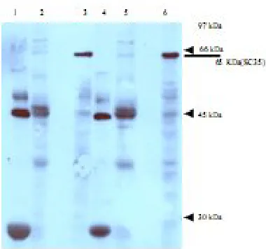
work to determine the interaction of ORF P with cellular and viral proteins. Using a GST pulldown assay, we previously showed that ORF P interacts with a number of cellular proteins (personal data). Therefore, the GST pulldowns were screened with a range of antibodies against proteins with some role in splicing and posttranscriptional processing and were of an appropriate molecular weight which could identified in the gel (pulldown experiments). Western-blotting of cellular extracts was carried out with these antibodies (Ab against above mentioned proteins). The only identified cellular protein interacting with ORF P detected in these experiments was SC35. Figure 1 shows Western-blotting of GST and GST-ORF P pulldown extracts with an antibody against SC35. As expected, the specific 65 kDa, SC35 band was detected in both mock infected (lane 6) and 17+(lane 3) cell extracts. The 65 kDa SC35 band was detected specifically in the GST-ORF P pulldowns (lanes 2 and 5) but not in GST pulldowns (lanes 1 and 4).
Fig. 1. Western-blotting of GST-ORF P pulldown with an antibody against SC35. GST pulldown extracts were run on a 12.5% SDS-PAGE and analyzed by Western-blotting using an antibody against SC35. Lanes 1–3: 17+ infected cell extracts; lane 1, 17+/GST pulldown; lane 2, 17+/GST-ORF P pulldown; lane 3, whole 17+ infected cell extracts; lanes 4-6, MI extract; lane 4, MI/GST pulldown; lane 5, MI/GST-ORF P pulldown; lane 6, whole MI cell extracts. SC35 band is indicated (). Molecular weights are marked ().
Colocalization: nuclear and cytoplasmic fractionation of SC35/ORF P. Our results have
demonstrated that ORF P interacts in vitro with
SC35. If this was representative of an in vivo
interaction, it would be expected that both proteins would be located in the same cellular compartment. To investigate the intracellular location of ORF P and SC35, BHK cells were infected with tsK, 17+
and 1716 or mock infected and their nuclear and cytoplasmic proteins were separately extracted and Western blotted with both antibody against SC35 and antiserum 128 against ORF P. Figure 2 shows Western-blotting of nuclear and cytoplasmic extracts with an antibody against SC35. SC35 is located in the nucleus of tsK (lane 1), 17+(lane 3),
1716 (lane 5) and mock infected (lane 7) and BHK cells with no protein were detected in the cytoplasm (lanes 2, 4, 6 and 8).
Figure 3 shows the Western-blotting of nuclear and cytoplasmic extracts with antiserum 128. ORF P located in approximately equal amounts in both the nucleus and cytoplasm of BHK cells were infected with tsK (lanes 1 and 2) and 17+ (lanes 3
and 4). As expected, no ORF P was detected in 1716 (lanes 5 and 6) or mock infected (lanes 7 and 8) extracts. In lane 9, a whole cell extract from ts
K-infected BHK cells was used as a positive control to show the 30 kDa ORF P protein.
Fig. 2. Western blotting of BHK nuclear and cytoplasmic extracts with SC35 antibody. BHK cells were infected with HSV-1 at a multiplicity of infection (m.o.i) of 20 pfu/cell and harvested at 24 h post infection (pi). Analysis of SC35 distribution in both BHK cell nuclear and cytoplasmic extracts was carried out by 7.5% SDS-PAGE and Western-blotting with SC35 antibody and anti IgG-HRP, reacted with ECL and exposed to autoradiography. Lanes 1, 3, 5 and 7, nuclear extracts; lanes 2, 4, 6 and 8, cytoplasmic extracts. Lanes 1 and 2, tsK; lanes 3 and 4, 17+; lanes 5 and 6, 1716; lanes 7 and 8,
MI. Molecular weight markers are indicated on the right and SC35 related bands are marked on the left ().
DISCUSSION
The only characterized cellular protein interacting with ORF P detected in these experiments was SC35. SC35 is an essential component of small non-nuclear ribonucleoprotein particles and is a splicing factor [21-23]. Previously was shown that ORF P interacts with a number of splicing factors and may play a role in splicing [21]. Having demonstrated that ORF P interacted in vitro with
SC35, we wished to determine if this was representative of an in vivo interaction. To occur
this, it would be expected that both proteins would be located in the same cellular compartment. It has previously been demonstrated that SC35 localizes mainly to the nucleus [17] and thus we wished to confirm this and determine if ORF P was also located in the nucleus. To investigate the intracellular location of ORF P and SC35, BHK cells were infected with tsK and 17+, their nuclear
and cytoplasmic proteins were extracted separately and Western-blotted with antisera against SC35 and ORF P.
As previously published, SC35 is located in the cell nucleus with no protein being detected in the cytoplasm [17]. ORF P is located in approximately equal amounts in both the nucleus and cytoplasm of
of Lagunoff et al. [24] who detected ORF P in the
nucleus of cells infected with a mutant which overproduced it using immunofluorescence and Western-blotting of fractionated cell extracts.
Antisera 128 did not work in immunofluorescence due to their high background and thus there was no attempt to investigate colocalization of ORF P and SC35 by immunofluorescence.
Overall, these results show that ORF P interacts with SC35in vitroand also colocalizes with ORF P
in the nucleus of infected cells. However, due to technical difficulties, we were unable to determine if an in vivointeraction was occurring. This data is
in agreement with the previously published work in which using immunofluorescence it was demonstrated that ORF P colocalizes with SM antigens and SC35 in the nuclei of infected cells, interacts with SM components in a GST pulldown assay and with p32 in a yeast-two-hybrid system [21]. Further works are needed to determine if ORF P interacts with SC35 or the other cellular proteins
in vivo.
Fig. 3.Western-blotting of BHK nuclear and cytoplasmic extracts with anti-ORF P serum 128. BHK cells were infected with HSV-1 at a m.o.i. of 20 pfu/cell and harvested at 24 h pi. Analysis of ORF P distribution in both BHK cell nuclear and cytoplasmic extracts was carried out by 12.5% SDS-PAGE and Western-blotting with antiserum 128 and protein-A-HRP, reacted with ECL and exposed to autoradiography. Lanes 1, 3, 5 and 7, nuclear extracts; lanes 2, 4, 6 and 8, cytoplasmic extracts. Lanes 1 and 2, tsK; lanes 3 and 4, 17+; lanes 5 and 6, 1716; lanes 7 and 8, MI; lane 9, tsK infected whole cell extract. Molecular weight markers are indicated on the right and ORF P related bands are marked on the left ().
REFERENCES
1. Perng, G.C., Chokephaibulkit, R.L., Thompson, N.M., Sawtell, S.M., Slanina, H., Ghiasi, A.B. and Wechsler, S.L. (1996) The region of the herpes simplex virus type 1 LAT gene that is colinear with the ICP34.5 gene is not involved in spontaneous
2. Stevens, J.G., Wagner, E.K., Devi-Rao, G.B., Cook, M.L. and Fedelman, L. (1987) RNA complementary to a herpesvirus alpha gene mRNA is predominant in latently infected neurons. Science 235: 1056-1059.
3. Lagunoff, M. and Roizman, B. (1994) Expression of a herpes simplex virus 1 open reading frame antisense to the (1)34.5 gene and transcribed by an RNA 3' coterminal with the unspliced latency-associated transcript. J. Virol. 68: 6021-6028.
4. Lagunoff, M. and Roizman, B. (1995) The regulation of synthesis and properties of the protein product of open reading frame P of the herpes simplex virus 1 genome. J. Virol. 69: 3615-3623.
5. Randall, G. and Roizman, B. (1997) Transcription of the derepressed open reading frame P of herpes simplex virus 1 precludes the expression of the antisense gamma(1)34.5 gene and may account for the attenuation of the mutant virus. J. Virol. 71: 7750-7757.
6. Randall, G., Lagunoff, M. and Roizman, B. (2000) Herpes simplex virus 1 open reading frames O and P are not necessary for establishment of latent infection in mice. J. Virol. 74: 9019-9027.
7. Chen, S.H. and Lee, L.Y. (2002) Neither LAT nor open reading frame P mutations increase expression of spliced or intron-containing ICP0 transcripts in mouse ganglia latently infected with herpes simplex virus. J. Virol. 76: 4764-4772.
8. Lee, L.Y. and Schaffer, P.A. (1998) A virus with a mutation in the ICP4-binding site in the L/ST promoter of herpes simplex virus type 1, but not a virus with a mutation in open reading frame P, exhibits cell-type-specific expression of gamma (1)34.5 transcripts and latency-associated transcripts.
J. Virol. 72: 4250-4264.
9. Chou, J., Kern, E.R., Whitley, R.J. and Roizman, B. (1990) Mapping of herpes simplex virus-1 neurovirulence to (1)34.5, a gene nonessential for
growth in culture. Science 250: 1262-1266.
10. McKie, E.A., Hope, R.G., Brown, S.M. and MacLean, A.R. (1994) Characterization of the herpes simplex virus type 1 strain 17+ neurovirulence gene RL1 and its expression in a bacterial system. J. G. Virol. 75: 733-741.
11. Chou, J., Poon, A.P., Johnson, J. and Roizman, B. (1994) Differential response of human cells to deletions and stop codons in the (1)34.5 gene of
herpes simplex virus. J. Virol. 68: 8304-8311.
12. Ward, S.L., Scheunder, D., Poppers, J., Kaufman, R.J. and Leib, D.A. (2003) In vivoreplication of an ICP34.5 second-site suppressor mutant following corneal infection correlates with in vitro regulation
of eIF2 alpha phosphorylation. J. Virol. 77: 4626-4634.
13. Macpherson, I. and Stoker, M. (1962) Polyoma transformation of hamster cell clones-an investigation of genetic factors affecting cell competence. Virology 16:147-151.
14. Brown, S.M., Ritchie, D.A. and Subak-Sharpe, J.H. (1973) Genetic studies with herpes simplex virus
type 1. The isolation of temperature sensitive mutants, their arrangement into compementation groups and recombination analysis leading to a linkage map. J. G. Virol. 18: 329-346.
15. MacLean, A.R., Ul-Fareed, M., Robertson, L., Harland, J. and Brown, S.M. (1991) Herpes simplex virus type 1 deletion variants 1714 and 1716 pinpoint neurovirulence-related sequences in Glasgow strain 17+ between immediate early gene 1 and the 'a' sequence. J. G. Virol. 72: 631-639.
16. Preston, C.M. (1979) Abnormal properties of an immediate early polypeptide in cells infected with the HSV type 1 mutant tsK. J. Virol. 32: 357-369. 17. Boe, S.O., Bjorndal, B., Rosok, B., Szilvay, A.M.
and Kalland, K.H. (1998) Subcellular localization of human immunodeficiency virus type 1 RNAs, Rev, and the splicing factor SC-35. Virology 244: 473-482.
18. McKay, E.M., McVey, B., Marsden, H.S., Brown, S.M. and MacLean, A.R. (1993) The herpes simplex virus type 1 strain 17 open reading frame RL1 encodes a polypeptide of apparent M(r) 37K equivalent to ICP34.5 of herpes simplex virus type 1 strain F. J. G. Virol. 74: 2493-2497.
19. Hutchinson, L., Browne, H., Wargent, V., Davis-Poynter, N., Primorac, S., Goldsmith, K., Minson, A.C. and Johnson, D.C. (1992) A novel herpes simplex virus glycoprotein, gL, forms a complex with glycoprotein H (gH) and affects normal folding and surface expression of gH. J. Virol. 66: 2240-2250.
20. Bryant, H., Wadd, S., Filhol, O., Scott, J.E., Hsieh, T.Y., Everett, R.D. and Clements, J.B. (1999) The multifunctional herpes simplex virus IE63 protein interacts with heterogeneous ribonucleoprotein K and with casein kinase 2. J. Biol. Chem. 274: 28991-28998.
21. Bruni, R. and Roizman, B. (1996) Open reading frame P of herpes simplex virus gene repressed during productive infection encodes a protein that binds a splicing factor and reduces synthesis of viral proteins made from spliced mRNA. Proc. Natl. Acad. Sci. USA 93: 10423-10427.
22. Sandri-Goldin, R.M., Hibbard, M.K. and Hardwicke, M.A. (1995) The C-terminal repressor region of herpes simplex virus type 1 ICP27 is required for the redistribution of small nuclear ribonucleoprotein particles and splicing factor SC35; however, these alterations are not sufficient to inhibit host cell splicing. J. Virol. 69: 6063-6076.
23. Zahler, A.M., Lane, W.S., Stolk, J.A. and Roth, M.B. (1992) SR proteins: a conserved family of pre-mRNA splicing factors. Gen and Develop. 6: 837-847.
24. Lagunoff, M., Randall, G. and Roizman, B. (1995) Phenotypic properties of herpes simplex virus 1 containing a derepressed open reading frame P gene.
J. Virol. 70: 1810-1817.