Cytotoxicity Effect of Cladribine on the MCF-7 Human Breast
Cancer Cell Line
Mohammad Hashemi, Fatemeh Karami-Tehrani
*and Saeed Ghavami
Cancer Research Lab, Dept. of Clinical Biochemistry, School of Medical Sciences, Tarbiat Modarres University, Tehran, Iran
Received 5 March 2003; revised 19 July 2003; accepted 6 August 2003
ABSTRACT
Cladribine, an analogue of deoxyadenosine, is highly toxic for both non-dividing and proliferating cells and has shown activity in the treatment of several malignancies. Therefore, the aim of the present study is to investigate the cytotoxicity effect of cladribine (2-CdA) on the breast cancer cell line,
MCF-7 (estrogen receptor positive, ER+). MTT assay, annexin V-Fluorescein/PI and Hoechst 33258 staining
were used to detect cytotoxicity and cell apoptosis. The activation of caspase-3 and -9 was assayed using caspase activation assay kits. Gel electrophoresis was performed to detect DNA fragmentation. Treatment of MCF-7 cells with different concentrations of 2-CdA resulted in a significant increase in the cell death. Annexin V-Fluorescein/PI and Hoechst 33258 staining revealed that the cell death was
mainly an apoptotic type. A significant (p<0.05) increase in the activity of caspase-9 was observed but
Caspase-3 activity was unchanged and DNA laddering profile was not obtained. Pre-treatment of the
cells with kinase inhibitor, 5-amino-5-deoxyadenosine inhibited the cytotoxicity effect of cladribine.
In conclusion, this study has shown that high dose of cladribine (higher than 25μM) has an apoptotic
effect on MCF-7 cells and that its intracellular phosphorylation is necessary. Iran. Biomed. J. 8 (1): 7-12,
2004
Keywords: Cladribine, Apoptosis, MCF-7, Caspase-3, Caspase-9
INTRODUCTION
rogrammed cell death is a fundamentally important process required maintaining the integrity and homeostasis of multicellular organisms. Interest in programmed cell death, or apoptosis was focussed originally on its role in tissue homeostasis [1]. More recently however, attention has centered on its importance in tumor development and treatment. It has been suggested that apoptosis plays an important role in the deletion of pre-neoplastic cells, thus preventing the progress of disease [2], and that inability to delete such cells via apoptosis may play an important role in fixation of genetic mutations and the development of tumors. The importance of apoptosis in the treatment of already established tumors was based on the observation that radiation and drugs used in the cancer chemotherapy induce cell death via apoptosis [3]. All aspects of apoptosis are now under intense investigation, from the effects of
drugs in both cultured and primary cancer cells of all types, to the study of specific genetic pathways controlling the process.
One such drug is 2-chloro-2-deoxyadenosine (cladribine), an analogue of deoxyadenosine, which is resistant to degradation by adenosine deaminase [4-7]. In general, adenosine analogues can trigger apoptosis or necrosis in various cell types and, in several instances, can inhibit cell growth and interfere with the cell cycle [8]. Cladribine has also shown activity in the treatment of several hematological malignancies, including hairy cell leukemia and chronic lymphocytic leukemia (CLL) [9]. Activation of extracellular adenosine receptors (A1, A2A, A2B and A3), as well as direct intracellular action has been claimed to play a role, depending upon the cell type and specific pathophysiological conditions [10]. Moreover, the intracellular phosphorylation of adenosine analogues to their corresponding ATP derivatives has been suggested to mediate the cytotoxic effects
P
of these compounds in lymphoid tumors [8] although additional intracellular pathways (e.g., interference with the cell cycle) may also play a role. Intracellularly, 2-chloro-2'-deoxyadenosine (2-CdA) is phosphorylated by deoxycytidine kinase to 2-CdAMP, and thereafter successively converted to 2-CdADP and 2-CdATP. The cytotoxicity of cladribine results from the inhibitory effect of 2-CdATP on various enzymes involved in DNA replication and repair, including ribonucleotide reductase and DNA polymerase [11] and thereby inducing DNA damage and cell death [12]. In view of the potential use of cladribine in the treatment of some cancers, the present study was undertaken to examine cladribine for its effect on the induction of apoptosis in estrogen receptor positive breast cancer cell line (MCF-7) which may prove its efficacy in the treatment of breast cancers as well.
MATERIALS AND METHODS
Cladribine, chemicals, culture media and related compounds were purchased from Sigma Co. (USA). Cell culture plastic ware was obtained from Nunc Co. (Denmark). Caspase-3 colorimetric assay kit (Cat. No. 101K4019) and DNA ladder marker, 1 Kb (61K1778) were obtained from Sigma (Germany). Caspase -9 colorimetric assay kit (Cat. No. BF10100) and annexin V-FITC apoptosis detection kit (Cat. No. TA4638) were purchased from R & D systems Co. (USA).
Cell culture. MCF-7 (NCBI C135) breast cancer
cell line was obtained from National Cell Bank of Iran (the Pasteur Institute of Iran, Tehran) and cultured in RPMI 1640 supplemented with 10% FCS, 100 U/ml penicillin and 100 μg/ml streptomycin. They were incubated in a humidified incubator with 5% CO2and 95% air at 37°C.
MTT assay. To evaluate the cytotoxicity effect of
cladribine on the MCF-7 breast cancer cell line, MTT colorimetric assay was applied [13]. Briefly, asynchronously growing cells (3 104 cells/ml) were transferred into 48-well culture plates containing 500 µl of medium and incubated for 24 h. The culture medium was replaced by fresh medium containing 25, 50, 100 and 500 μM cladribine, and incubated for 24 and 48 h. Then, MTT assay was performed and percent of the cell viability was calculated using the equation: (mean OD of treated cells/mean OD of control cells)
100.
Pretreatment with receptor antagonist and kinase inhibitor. To examine the role of
extracellular adenosine receptors in the cladribine-induced cell death, cells were pretreated with 10 µM of 8-phenyltheophylline, a receptor antagonist, for 30 min prior to the treatment with cladribine. In order to provide evidence for the intracellular phosphorylation of cladribine, a 30-min pre-exposure to the kinase inhibitor, 5’-amino-5’-deoxyadenosine (20 µM) was performed.
Analysis of nuclear morphology. Cells were
plated in 8-well chamber slides and allowed to adhere. Cladribine treated cells were fixed with methanol-acetic acid 3:1 (v/v) for 10 min after that staining was carried out with Hoechst 33258 (10 µg/ml) in dark (10 min) at 37°C. Slides were then washed with PBS (pH 7.4) and examined by a fluorescence microscope (Micros, Austria). Apoptotic cells were defined on the basis of nuclear morphology changes such as chromatin condensation and fragmentation. Annexin V/PI staining was done according to kit manual. Early apoptotic cells show green fluorescence.
Caspase-3 and -9 activation assay. Caspase-3
and -9 colorimetric assay kits were used to investigate caspase-3 and -9 activation in the treated MCF-7. Briefly, to estimate caspase-3 activity, cells were lysed by incubation with cell lysis buffer on ice for 15 minutes and then centrifuged at 20,000g at 4°C for 10 minutes. For caspase-9 activation assay, cells were lysed by incubation with cell lysis buffer on ice for 10 minutes and then centrifuged at 10,000g at 4°C for 1minute. Enzymatic reactions were carried out in a 96-well flat-bottom microplate. To each sample, 5 and 50 μl of cell lysate (100-200 μg total protein) were added for caspase-3 and -9, respectively. Additional controls, one frees from cell lysate and the other lacking substrate as well as caspase-3 positive control has been included. Protein was estimated by Bradford method [14]. The activities were expressed as nmole/min/mg protein.
DNA laddering. Cells, treated with different
concentrations of cladribine for different times, were trypsinized, washed twice with ice-cold PBS and centrifuged. The pellets were lysed using lysis buffer containing 50 mM Tris-HCl (pH 8.0), 20 mM EDTA, 10 mM NaCl, 1% (w/v) sodium dodecyl sulfate (SDS). The lysate was incubated sequentially with 20 µg/ml RNase (at 37°C for 60 min) and 100 µg/ml proteinase K (at 37°C for 3-5
h). DNA was extracted with an equal volume of phenol/chloroform/isoamyl alcohol (25:24:1, v/v/v) and precipitated with ethanol overnight at -20°C.
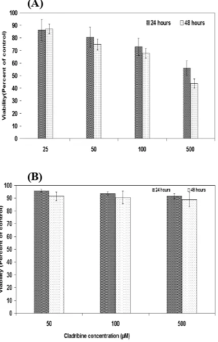
Fig. 1. Effect of cladribine on the growth of breast cancer cell line. MCF-7 cells were treated with different concentrations of cladribine for 24 and 48 h, and the viability was assessed by MTT assay. (A), pretreatment with 5’-amino-5-deoxyadenosine (kinase inhibitor) inhibited the cytotoxicity effect of cladribine in the MCF-7 cells (B). Results are expressed as percent of corresponding control and represent the mean ± SD of 6 repeats.
The precipitated DNA was washed once in 70% ethanol, resuspended in TAE buffer and then was applied on 1.2% agarose gel containing 0.5 µg/ml ethidium bromide and electrophoresis was performed for 2 h at 100 V using TAE running buffer. A ladder of 1 Kb was used as a marker.
Statistical analysis. The results were expressed
as the mean S.E.M and statistical differences were evaluated by one way ANOVA P<0.05 was considered significant.
RESULTS
Cytotoxicity assay. To evaluate the cytotoxicity
effect of cladribine, viability tests were applied using MTT assay. As it is shown in Figure 1, treatment of the ER+MCF-7 cells with 25, 50, 100 and 500 μM concentrations of cladribine resulted in a significant (p<0.05) increase in the cell death (Fig. 1A). Cladribine at concentrations bellow 25 μM had no significant effect. Pretreatment of the cells with kinase inhibitor, 5’-amino-5’-deoxyadenosine resulted in the inhibition of cytotoxicity (Fig. 1B). Pretreatment with 8-phenyltheophylline (a receptor antagonist), however, did not affect the cladribine-induced cytotoxicity.
Detection of apoptosis by annexin V/PI staining and Hoechst 33258. Fluorescein-conjugated
annexin V (FL1-H) and PI (FL2-H) staining (detected by epifluorescence microscope) were used to distinguish apoptotic cells. The cell death that induced by cladribine is mainly an apoptotic type (Fig. 2A). Furthermore, The apoptotic changes in the nuclear morphology, using Hoechst 33258 staining, are also observed (Fig. 2B) in comparison to control (Fig. 2C).
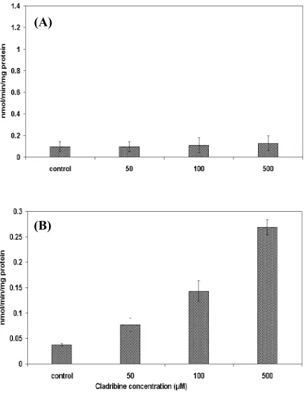
Caspase -3 and -9 activation. To explore the
possible biochemical mechanisms underlying cladribine-induced apoptosis, the activation of caspase-3 and -9 were assayed. The results demonstrated that the activity of caspase-3-protease remained unchanged (Fig. 3A). However, the activity of caspase-9 was significantly elevated in the treated ER+ MCF-7 breast cancer cells (Fig. 3B).
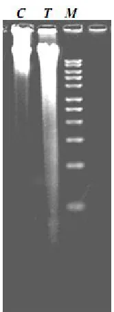
DNA laddering. Electrophoretic analysis of DNA
extracted from untreated and treated cells are shown in Figure 4. The DNA from untreated cells was unfragmented (C). Addition of 500 µM cladribine to the MCF-7 cells resulted in a smear, characteristic of a random DNA degradation (T).
DISCUSSION
In the present study, we showed, for the first time, that cladribine induces apoptosis in ER+MCF-7 breast cancer cell line. Cell cytotoxicity was evaluated by MTT method. Apoptosis was demonstrated by morphological analysis of treated cells using annexin-V/PI and Hoechst staining. (A)
(B)
(A)
(B)
(C)
Fig. 2.Morphological changes using annexin V/PI staining (A)shows that non-apoptotic cells are annexin V-/PI-whereas
early apoptotic cells are annexin V+/PI-and late apoptotic cells
are annexin V+/PI+. Hoechst 33258 staining (C)shows nuclear
condensation characteristic of apoptotic cells.
The concentrations at which significant cell death was observed in the MCF-7 cells were equal or greater than 25 µM. These findings are different
from those obtained for other cancer cell lines including human astrocytoma [15], colorectal carcinoma HCT116 [12], K562 cells [16] and human leukemia cell lines HSB2 and Jurkat [14] indicating that these cells are more sensitive to the cytotoxicity effect of cladribine. Enzymatic factors important in cladribine biotransformation include deoxycytidine kinase (dCK), deoxyguanosine kinase (dGK) and 5’nucleotidase (5’NT) [17]. Therefore, the lack or low expression/activity of the enzymes responsible for the intracellular activation of this adenosine analogue in the MCF-7 breast cancer cells, with respect to the above mentioned cell lines, may be responsible for the differential effect exerted by cladribine. To examine this possibility, the cells were pretreated with 5’-amino-5’-deoxyadenosine, which inhibits the phospho-rylation of cladribine by dCK. The results revealed that dCK inhibitor is able to prevent the induction of cell death by cladribine, indicating the involvement of dCK in the phosphorylation which is responsible for the observed cytotoxicity effect of cladribine. Furthermore, high dose of cladribine therapy has been used for chronic myelogenous leukemia [18].
Fig. 2.Morphological changes using annexin V/PI staining (A) shows that non-apoptotic cells are annexin V-/PI-whereas early
apoptotic cells are annexin V+/PI-and late apoptotic cells are
annexin V+/PI+. Hoechst 33258 staining (B) shows nuclear
condensation characteristic of apoptotic cells.
Fig. 3.Activity of caspase-3 (A)and -9(B)in the MCF-7 breast cancer cells after treatment with cladribine for 48 h. Results are expressed as activity of the enzyme and represent the mean ± SD of 6 repeats.
(A)
(B)
These investigators treated patients with doses (21.5 mg/m2daily) higher than the usual dose, and our results are highly relevant to this cladribine therapy trial. In addition, other investigators using 2-chloroadenosine-treated rheumatoid fibroblasts found a dose response greater than 50 µM [19].
Fig. 4.Agarose gel electrophoresis of DNA from untreated (C), treated (T) MCF-7 as well as a 1 Kb DNA marker (M).
Activation and involvement of extracellular adenosine receptors was ruled out, since pretreatment of the cells with 8-phenyltheophylline, a receptor antagonist, did not affect the apoptotic effect induced by cladribine. Therefore, cladribine needs to be phosphorylated inside the cells to induce cell death.
Activation of caspases is generally considered to be a requisite event during apoptosis. Our results also demonstrate that apoptosis induced by cladribine was dependent on the activation of caspase-9, suggesting a possible mitochondrial apoptotic pathway, e.g. through calcium-dependent cytochrome c release or a change in Bax/
Bcl-2ratio. These findings are in agreement with those obtained in the cladribine-treated CCRF-CEM cell line [17].
Concerning caspase-3 activation, MCF-7 cells did not show any significant elevation following treatment with cladribine. We have also previously reported that caspase-3 activation did not occurred upon treatment with tamoxifen, suggesting that the induction of apoptosis in the ER+ MCF-7 breast cancer cells is not dependent on caspase-3 [20]. In addition, other studies have revealed that MCF-7 cells lack pro-caspase-3 polypeptide as a
consequence of 47-bp deletion within exon 3 of the pro-caspase-3 gene that alters the reading frame of the message and results in an unstable truncated polypeptide [21].
In conclusion, this is the first study demonstrating the efficacy of the cladribine in the induction of apoptosis in ER+MCF-7 breast cancer cells through the intracellular phosphorylation reaction that may lead to a mitochondrial apoptotic pathway.
REFERENCES
1. Vermes, I. and Haanen, C. (1994) Apoptosis and programmed cell death in health and disease. Adv. Clin. Chem. 31: 177-246.
2. Kerr, J.F.R., Winterford, C.M. and Harmon, B.V. (1994) Apoptosis-its significance in cancer and cancer therapy. Cancer 73: 2013-2026.
3. Pottern, C.S., Merrit, A., Hickman, J., Hall, P. and Faranda, A. (1994) Characterization of radiation-induced apoptosis in the small intestine and its biological significance. Int. J. Radiat. Biol. 65: 71-78.
4. Carson, D.A., Wasson, D.B., Kaye, J., Ullman, B., Martin, D.W. and Robins Montgomery, J.A. (1980) Deoxycytidine kinase-mediated toxicity of deoxyadenosine, R.K. and analogs toward malignant human lymphoblasts in vitro and toward murine L1210 leukemia in vivo. Proc. Natl. Acad. Sci. 77: 6865-6869.
5. Seto, S., Carrera, C.J., Wasson, D.B. and Carson, D.A. (1986) Biochemical basis for deoxyadenosine and 2-chlorodeoxyadenosine toxicity to resting human lymphocytes. Adv. Exp. Med. Biol. 195 Pt B: 577-582.
6. Seto, S., Carrera, C.J., Kubota, M., Wasson, D.B. and Carson, D.A. (1985) Mechanism of deoxyadenosine and 2-chlorodeoxyadenosine toxicity to nondividing human lymphocytes. J. Clin. Invest. 75: 377-383.
7. Avery, T.L., Rehg, J.E., Lumm, W.C., Harwood, F.C., Santana, V.M. and Blakley, R.L. (1989) Biochemical pharmacology of 2-chloro-deoxyadenosine in malignant human hematopoietic cell lines and therapeutic effects of 2-bromo-deoxyadenosine in drug combinations in mice. Cancer Res. 49: 4972-4978.
8. Ceruti, S., Giammarioli, A.M., Camurri, A., Falzano, L., Rufini, S., Frank, C., Fiorentini, C., Malorni, W. and Abbracchio, M.P. (2000) Adenosine- and 2-chloro-adenosine-induced cytopathic effects on myoblastic cells and myotubes: involvement of different intracellular mechanisms. Neuromuscul. Disord. 10:436-446.
9. Delannoy, A. (1996) 2-chloro-2'-deoxyadenosine: clinical applications in hematology. Blood Rev. 10: 148-166.
10. Abbracchio, M.P., Ceruti, S., Brambilla, R., Franceschi, C., Malorni, W., Jacobson, K.A., Von Lubitz, D.K. and Cattabeni, F. (1997) Modulation of apoptosis by adenosine in the central nervous system: a possible role for the A3 receptor. Pathophysiological significance and therapeutic implications for neurodegenerative disorders. Ann. N.Y. Acad. Sci. 825:11-22.
11. Bontemps, F., Delacauw, A., Cardoen, S., Van Den Neste, E. and Van Den Berghe, G. (2000) Metabolism and cytotoxic effects of 2-chloroadenosine, the major catabolite of 2-chloro-2'-deoxyadenosine. Biochem. Pharmacol. 59:1237-1243.
12. Galmarini, C.M., Voorzanger, N., Falette, N., Jordheim, L., Cros, E., Puisieux, A. and Dumontet, C. (2003) Influence of p53 and p21 (WAF1) expression on sensitivity of cancer cells to cladribine. Biochem. Pharmacol. 65: 121-129. 13. Carmichael, J., Mitchell, J.B., DeGraff, W.G.,
Gamson, J., Gazdar, A.F., Johnson, B.E., Glatstein, E. and Minna, J.D. (1988) Chemosensitivity testing of human lung cancer cell lines using the MTT assay. Br. J. Cancer 57: 540-547.
14. Bradford, M.M. (1976) A rapid and sensitive method for the quantities of microgram of protein utilizing the principle of protein-dye binding. Anal. Biochem. 72: 248-254.
15. Ceruti, S., Franceschi, C., Barbieri, D., Malorni, W., Camurri, A., Giammarioli, A.M., Ambrosini, A., Racagni, G., Cattabeni, F. and Abbracchio, M.P. (2000) Apoptosis induced by 2-chloro-adenosine and 2-chloro-2'-deoxy-adenosine in a human astro-cytoma cell line: differential mechanisms and
possible clinical relevance. J. Neurosci. Res. 60: 388-400.
16. Wyczechowska, D. and Fabianowska-Majewska, K. (2003) The effects of cladribine and fludarabine on DNA methylation in K562 cells. Biochem. Pharmacol. 65: 219-225.
17. Chandra, J., Mansson, E., Gogvadze, V., Kaufmann, S.H., Albertioni, F. and Orrenius, S. (2002) Resistance of leukemic cells to 2-chloro-deoxyadenosine is due to a lack of calcium-dependent cytochrome c release. Blood 99: 655-663.
18. Dann, E.J., Anastasi, J. and Larson, R.A. (1998) High-dose cladribine therapy for chronic myelogenous leukemia in the accelerated or blast phase. J. Clin. Oncol. 16: 1498-1504.
19. Koshiba, M., Kosaka, H., Nakazawa, T., Hayashi, N., Saura, R., Kitamura, N. and Kumagai, S. (2002) 2-Chloroadenosine but not adenosine induces apoptosis in rheumatoid fibroblasts independently of cell surface adenosine receptor signaling. Br. J. Pharmacol. 135:1477-1486.
20. Salami, S. and Karami-Tehrani, F. (2003) Biochemical studies of apoptosis induced by tamoxifen in estrogen receptor positive and negative breast cancer cell lines. Clinical Biochemistry. (in press).
21. Kottke, T.J., Blajeski, A.L., Meng, X.W., Svingen, P.A., Ruchaud, S., Mesner, P.W., Boerner, S.A. Jr., Samejima, K., Henriquez, V., Chilcote, T.J., Lord, J. Salmon, M., Earnshaw, W.C. and Kaufmann, S.H. (2002) Lack of correlation between caspase activation and caspase activity assays in paclitaxel-treated MCF-7 breast cancer cells. J. Biol. Chem. 277: 804-815.