ــــــــــــــــــــــــــــــــــــــــــــــــــــــــــــــــــــ
Original Articleـــــــــــــــــــــــــــــــــــــــــــــــــــــــــــــــــــ
ـ
35 Copyright© 2016, Published by Mazandaran University of Medical Sciences on behalf of Iranian Society of Medical Mycology and Invasive Fungi Research Center. This is an open-access article distributed under the terms of the Creative Commons Attribution-Non Commercial 4.0 International License (http://creativecommons.org/licenses/by-nc/4.0/) which permits copy and redistribute the material just in noncommercial usages, provided the original work is properly cited.
A real time PCR assay on blood for diagnosis of invasive candidiasis in
immunocompromised patient
Ashrafi M1, Nabili M1,2, Shokohi T3*, Janbabaie G4, Hedayati MT3, Ali-Moghaddam K5
1 Student research committee, Mazandaran University of Medical Sciences, Sari, Iran 2 Social Security Organization, Golestan, Iran
3 Invasive Fungi Research Center (IFRC), and Department of Medical Parasitology and Mycology, Mazandaran University of Medical Sciences, Sari, Iran
4Department of Internal Medicine, Cell and Molecular Biology Research Center, Mazandaran University of Medical Sciences, Sari, Iran
5 Hematology-Oncology Research Center and Stem Cell Transplantation Research Center (HORCSCT), Tehran University of Medical Sciences, Iran
*Corresponding author: Tahereh Shokohi, Department of Medical Mycology and Parasitology, Sari Medical School, Km 18 Khazarabad Road, Iran. Tel: +98 11 33543781; Fax: +98 11 33543781; Email: shokohi.tahereh@gmail.com
(Received: 7 June 2014; Revised: 25 July 2014; Accepted: 5 August 2014)
Abstract
Background and Purpose: Invasive candidiasis (IC) is a significant cause of morbidity and mortality in patients with hematologic disorders and bone marrow transplant recipients. Rapid, specific and sensitive test for the timely accuracy in immunocompromised patients to reduce mortality rates and prevent IC progress is necessary. We established a real-time PCR assay on blood for the diagnosis and differentiation of the causative Candida species.
Materials and Methods: Whole blood samples were collected twice, from 72 patients for Real Time PCR and blood
culture assays. The primers and hybridization probes were designed to potentiate the specific sequence of 18S rRNA genes using Light Cycler system and Fluorescence Resonance Energy Transfer (FERT). The patients with hematologic malignancies and bone marrow transplant recipients were evaluated for IC based on the revised European Organization for Research and Treatment of Cancer/ Mycoses Study Group (EORTC/MSG) criteria.
Results: From 2009 to 2011, 72 patients with hematologic malignancies and bone marrow transplant recipients were evaluated for IC. The female to male ratio was 27:45; the mean age was 32.1 years. The most common malignancy in this patient was acute myeloid leukemia (AML) (27.8%) and acute lymphoblastic leukemia (ALL) (26.4%). Out of 72 patients, 11 patients (15.3%) had positive real time PCR /probe results. Based on the melting temperature (Tm) analysis, 5 (45.4%) C. krusei, 3 (27.2%) C. tropicalis, 2 (18.1%) C. parapsilosis and 1 C. albicans (9%) were identified. According to the revised EORTC/MSG, 1 patient (9%) and 10 patients (91%) were defined as proven and possible groups of IC, respectively.The mortality rate in proven and possible IC patient was found 54.5%.
Conclusion: The established Real-time PCR/FRET probe assay is an appropriate diagnostic tool for the detection of Candida species DNA and the management of patients suffering from hematologic malignancies and bone marrow recipient are at risk for IC.
Keywords: Candida, hematological malignancy, identification, invasive, Real-Time PCR
How to cite this paper:
Ashrafi M, Nabili M, Shokohi T, Janbabaie G, Hedayati MT, Ali-Moghaddam K. A real time PCR assay on blood for diagnosis of invasive candidiasis in immunocompromised patient. Curr Med Mycol. 2015; 1(1): 35-41.DOI: 10.18869/acadpub.cmm.1.1.35
Introduction
he proportion of cancer patients with deep fungal infections has increased dramatically in recent decades [1]. Invasive candidiasis (IC) is a serious opportunistic infections that often in persons with impaired immunity, especially in the bone marrow and organ transplant recipients, or undergoing chemotherapy for the treatment of hematologic malignancies and severe neutropenia have occurred [2, 3] The mortality rates of IC have increased to 80%, which is currently the fourth cause of nosocomial bloodstream infection [4,5]. Despite the
emergence of new antifungal drugs, the mortality rate of IC in patients with acute leukemia and bone marrow transplant recipients is high (50% and 90% of cases, respectively) [6]. The well-known risk factors for IC include chemotherapy, immunosuppres-sive disease, hematopoietic stem cell or solid organ transplantation, parenteral nutrition, and indwelling catheters or prosthetic device [7]. Although Candida albicans is the most common cause of IC and two-thirds of all
Candida krusei, C. tropicalis and C. parapsilosis are isolated from IC [8]. Current conventional diagnostic tools for IC include direct microscopy examination of tissue sections or bronchoalveolar lavage fluid, blood culture, detection of surface proteins (e.g. glucomannan or (1, 3)-beta-D-glucan) and detection of antibodies [9-12]. Earlydiagnosis of IC is difficult. Diagnosis based on clinical symptoms and cultures are non-specific and lack of sufficient sensitivity for the early diagnosis of IC. Serologic evaluation of IC by antibodies and circulating antigens show variable sensitivity and specificity [9,13]. Different molecular techniques for the early diagnosis of IC are present but these current PCR protocols have been criticized because of poor sensitivity, lack of standardization and excessive time and labor efforts. Some molecular analytical systems for the identification of various Candida species on blood samples were developed. Recently, LightCycler PCR Fluorescence Resonance Energy Transfer (FRET) techniques are increasingly being considered for early detection and identification. This technique has a high sensitivity and specificity [14]. LightCycler PCR technique is able to detect low fungal burden in blood and serum samples [15]. In this study, we aimed to extend a real-time PCR assay which was previously optimized for the detection of C. albicans on blood [16] for the diagnosis of invasive candidiasis in immunocompromised patients and subsequently differentiating causative
Candida species using oligohybridization probe melting temperature (Tm) analysis.
Material and Methods
From 2009 to 2011, this descriptive study was designed to determine IC and candidemia in patients with hematologic malignancies in the oncology ward at Imam Khomeini Hospital in Sari, and bone marrow transplant recipient in Shariati Hospital in Tehran. Eighty patients met the inclusion criteria, 8 patients were excluded due to poor patient satisfaction and ultimately 72 patients were studied.The study protocol was approved by the Medical Research Ethics Committee of the Mazandaran University of
Medical Sciences (Ethical no. 89-5-13) and all patients gave their informed consent. The patients who were not willing to participate were excluded from the study.
Clinical samples
Clinical samples were obtained from patients thought to be at high risk from fungal infection based on EORTC/MSG [17].These criteria included host factors, clinical signs and the mycological study of clinical samples in patients. Ten ml of whole blood, were taken twice a week, as well as 5ml for biphasic brain heart infusion medium bottle, 5ml for Ethylenediaminetetraacetic acid (EDTA) tubes. Blood culture bottles were incubated at 37 °C for two weeks and examined for fungal growth every day. All clinical samples were cultured on sabouraud dextrose agar with chloramphen-icol and direct microscopic examination was performed. Candida obtained from blood culture were identified according to morphological (germ tube, chlamydospore test and CHROM agar Candida medium) and biochemical test using API 20C Aux Candida kits (Biomerieux, France).
DNA extraction
DNA extracted from whole blood was previously described [18,19]. Briefly, erythrocytes were hypotonically lysed in Red Cell Lysis Buffer (RCLB) (10mM Tris [PH=7.6], 5mM MgCl2. 10mM NaCl) for 10 min at 37oC. The samples were pelleted at 5000
g for 10 min. The pellets were vortexed using glass beads (425-600µm, Sigma-Aldrich Corp, St. Louis, MO USA) for 3 min and the supernatant were treated with 180 U lyticase (Sigma-Aldrich Corp, St. Louis, MO USA) at 37 oC for 30 min. Two hundred microliter of
Primer and Probe design
The primers and hybridization probes were designed to potentiate the specific sequence of 18S rRNA genes using LightCycler system and Fluorescence Resonance Energy Transfer (FERT). Primers and hybridization probes were designed specifically for the detection of 18S rRNA gene of Candida spp. [14, 20] (Table 1).
Table 1. Primers and probes used in DNA amplification
Primers:
CAN-F (5´-ATT GGA GGG CAA GTC TGG TG) CAN-R (5´-ATC CCT TAG TCG GCA TAG-3´) (Eurofins MWG Operon, Ebensburg, Germany,) Probes:
LC 640 5′ TGG CGA ACC AGG ACT TTT ACT TTG A- phosphate 5′ AGC CTT TCC TTC TGG GTA GCC ATT-LC fluorescein (Tibmolbiol, Berlin, Germany)
Light Cycler-based PCR assay
Real-time PCR was performed on a Roche LightCycler (Roche Diagnostics, Mannheim, Germany) in a 20 µl reaction mix containing 2 µl of the extracted DNA, 15.4 µl Master mix (Light cycler DNA Master Mix, Roche Kit), 0.6 µl of HS prime Taq DNA polymerase 250 unit (GeNet Bio), 1 µl of each Primer (10 pmol/µl of primers, MWG-Operon), 1 µl of each hybridization probe (10 pmol/µl of probes, MWG-Operon). Seventy-two samples were run in parallel by performing 45 cycles of repeated denaturation (5s at 95°C), annealing (15 s at 62°C), and enzymatic chain extension (25 s at 72°C). The PCR was followed by melting temperature analysis cycle compromising 90°C for 20 s (TTR of 20°C s-1), 45°C for 20 s (TTR of 20°C s-1) and 90°C for zero s (TTR of 0.2°C s-1) to check the specificity of the PCR product. And for the end stage, a cooling rate of 40°C to 30 s for one cycle was defined. To monitor contamination, aliquots of saline and human fibroblast DNA were prepared concurrently as negative extraction and amplify-cation controls. All these controls were tested in the same procedure as described above.
Results
Of the 72 patients, 45(62.5 %) males and 27(37.
5 %) were females. The age ranged between 2
years and 74 years. Their median age of patients in this study was 32.17 years. The mortality rate in present study was 33.3%. The highest mortality
rate in patients with Acute Lymphoblastic Leukemia (ALL) 29.7%, Acute Myeloid Leukemia (AML) 27%, non-Hodgkin's lymphoma and Hodgkin's lymphoma 11.3%, were seen. Sixty-six patients (91.7%) had neutropenia and
(96.8%) patients were receiving chemotherapy.
Regarding the clinical criteria, most of the symptoms include persistent high fever (86.1%), lethargy and weakness (56.9 %), cough (20.8%) and high fever and falling (12.5 %) were seen. One culture positive blood sample yielded C. albicans
(1.4% of the patients) according to morphological and biochemical tests.
Antifungal agents were prescribed in 53 patients (87.5%). The most commonly used antifungal were fluconazole (34 patients; 54%) followed by amphotericin (10 patients; 40%), and combination therapy with amphotericin and fluconazole (4 patients; 5.6 %).
Quantification and Optimization of the assay
The determination sensitivity, specificity, reproducibility and assessment of detection limit of the assay of Candida albicans in whole blood were described in detailed previously by Nabili et.al, [16]. Briefly to determine specificity, the oligonucleotide specific for C. albicans
hybridized only with DNA extracted from C.
albicans cultures. All PCR reactions with DNA from other fungal species such as
Aspergillus spp. and human genomic DNA were negative, indicating the specificity was 100% for this method. Melting temperature analysis of C. albicans was indicated in a single point of maximum temperature signal with the probe. Tm analysis subsequent to real time PCR amplification using standard strain revealed a specific melting peak signal with C.albicans probing setting and allowing the differentiation of C. krusei, C. tropicalis, C. parapsilosis and C. albicans. The specific melting temperature (Tm) patterns are shown in Table 2. Reproducibility of the FRET assay was evaluated by using inter-assay method previously [16]. Briefly, the amplification of serially diluted C. albicans (100 to 106 CFU) was repeated 6 times
previously published manuscript [16] for each PCR reaction, which indicates the high sensitivity of this method.
Real-Time-PCR results in clinical samples
We analyzed 110 blood samples from 72 patients, for testing Real-Time PCR. Of the 72 patients, 11 patients (15.3 %) were positive for the presence of Candida DNA on blood by Real-Time PCR/probe. Based on the melting temperature (Tm) analysis, 5 (45.4 %) C. krusei,
Table 2. Melting temperatures (Tm) for standard strains of
Candida species with Candida albicans hybridization probe
set
Candida species/ Standard Strain Tm ˚C
C. krusei (CBS 573) 55.66
C. tropicalis (CBS 94) 63.15
C. parapsilosis (CBS 604) 62.47
C. albicans (CBS 562) 65.76
3 (27.2%) C. tropicalis, 2 (18.1 %) C. parapsilosis and 1 C. albicans (9%) were identified. According to host factors, clinical and mycology criteria on the revised EORTC/ MSG,1 patient (9%) and 10 patients (91%) were defined as proven and possible IC, respectively (Table 3).The mortality rate in proven and possible IC patient was found 54.5%.
Discussion
Candida species are the cause of nosocomial bloodstream infection in 8-9% patients admitted to the intensive care unit (ICU). Cancer patients are at a high risk of developing bloodstream infection [7,21, 22]. Serious risk factors for invasive candidiasis are chemotherapy in patients with hematologic malignancies and immunosuppressive therapy in patients receiving hematopoietic stem cell
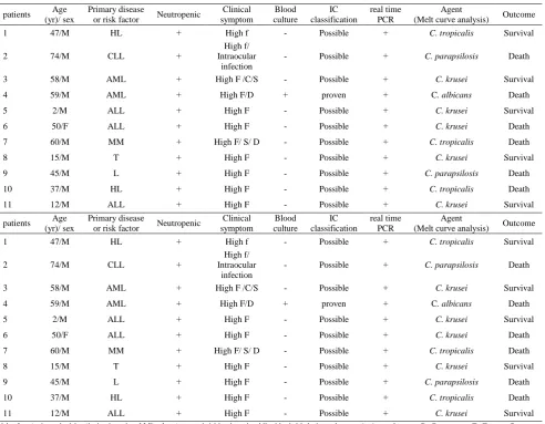
Table 3. Demographic, characteristics and clinical data of eleven patients with proven or possible IC
Outcome Agent
(Melt curve analysis) real time PCR IC classification Blood culture Clinical symptom Neutropenic Primary disease or risk factor Age (yr)/ sex patients Survival C. tropicalis + Possible -High f + HL 47/M 1 Death C. parapsilosis + Possible -High f/ Intraocular infection + CLL 74/M 2 Survival C. krusei + Possible -High F /C/S +
AML 58/M
3
Death C. albicans
+ proven + High F/D + AML 59/M 4 Survival C. krusei + Possible -High F + ALL 2/M 5 Death C. krusei + Possible -High F + ALL 50/F 6 Death C. tropicalis + Possible -High F/ S/ D + MM 60/M 7 Survival C. krusei + Possible -High F + T 15/M 8 Death C. parapsilosis + Possible -High F + L 45/M 9 Death C. tropicalis + Possible -High F + HL 37/M 10 Survival C. krusei + Possible -High F + ALL 12/M 11 Outcome Agent
(Melt curve analysis) real time PCR IC classification Blood culture Clinical symptom Neutropenic Primary disease or risk factor Age (yr)/ sex patients Survival C. tropicalis + Possible -High f + HL 47/M 1 Death C. parapsilosis + Possible -High f/ Intraocular infection + CLL 74/M 2 Survival C. krusei + Possible -High F /C/S +
AML 58/M
3
Death C. albicans
+ proven + High F/D + AML 59/M 4 Survival C. krusei + Possible -High F + ALL 2/M 5 Death C. krusei + Possible -High F + ALL 50/F 6 Death C. tropicalis + Possible -High F/ S/ D + MM 60/M 7 Survival C. krusei + Possible -High F + T 15/M 8 Death C. parapsilosis + Possible -High F + L 45/M 9 Death C. tropicalis + Possible -High F + HL 37/M 10 Survival C. krusei + Possible -High F + ALL 12/M 11
transplant [23]. Several conventional micro-biologyical techniques such as blood cultures for the diagnosis of invasive candidiasis are used which has a very low sensitivity. The detection of immunological and molecular markers has provided an alternative for early diagnosis of invasive candidiasis [21]. Several PCR based techniques have been developed that their sensitivity and specificity are above 90% [24]. The 18S rRNA genes are highly conserved regions of the genome and used to detect Candida species [25]. Real time PCR in conjugation with fluorescent detection of amplicon using FRET has several advantages to culture-based method for the diagnosis of invasive candidiasis. This method is highly sensitive, rapid, and allows differentiating various species of Candida and quantifying the fungal load in blood samples [14]. In the present study, the same gene region was used in real time PCR technique for the detection of Candida species. This method has high sensitivity and able to detect at least two copies of the genome C. albicans in serum samples [26]. In Klingspor's study [27], the regions 18S rDNA and in Hsu study [28], the regions 28S rDNA were used as target for detection. The researchers showed that these two gene regions have a high sensitivity for detection of Candida species [27, 28]. In the present study, to assess the sensitivity of the technique using FRET, prepared from serial dilutions (100 to 106 C. albicans standard
strain) were mixed with the blood of healthy volunteers. Serial dilutions of all samples successfully amplified with the increase in fluorescence signal probes were amplified by real time PCR method. In our study, the minimum amount of DNA, 10 CFU was determined. In Loffler et al. study, PCR-ELISA assay was performed at the detection limit of the 5 CFU Candida in 1 ml whole blood [29]. In a study using two fluorogenic probes in a TaqMan-based PCR assay, the low limit of detection of 1-20 CFU/ml blood have been reported [30]. Horn et al. in a study conducted in 2011 by PCR, the sensitivity of 10 CFU/ml was reported [31]. Schabereiter-Gurtner et al., in a study conducted in 2007, using SYBR Green
technique, the sensitivity of 5 to 10 CFU/ml blood for detecting Candida in whole blood was reported, which is consistent with the current study [32]. The differences of sensitivity depend on to certain factors such as applying different methods of DNA extraction, using different primers and probes, using different devices and techniques of the implementation.
The study also revealed that the FRET technique has high specificity and good reproducibility. In this study, Melting temperatures (Tm) for C. albicans 65.76 °C, C. krusei 55.66 °C, C. tropicalis 63.15 °C and C. parapsilosis 62.47 °C was identified. Fricke et al. in a study conducted in 2010 with the FRET technique to analyze the melting temperature of
C. albicans 67 °C, C. tropicalis 63.9 °C, C. parapsilosis 61°C and C. glabrata 62.3°C [33]. The differences between these studies may result from different materials, device and operating system. The prevalence of invasive candidiasis in patients with hematologic malignancy and bone marrow transplant recipients, by real time PCR/FRET method was 15.2 %, which indicated a high prevalence. Hachem et al. reported, the prevalence of the disease to be as 12 % in patients with hematologic malignancies in [34]. In our study, based on melting curve analysis C. krusei in 5 cases (45.4%), C. tropicalis in 3 cases (27.2 %), C. parapsilosis in 2 cases (18.1 %) and C. albicans in 1 case (9.3 %) were identified. The majority of Candida species isolated from patients was non-albicans Candida species (90.7%). Abi-said et al. [35] demonstrated that
C.albicans, C.tropicalis and C. parapsilosis
were the most common species obtained from blood samples of patients with hematological malignancies and transplant recipients whereas in our study, C. albicans is the less common causative agent.
before starting treatment to prevent drug interactions, is important because non- albicans
species of Candida often is resistant to azoles and polyene antifungal drugs.
Conclusion
The established Real-time PCR/FRET probe assay is an appropriate diagnostic tool for the detection of Candida species DNA and the management of patients suffering from hematologic malignancies and bone marrow recipients are at risk for IC.
Acknowledgements
We are grateful to Dr. Shahram Samiei for his excellent real time PCR support.
Authors’ contributions
T.S. designed and managed the research, wrote the draft manuscript and edited the final manuscript. G.J. and K.AM. referred the patients, M.A.,M.N.applied all tests and wrote the draft, and MT.H. managed the research.
Conflicts of interest
The authors state no conflict of interest.
Financial Disclosure
No financial interests related to the material of this manuscript have been declared.
References
1. De Marie S. New developments in the diagnosis and management of invasive fungal infections. Haematologica. 2000; 85(1): 88-93.
2. DiNubile MJ, Hille D, Sable CA, Kartsonis NA.Invasive candidiasis in cancer patients: observations from a randomized clinical trial. J Infect. 2005; 50(5): 443-9.
3. Verfaillie C, Weisdorf D, Haake R, Hostetter M, Ramsay N, McGlave P. Candida infections in bone marrow transplant recipients.Bone marrow transplant. 1991; 8(3): 177-84.
4. Richardson MD, Warnock DW. Fungal infection: diagnosis and management. John Wiley & Sons; 2012.
5. Eggimann P, Bille J, Marchetti O. Diagnosis of invasive candidiasis in the ICU. Ann Intensive Care. 2011; 1(1): 1-10.
6. Herbrecht R, Flückiger U, Gachot B, Ribaud P,Thiebaut A, Cordonnier C. Treatment of invasive Candida and invasive Aspergillus infections in adult haematological patients. European J Cancer Suppl. 2007; 5(2): 49-59.
7. Concia E, Azzini AM, Conti M. Epidemiology incidence and risk factors for invasive candidiasis in high-risk patients. Drugs. 2009; 69(1): 5-14.
8. Chow JK, Golan Y, Ruthazer R, Karchmer AW, Carmeli Y, Lichtenberg D, et al. Factors associated with candidemia caused by non-albicans Candida species versus Candida albicans in the intensive care unit. Clin Infect Dis. 2008; 46(8): 1206-13.
9. Arancia S, Carattoli A, La Valle R, Cassone A, De Bernardis F. Use of 65kDa mannoprotein gene primers in real time PCR identification of Candida albicans in biological samples. Mol Cell Probes. 2006; 20(5): 263-8.
10. Jones J. Laboratory diagnosis of invasive candidiasis. Clin Microbiol Rev. 1990; 3(1): 32-45. 11. Mannarelli B, Kurtzman C. Rapid identification of
Candida albicans and Other human pathogenic yeasts by using short oligonucleotides in a PCR. J Clin Microbiol.1998; 36(6): 1634-41.
12. Guery BP, Arendrup MC, Auzinger G,Azoulay É, Sá MB, Johnson EM, et al. Management of invasive candidiasis and candidemia in adult non-neutropenic intensive care unit patients: PartII. Treatment. Intensive Care Med. 2009; 35(2): 206-14.
13. Ellepola A, Morrison CJ. Laboratory diagnosis of invasive candidiasis. J Microbiol. 2005; 43(5): 65-84. 14.Loeffler J, Henke N, Hebart H, Schmidt D, Hagmeyer L, Schumacher U, et al.Quantification of fungal DNA by using fluorescence resonance energy transfer and the light cycler system.J Clin Microbiol. 2000; 38(2): 586-90.
15.Dunyach C, Bertout S, Phelipeau C, Drakulovski P, Reynes J, Mallié M. Detection and identification of Candida spp. in human serum by LightCycler® real-time polymerase chain reaction. Diagn Microbiol Infect Dis. 2008; 60(3): 263-71.
16.Nabili M, Ashrafi M, Jan Babaei Gh, Hedayati MT, Ali Moghaddam K, Shokohi T. Quantification and optimization of Candida albicans DNA in blood Samples. Reports Biochem Mol Biol. 2013; 2(1): 1-6. 17.De Pauw B, Walsh TJ, Donnelly JP, Stevens DA, Edwards JE, Calandra T, et al. Revised definitions of invasive fungal disease from the European organization for research and treatment of cancer/invasive fungal infections cooperative group and the national institute of allergy and infectious diseases mycoses study group (EORTC/MSG) consensus group.Clin Infect Dis.2008; 46(12): 1813-21.
18.Nabili M, Shokohi T,Janbabaie G, Hashemi-Soteh MB, Ali-Moghaddam K, Aghili SR. Detection of invasive aspergillosis in bone marrow transplant recipients using real-time PCR.J Glob Infect Dis. 2013; 5(2): 68-75.
20.Einsele H, Hebart H, Roller G, Löffler J, Rothenhofer I, Müller C, et al. Detection and identification of fungal pathogens in blood by using molecular probes. J Clin Microbiol.1997; 35(6): 1353-60.
21.Mikulska M, Furfaro E, Viscoli C. Biomarkers for Diagnosis and Follow-Up of Invasive Candidiasis: A Brief Review of the ECIL Recommendations.Curr Fungal Infect Rep. 2012; 6(3): 192-7.
22.Kuhn DM, Mukherjee PK, Clark TA, Pujol C,Chandra J, Hajjeh RA, et al. Candida parapsilosis characterization in an outbreak setting. Emerg Infect Dis. 2004; 10(6):1074.
23.Castagnola E, Faraci M, Moroni C, Bandettini R, Granata C, Caruso S, et al. Invasive mycoses in children receiving hemopoietic SCT. Bone Marrow Transplant. 2008; 41: S107-S11.
24.White PL, Perry MD, Barnes RA. An update on the molecular diagnosis of invasive fungal disease. FEMS Microbiol Lett. 2009; 296(1): 1-10.
25.O'Sullivan CE, Kasai M, Francesconi A, Petraitis V, Petraitiene R, Kelaher AM, et al. Development and validation of a quantitative real-time PCR assay using fluorescence resonance energy transfer technology for detection of Aspergillus fumigatus in experimental invasive pulmonary aspergillosis. J Clin Microbiol. 2003; 41(12): 5676-82.
26.White PL, Archer AE, Barnes RA. Comparison of non-culture-based methods for detection of systemic fungal infections,with an emphasis on invasive Candida infections. J Clin Microbiol. 2005; 43(5): 2181-7.
27.Klingspor L, Jalal S. Molecular detection and identification of Candida and Aspergillus spp. from clinical samples using real‐time PCR.Clin Microbiol Infect. 2006; 12(8): 745-53.
28.Hsu M-C, Chen K-W, Lo H-J, Chen Y-C, Liao M-H, Lin Y-H, et al. Species identification of medically important fungi by use of real-time LightCycler PCR. J Med Microbiol. 2003; 52(12): 1071-6.
29.Loffler J, Hebart H, Sepe S, Schumcher U, Klingebiel T, Einsele H. Detection of PCR-amplified fungal DNA by using a PCR-ELISA system. Med Mycol. 1998; 36(5): 275-9.
30.Maaroufi Y, Heymans C, De Bruyne J-M, Duchateau V, Rodriguez-Villalobos H, Aoun M,et al.Rapid detection of Candida albicans in clinical blood samples by using a TaqMan-based PCR assay. J Clin Microbiol. 2003; 41(7): 3293-8.
31.Horn DL, Neofytos D, Anaissie EJ, Fishman JA, Steinbach WJ, Olyaei AJ, et al. Epidemiology and outcomes of candidemia in 2019 patients: data from the prospective antifungal therapy alliance registry. Clin Infect Dis. 2009; 48(12): 1695-703.
32.Schabereiter-Gurtner C, Selitsch B, Rotter ML, Hirschl AM, Willinger B. Development of novel real-time PCR assays for detection and differentiation of eleven medically important Aspergillus and Candida species in clinical specimens. J Clin Microbiol. 2007; 45(3): 906-14.
33.Fricke S, Fricke C, Schimmelpfennig C, Oelkrug C, Schönfelder U, Blatz R, et al.A real‐time PCR assay for the differentiation of Candida species. J Appl Microbiol. 2010; 109(4): 1150-8.
34.Hachem R, Hanna H, Kontoyiannis D, Jiang Y, Raad I. The changing epidemiology of invasive candidiasis. Cancer. 2008; 112(11): 2493-9.