Professor of Neurology, Shahid Beheshti
University of Medical Sciences,
Tehran, Iran
Associate Professor of Neurology,
Neurology Department, Tehran University of
Medical Sciences, Tehran, Iran
Assistant Professor of Neurology, Neurology Department, Tehran University of
Medical Sciences, Tehran, Iran
,
Tehran
University of Medical Sciences
Tehran University of
Medical Sciences
Email: ijnl@tums.ac.ir
http://ijnl.tums.ac.ir
Copy Edit, Layout Edit, Proof Reading and Design: Farzanegan Radandish Co.Postal Code: 81465-1798, Isfahan, Iran; Telefax: +98 311 6686302 www.farzaneganco.ir; Email: f.radandish@gmail.com
Indexed in
PubMed, PubMed Central, Academic Keys,
Cite Factor (Directory Indexing of International Research Journals),
Directory of Open Access Journals (DOAJ), Directory of Research Journal Indexing (DRJI), Ebsco,
Electronic Journals Library,
Google Scholar, InfoBase Index,
Islamic World Science Citation Center (ISC), LocatorPlus,
Scientific Information Database (SID), Ulrichsweb Global Serials Directory, Universal Impact Factor,
Iranian Journal of Neurology © 2015
II Iran J Neurol 2015; 14(1)
The Iranian Journal of Neurology
is dedicated to the
Iranian Neurological Association. The journal is a
peer-reviewed journal published quarterly and publishes
neurological experiences in basic or clinical fields in
English Language
.
The Iranian Journal of Neurology
aims
to publish manuscripts of a high scientific quality
representing original clinical, diagnostic or experimental
works or observations in neurological sciences. Papers in
English
are welcomed, particularly those which bring
novel information and researches in clinical or basic fields
from the neurological disorders. All received manuscripts
coving the scope of the journal will be evaluated by
properly competent referees .
Submissions should be accompanied by a cover letter
including a declaration by the first author on behalf of the
others to the effect that
(1) The paper has not been published to date (except
for abstracts of conference materials).
(2) The paper has not been accepted for publication
elsewhere.
(3) All persons listed as the authors have read it and
approved it for publication. The cover letters should be
submitted in section "Comments for the Editor".
Articles must be written in accurate scientific English
appropriate for publication. The articles are subject to
review and editing; however, the authors are responsible
for the correctness the manuscript's English language.
The
articles
must
be
submitted
only
online:
ijnl.tums.ac.ir
The Editorial Board reserves the right to reject a paper
without seeking reviewers’ opinion provied the content or
the form of the paper does not meet minimum acceptance
criteria or if the subject of the paper is beyond the aims
and scope of the journal.
Everyone listed as the author of a paper is responsible
for the reliability and completeness of data presented in
the paper.
Do not submit papers that copy fully or partially
previously published papers.
Indicate that this submission is ready to be considered
by this journal by checking off the following:
The submission has not been previously published,
nor is it before another journal for consideration (or an
explanation has been provided in Comments to the Editor).
The submission file is in Microsoft Word document
file format.
Where available, URLs for the references have
been provided.
The text is double-spaced; uses an Arial 12-point
font; and all illustrations, figures, and tables are placed
within the text at the appropriate points, rather than at the
end.
The text adheres to the stylistic and bibliographic
requirements outlined in the Author Guidelines, which is
found in About the Journal.
If the Editorial Board is not notified in advance and the
paper is found to have been copied during editorial work,
the paper shall be rejected.
We expect that all studies reported in the journal
conform to the requirements of the Declaration of Helsinki
(1989). Information on the consent of a relevant ethics
committee to perform the trial and the informed consent of
the patients to participate in the trial should be given in the
Material and methods section of each paper in which
diagnostic or therapeutic intervention does not follow from
the standard procedure. Authors of case reports must not
disclose personal data of patients described.
The journal publishes:
Original Article
Review Article
Case Report
Short Communication
Clinical Notes
Editorial
Letters to Editor
Neurological Images
Neurological Videos
Iranian Neurological Events
Clinical Quiz
Details
Original and review papers:
The maximum length of
original and review papers (including tables and figures
materials) is 3000 words.
Case reports:
Case reports will be accepted only as Letter
to the Editor.
Short communications: The
maximum word number of
short communications should be below 1200 words with
maximum one table or figure and 10 references. The
manuscript should be structured including introduction,
materials and methods, results, discussion, and conclusion
with a structured abstracts as original articles.
Iranian Journal of Neurology © 2015
Iran J Neurol 2015; 14(1) III
unstructured. The videos should be uploaded as
supplementary files.
Letter to the Editor:
May concern short scientific reports
and comments. The maximum number of words should be
below 800 words with maximum 5 references, no abstract,
no table or figure, and unstructured.
Clinical notes:
Refer to important interesting observations
which are imperative for reminders in clinical practice.
The maximum number is 1000 words with maximum 5
references, 1 table and 1 figure with no abstract.
Iranian neurological events :
Include the brief description
of major regional events (congresses or seminar)
implemented in Iran.
Manuscripts should be submitted in 12 points, Arial
font, with double line spacing and sufficient margins of
2.5 cm.
The text should not be formatted.
Each section of the paper should begin on a new page
Page 1: Title Page
Page 2: Abstract and Key Words
Page 3 and subsequent pages: manuscript body
including Introduction, Materials and Methods, Results,
Discussion, Conclusion, References, Tables, Figures
1. Title page:
Title page
should contain paper title, full names of
authors, authors’ place of work, full name and address of
the corresponding author (including e-mail address and
telephone number), given in that order.
2. Abstract page:
The length of the abstract should be at least 200
and not more than 250 words for original papers and not
more than 150 words for review papers and case reports.
Abstracts of original papers should be structured to
include the background, methods, results and conclusion.
Below the abstract authors should provide between
three and six keywords conforming to Medical Subject
Headings (Index Medicus).
3. Page three and subsequent pages
of the original paper
and short communication should include the text arranged
in the following order (for other mansucript type, see
above):
1.
Introduction:
The introduction should be as
concise as possible and introduce the context of the paper
to the reader; the paper should clearly state the research
hypothesis and the objective of the study.
2.
Materials and Methods:
Description of the
studied population or material should be detailed and
include all information necessary to assess the reliability
of results obtained in the study and/or allow the
experiment to be repeated by other researchers; the section
related to statistical analysis should have information on
applied statistical tests and programs.
3.
Results:
Present results directly related to the topic
of the paper only; tables and/or figures are recommended.
4.
Discussion
5.
Conclusions:
These should be brief, follow directly
from results presented above and correspond to the aim of
the paper outlined in the introduction.
6.
Acknowledgements:
Should comprise information
on sources of funding (grant numbers); acknowledgements
should concern those who made a significant contribution
to the paper, but who did not meet the criteria to be listed
as authors.
7.
References:
References should be listed in the
order quoted in the paper. Please cite source and major
papers that offer interested readers an opportunity to
obtain more detailed information. Avoid citin g review
papers and conference reports, if they are not the only
materials on a given topic.
In the paper references should be given in
superscripts
with no space between the comma and the consecutive
number.
Authors are advised to carefully verify citation details.
Give names of first
six
authors; if there are more
authors, add “et al.“. Use Index Medicus abbreviations for
journal titles. Then mention the volume and the issue of
the journal.
The recommended style for journal references is as
follows:
[Reference number][Authors]. [Article title]. [Journal
Name] [Year of publication]; [volume](issue): [Pages
range].
For Journal Example:
1. Janghorbani M, Amini M, Willett WC, Mehdi Gouya
M, Delavari A, Alikhani S, et al. First nationwide survey
of prevalence of overweight, underweight, and abdominal
obesity in Iranian adults. Obesity (Silver Spring) 2007;
15(11): 2797-808.
For Books Example:
2. Ropper AH, Brown RJ. Adams and Victors principles
of neurology. 8
thed. New York, NY: McGraw Hill
Professional; 2005. p. 271.
Tables:
Each table should be placed on a separate page.
Tables should be numbered with Arabic numerals in the
order in which they appear in the text. Authors should
indicate the position of tables in the paper. Titles and
headings of tables should be given in English. Information
given in tables should not be repeated in the body of the
text. Explanations concerning tables, e.g. full names of
abbreviations should be given in footers below tables and
should be consecutively marked: “*”,“**”,“***” etc.
Figures:
Figures and photographs should be numbered
with Arabic numerals and attached as separate printouts
(in the electronic version, as separate files). Figures should
be saved in one of the following formats: .jpg.
Iranian Journal of Neurology © 2015
IV Iran J Neurol 2015; 14(1)
Abbreviations
should be always clarified when used for
the first time in the text (including the abstract).
Abbreviations should not be used in paper titles, unless in
exceptional circumstances.
Review process:
All papers submitted for publication in
the journal are assessed by two independent reviewers
with the mutual anonymity rule as to the names of
reviewers and authors observed.
Plagiarism policy:
According to the plagiarism policy of
Iranian Journal of Neurology, plagiarism is defined as a
paper which replicates another publication with as a
minimum 25% resemblance and devoid of citation.
In any time the evidence of plagiarism is detected, the
manuscript will be withdrawn and the author will be
sanctioned from publishing papers permanently.
Noscapine and ischemic stroke Iran J Neurol 2015; 14(1) V
Table of Contents
Review Article
Epileptic syndromes: From clinic to genetic
Abbas Tafakhori, Vajiheh Aghamollaii, Sara Faghihi-Kashani, Payam Sarraf, Laleh Habibi ... 1-7
Original Articles
The prevalence of female sexual dysfunction among migraine patients
Mohammad Abdollahi, Mansoureh Toghae, Firoozeh Raisi, Elaheh Saffari ... 8-11
A novel effect of Noscapine on patients with massive ischemic stroke: A
pseudo-randomized clinical trial
Massoud Mahmoudian, Mohammad Rezvani, Mohammad Rohani, Foozya Benaissa,
Mehdi Jalili, Shadi Ghourchian ... 12-16
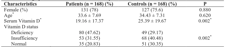
Serum 25(OH) Vitamin D levels is not associated with disability in multiple sclerosis
patients: A case-control study
Masoud Nikanfar, Ali Akbar Taheri-Aghdam, Maria Yazdani, Sheida Shaafi,
Nooshin Masoudian, Hossein Akbari, Parisa Youhanaee, Hamzeh Abbaszadeh ... 17-21
Comparison of endovascular coiling and surgical clipping for the treatment of
intracranial aneurysms: A prospective study
Zeinab Taheri, Mohammad Hosein Harirchian, Hosein Ghanaati, Alireza Khoshnevisan,
Payman Salamati, Mojtaba Miri, Kavous Firouznia, Mina Saeednejad, Madjid Shakiba,
Vafa Rahimi-Movaghar ... 22-28
Accuracy of magnetic resonance spectroscopy in distinction between radiation necrosis
and recurrence of brain tumors
Mousa Reza Anbarloui, Seyed Mohammad Ghodsi, Alireza Khoshnevisan, Masoud Khadivi,
Sina Abdollahzadeh, Ahmad Aoude, Soheil Naderi, Zeynab Najafi, Morteza Faghih-Jouibari
..
29-34
Elevated troponin T after acute ischemic stroke: Association with severity and location
of infarction
Siamak Abdi, Shahram Oveis Gharan, Farnaz Sinaei, Askar Ghorbani ... 35-40
Proposed equation between flexor carpi radialis H-reflex latency and upper limb length
Saeid Khosrawi, Parisa Taheri, Seyad Hasan Hashemi ... 41-46
Clinical Note
Mixed movement disorders revealing an atypical form of creatine deficiency syndrome
Fahmi Nasrallah, Hanene Benrhouma, Ichraf Kraoua, Gilbert Briand, Souhiel Omar,
Ilhem Turki Ben Youssef, Naziha Kaabachi ... 47-49
Letter to Editor
Patent foramen ovale and stroke: Does presence of a migraine headache or any
character of patent foramen ovale increase the risk of stroke?
Abdolhamid Shariat, Ehsan Yaghoubi, Kamran Aghasadeghi, Abbas Rahimi, Reza Nemati,
Nahid Ashjazadeh ... 50-51
Neurological Video
Eating dystonia in a case of neuroacanthocytosis
Mohammad Rohani, Gholamali Shahidi ... 52
Special Article
Neurolaw: A brief introduction
Iranian Journal of Neurology © 2015 Corresponding Author:Vajiheh Aghamollaii
Email: ijnl@tums.ac.ir Email: vajiheh102@gmail.com
Review Article
Iran J Neurol 2015; 14(1): 1-7
Epileptic syndromes: From clinic to
genetic
Abbas Tafakhori
1, Vajiheh Aghamollaii
2, Sara Faghihi-Kashani
3, Payam Sarraf
1, Laleh Habibi
41
Department of Neurology, School of Medicine, Imam Khomeini Hospital AND Iranian Center of Neurological Research, Tehran
University of Medical Sciences, Tehran, Iran
2
Department of Neurology, School of Medicine, Roozbeh Hospital AND Iranian Center of Neurological Research, Tehran University of
Medical Sciences, Tehran, Iran
3
Department of Neurology, School of Medicine, Tehran University of Medical Sciences, Tehran, Iran
4Department of Medical Genetics, School of Medicine, Tehran University of Medical Sciences, Tehran, Iran
Keywords
Epilepsy, Genetic, Inheritance, Chromosomal
Abnormalities, Mutation
Abstract
Epilepsy is one of the most common neurological
disorders. Studies have demonstrated that genetic
factors have a strong role in etiology of epilepsy.
Mutations
in
genes
encoding
ion
channels,
neurotransmitters and other proteins involved in the
neuronal biology have been recognized in different
types of this disease. Moreover, some chromosomal
aberration including ring chromosomes will result in
epilepsy. In this review, we intend to highlight the role
of molecular genetic in etiology of epilepsy
syndromes, inspect the most recent classification of
International League against Epilepsy and discuss the
role of genetic counseling and genetic testing in
management of epilepsy syndromes. Furthermore, we
emphasize on collaboration of neurologists and
geneticists to improve diagnosis and management.
Introduction
Epilepsy
Definition
“Epilepsy” is derived from the Latin term meaning,
“to be attacked.” In medicine, epilepsy is defined as
recurring episodes of seizures due to excessive and
abnormal synchronous neural activity of the cerebral
cortex, which could be induced by cellular or
molecular defects in cerebral tissues.
1In cases of an
altered endocrine or metabolic state, it would be
categorized
as
structural/metabolic
epilepsy.
However, on occasions the underlying disorder could
not be recognized, and it would be classified under
unknown category. Epilepsies attributed to known
genetic disorders are classified as genetic epilepsies
2(refer to the next section).
Annual incidence of seizures in the general
population is estimated to be 61/100,000 persons
3with higher occurrence in both extremes of life.
4Clinical diagnosis of epilepsy is carried out mainly
by evaluation of patient’s detailed history. Positive
electroencephalogram (EEG) results are supportive for
confirmation of epilepsy. Nevertheless, negative
findings might not exclude the diagnosis of
epilepsy.
5,6Clinical presentation of epilepsy may
easily be mistaken with conditions mimicking
seizure’s features, including hypoglycemia, sleep
disorders, migraines, transient ischemic attacks and
transient global amnesia.
7,8Classification
Based on the 2010 report of International League
Against Epilepsy (ILAE),
9the etiology of epileptic
seizures is divided into three major classes as
discussed below.
Genetic epilepsy: this category (previously known
as idiopathic) implies epileptic disorders that are a
direct consequence of either known single gene
defects or complex inheritances in which the epilepsy
is
the
essential
symptom.
Nonetheless,
the
Iranian Journal
of Neurology
2 Iran J Neurol 2015; 14(1) Tafakhori et al.
contribution of environmental factors in disease
expression cannot be disregarded.
2,9The recent
alternation of “idiopathic” to “Genetic” has the
advantage of highlighting the genetic predisposition,
and it no longer conflate other concepts (e.g.
prognosis). Most cases display clinical features
during childhood or adolescence. Although some
suffers from a variety of subtle cognitive and
behavioral challenges, the affected patients may have
normal intelligence, and EEG might also show
generalized discharges.
9,10Genetic epilepsy is further
divided into generalized and partial epilepsy.
Childhood absence epilepsy, juvenile myoclonic
epilepsy and epilepsy with grand-mal seizures on
awakening are examples of genetic generalized and
benign focal epilepsy of children is an instance of
partial genetic epilepsy.
2,9Structural/metabolic epilepsy: Epilepsies classified
under
this
category
(previously
known
as
symptomatic) require specific structural or metabolic
defects that have been demonstrated to be associated
with considerably higher risk of epilepsy. Genetic
abnormalities, including mutations and chromosomal
abnormalities (e.g. tuberous sclerosis) might be the
origin of this category of epilepsy with a particular
metabolic or structural disorder inserted between
genetic defect and occurrence of epilepsy.
9Unknown epilepsy: this category has replaced the
previous classification known as cryptogenic. It should
be noted that this category contains epileptic disorders,
which the underlying cause is not yet determined and
could be a consequence of a genetic or separate defect.
9,11Considering the enhancement of genetic methods and
improved neuroimaging techniques, the prevalence of
unknown epilepsy is decreasing.
11Role of genetic in etiology of epilepsy
Since establishment of Mendelian Inheritance laws in
1865,
modern
science
launched
numerous
investigations to discover the role of genetics in
pathology of human diseases. The recognized
scholarly debate of nature versus nurture, a popular
concept in epilepsy disorders, have influenced
research agendas for a century and many pioneers
tried to unveil this mystery by studying monozygote
(MZ) vs. dizygote twins (DZ).
12-15These approaches
provide an opportunity to decompose the variables
into genetic and environmental factors. Identical
twins share about 100% of their genes, while
fraternal twins share nearly 50%, and both share
many aspects of the environment by virtue of being
born in the same place and time. Detection of a
particular trait to be substantially more common in
MZ twins implicates the importance and strength of
genetic determinants in expression of the specific
trait.
16Concordance of epilepsy has been estimated 62%
in MZ pairs compared with 18% in DZ twins.
13Large
twin population studies suggest a higher rate of
epilepsy syndromes in MZ pairs,
13-15specifically
generalized epilepsy.
13These findings propose the
involvement
of
syndrome-specific
genetic
determinants in pathology of this group of disorders.
It has been estimated that genetic epilepsy affects
0.3-0.5% of general population.
1,17Children of one
parent with genetic epilepsy have a 4-6% risk, while
children of both parents with genetic epilepsy have a
12-20% risk.
1Recent reports have highlighted the importance of
genetic predisposition in epilepsy syndromes, as ILAE
has altered the previous “idiopathic” category to
“genetic” and has approved of genetic testing for
patients and families affected by epileptic syndromes
including X-linked infantile spasm, Dravet syndrome,
Ohtahara
syndrome,
and
early-onset
absence
epilepsy.
18Furthermore, the new approaches to
sequence DNA is revealing specific gene defects and
linking them to distinct clinical features of genetic
epilepsies.
8Genetic epilepsies could further divide into four
subgroups according to the mechanism of
inheritance: (1) genetic epilepsy with Mendelian
inheritance, (2) epilepsies with complex inheritance,
(3) genetic epilepsies associated with cytogenetic
abnormalities and (4) Mendelian disorders in which
epilepsy is one of the manifestations. The former
class is thought to account for a small number of
epilepsies, and the disease occurrence could be
tracked through generation. A proper pedigree
analysis will affirm whether the phenotype is
dominant or recessive, autosomal or X-linked
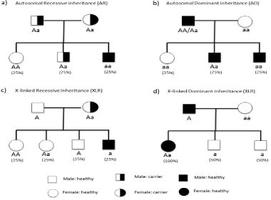
(Figure 1).
11Epilepsies with complex inheritance are
believed to be involved in 50% of epilepsies.
11,19Although familial aggregation is seen through
generations, the mode of inheritance cannot easily
be identified.
Detection of a specific chromosomal abnormality
(either structural or numerical) would be categorized
under
genetic
epilepsies
with
cytogenetic
abnormalities.
11This subgroup is mostly associated with
other neurological disorders and facial anomalies.
Mendelian disorders in which epilepsy is one of
the manifestations indicate multisystem disorders
with epilepsy as one of the characteristics. These
syndromes
include
neurocutaneous
and
Epileptic syndromes Iran J Neurol 2015; 14(1) 3
Figure 1. Mendelian modes of Inheritance (a) autosomal recessive inheritance. In this case
“a” is the mutated allele of the gene and “A” is non-mutated. Individual who receives
mutated allele from both parents (aa) would be affected with disease. Another persons
“AA” and “Aa” do not show phenotypes of disease. (b) Autosomal dominant inheritance. In
this model “A” (the dominant allele) is mutated allele and can cause disease, so any
individual who receives just one mutated allele (AA, Aa) would be affected. (c) X-linked
recessive inheritance. This mode has sex-based transmission because the gene is located on
X chromosome, therefore females have two alleles of the gene and males have just one
allele. If the mother is carrier, 50% of her boys will be affected and none of the girls in such
pedigrees would show the phenotype of disease. (d) X-linked Dominant inheritance. In this
example, the disease is caused by dominant mutated allele located in chromosome X. So if
the father is affected, all the girls would be affected and no boys would show the disease
phenotype. If the mother was affected too (Aa or AA) so the boys would have shown the
phenotype of disease with different percentage
The following section is dedicated to reviewing the
role of chromosomal abnormalities and gene
mutations in etiology of epileptic syndromes with
some examples. We are referring interested readers
for a complete list of genes mutated in epilepsies to
two reviews written by Garofalo et al.
20and
Kaneko et al.
21Chromosomal aberrations
Chromosomal aberration is characterized by atypical
number or structural abnormality of at least one
chromosome that usually leads to genetic disorders. In
numerical group, aneuploidy is usually due to
abnormal gametogenesis in parents.
22Considering
aneuploidy is accompanied with gaining or losing
considerable amount of genetic materials, apart from
sex chromosome disorders it is a fatal.
23However,
there are few cases of live birth. These patients usually
suffer from facial dysmorphisms and mental
retardation as well as seizure.
24,25Conventional
karyotyping
could
easily
identify
numerical
chromosomal aberrations.
26There are several forms of structural chromosomal
abnormalities including deletion, duplication and
translocation of portion of a chromosome. These
defects are not generally fatal, and newborns with
structural abnormalities may have developmental
delay and facial dysmorphism.
27Epilepsy is one of the
widespread features in this group of anomalies.
4 Iran J Neurol 2015; 14(1) Tafakhori et al.
ring chromosome 20 has a distinct feature of
prolonged high-voltage slow waves and seizures are
resistant to medications.
28Ring chromosome 14 has
also been reported to be resistant to antiepileptic
therapy.
32The onset of epilepsy in this chromosomal
disorder is often during the first year, mental
retardation would be a constant character and the
majority of cases have dysmorphic facial features.
EEG frequently reveals focal abnormality.
32Chromosome
6q
deletion
(Long
arm
of
chromosome 6) and chromosome 22q duplication
have been shown to be associated with dysmorphic
facial
abnormalities,
mental
retardation
and
epilepsy.
33,34As a result of high-resolution karyotyping, many
epileptic seizures have been linked to chromosomal
abnormalities.
35-37Aberrations such as microdeletions
and microduplications (microchromosomal defects)
that could not be detected by conventional karyology
might be identified by molecular cytogenetic
approaches
including
comparative
genomic
hybridization (CGH) array and
multiplex
ligation-dependent probe amplification (MLPA).
38-41Exploring
the nature of the human genome with high repetitive
DNA sequences lead to discovering recurrent
rearrangements of regions in some chromosomes such
as 15q and 16p that are involved in epilepsy could
result in recurrent heritable microdeletions and
microduplications.
42Gene mutations
In addition to chromosomal abnormalities, gene
mutations also could be associated with epilepsy
syndromes. A good example would be genes
encoding ion channel subunits.
43,44Excitatory or
inhibitory neurotransmitters in central nervous
system
45have also been recognized in Mendelian
forms of epilepsy
11,46,47and thus, following simple
Mendelian
mode
of
inheritance.
46,47Genetic
counseling could help identifying these disorders
through a prodigy and risk of disease could be
estimated for the next generation.
CHRNA4 gene encodes neural acetylcholine
receptor subunit α4.
48It was the first gene to be
associated with epilepsy syndromes. Mutation in this
gene has been linked to Autosomal dominant
nocturnal frontal lobe epilepsy.
49,50KCNQ2 and
KCNQ3 genes that encode voltage-gated potassium
channels were identified in families affected with
benign familial neonatal seizures.
51,52At least 37 genes for generalized myoclonic
epilepsy and febrile seizures, 47 genes for
symptomatic (structural/metabolic) epilepsy and 30
genes for epileptic encephalopathies have been
recognized.
20In a recent study of pediatric patients
affected with infantile spasms and Lennox-Gastaut
syndrome, two forms of epileptic encephalopathies,
and their parents, researchers found 329 de novo
mutations.
53These mutations are significantly more
prominent in genes sets regulated by fragile X protein.
Mutation of fragile X protein has been extensively
discussed in autism spectrum disorders as it is the most
widespread single-gene cause of autism
54. Further
genetic defects involved in epileptic encephalopathies
include MTOR, GABRA1 and FLNA.
53Mutation in SCN1A, a gene encoding
voltage-gated sodium channel, has been demonstrated to be
involved in Dravet syndrome. The affected patients
suffer from severe myoclonic epilepsy during infancy
with poor prognosis, as seizures are frequent,
prolonged and resistant to treatment. Developmental
delay will appear and some would have cognitive
impairment. There are reports of mutation in
PCDH19, a gene that encodes a calcium-dependent
cell-adhesion protein and is located on chromosome
X, in female patients with clinical symptoms related to
Dravet syndrome.
55,56Interestingly, 11-12% of affected
patients, who did not show any mutation in the
mentioned genes, had pathogenic copy number
variations (CNVs) in SCN1A gene. These CNVs might
be detected by array CGH and MLPA assay.
42,57Marini et al. showed that deletion of 9.3 Mb (49 genes)
of chromosome 2q without harming SCN1A gene
could also result in Dravert phenotype.
57The prevalence of Unvericht-lunderborg disease or
Baltic myoclonic epilepsy, a rare inherited form of
epilepsy with progressive myoclonus, is higher in
some regions (e.g. Sweden). This disorder has been
associated with mutation of CSTB, a gene encoding
cystatin B protein responsible for reducing the activity
of cathepsins enzymes (protease)
58and is inherited in
an autosomal recessive (AR) pattern.
59Furthermore,
different type of gene mutations including CHRNA4
gene (frontal lobe epilepsy) have been reported in
different populations.
49,50,60It seems necessary to
identify specific mutations in distinct population to
provide better genetic counseling for epilepsy.
21There are disorders that although epilepsy is one of
the symptoms, it is not the core sign. Some examples
are discussed below.
Lafora body disease, a neurodegenerative disorder,
is a fatal glycogen metabolism disorder with AR
inheritance
61,62and has been linked to EMP2A gene
mutation (Lafarin protein).
63Epileptic syndromes Iran J Neurol 2015; 14(1) 5
Myoclonus epilepsy and ragged-red fibers are a
rare mitochondrial disorder involving usually
mutation of MT-TK gene located on mitochondrial
DNA. It would lead to progressive neurological
symptoms, including blindness and myoclonic
epilepsy.
65Mitochondrial pattern of inheritance is
relatively complex (Figure 2) as maternal mutated
mitochondria affects zygote formation.
66Figure 2. Transmission of mitochondrial DNA mutation in
a hypothetical pedigree. This mode of inheritance is
categorized as non-mendelian transmission because the
mutated gene is not located in nuclear DNA. Mitochondria
and its DNA (mtDNA) will transmit to next generation
through oocyte cytoplasm so just mutated mtDNA from
mother could cause the disease. Since, we have too many
mitochondria and copies of mtDNA in a cell, the presence
of disease and severity of its phenotype will be depended on
the amount of mutated mtDNA inside individual’s cells.
Heteroplasmy means both mutated and non-mutated
mtDNA is present in a cell. Homoplasmy means the whole
mtDNAs in a cell are mutated or non-mutated
Malformation of cortical development disorders
represents a major spectrum of mental disabilities
with severe epilepsies caused by defective neuronal
migration.
Mutation
of
LIS1
gene
encoding
microtubule-associated protein is one of the several
genetic
defects
linked
to
these
disorders.
Lissencepahly with X-link gene mutation (xLIS)
67is
another defective neuronal migration disorder that
result in the lack of cerebral folds. Both genetic and
non-genetic factors (e.g. viral infections of the fetus)
are involved in etiology of these disorders.
68The introduction of new techniques of DNA
sequencing has helped identifying point mutations,
small insertions, and deletions.
38Genetic counseling and genetic testing in epilepsy
management
Epilepsy is a multifactorial disease, and both genetic
and environmental components are involved in
etiology (Table 1). Various investigations, particularly
twin studies, have contributed to detection the role of
genetic elements in epilepsy syndromes. These
findings will help predicting the clinical symptoms of
the affected individual through genotype-phenotype
correlation
69and conduct follow-up of high-risk
pregnancies or an infant born in a family with
increased rate of epilepsy.
70It will also aid the
clinician in anticipating the clinical features in
advance and manage in accordance.
Detection of specific genetic disorders will improve
our understanding of the inheritance pattern. Thus,
genetic counseling could better help families by
estimating the risk of disease in next generation and
family members of epileptic probands, who might be
at greater risk for epilepsy syndromes.
69Interestingly,
the same phenotype of epilepsy in different members
of a pedigree could be due to different genetic
defects.
67Consanguineous marriage will increase the
risk of epilepsy syndrome, especially childhood onset
of epilepsy
71-73and is a remarkable challenge for
clinicians and geneticists in societies where it is a
common tradition.
Conclusion
We emphasize on cooperation of clinicians (particularly
neurologist) and medical genetic experts in eastern
societies like Iran, where consanguineous marriage is a
common practice. This assistance is highlighted in
high-risk families. It should be noted that prior to any
genetic testing, patient and family members should be
pre-tested in genetic counseling sessions.
18The genetic testing now commercially available for
epilepsy includes analysis of 70 genes for detection of
point mutations and deletion/duplications using
DNA sequencing, CGH array, and MLPA techniques.
The specimen used for genetic testing could be whole
blood or any other body tissue appropriate for DNA
extraction, for example, amniotic fluid, and chorionic
villi samples are required for prenatal diagnosis.
Hence, collaboration of neurologist with geneticist
in the case of genetic epilepsy will help the diagnosis
and in some cases will improve management
20.
Conflict of Interests
The authors declare no conflict of interest in this study.
Acknowledgments
We appreciate the help of our department’s staff and
supports of the Tehran University of Medical
Sciences, Iran.
6 Iran J Neurol 2015; 14(1) Tafakhori et al.
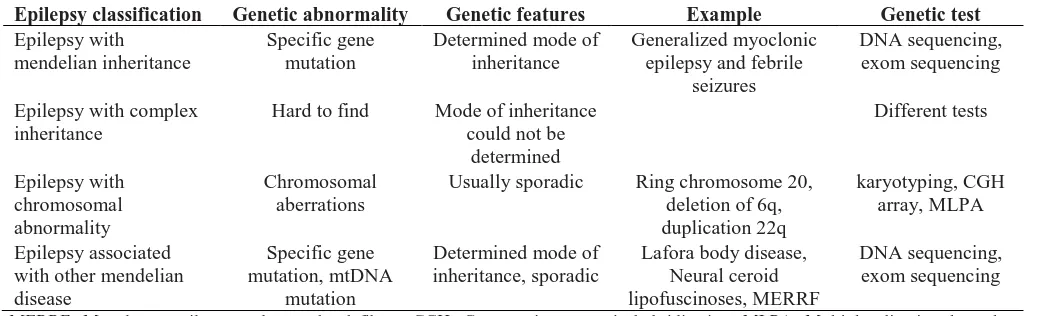
Table 1. Summary of genetic abnormalities in different forms of epilepsies
Epilepsy classification
Genetic abnormality
Genetic features
Example
Genetic test
Epilepsy with
mendelian inheritance
Specific gene
mutation
Determined mode of
inheritance
Generalized myoclonic
epilepsy and febrile
seizures
DNA sequencing,
exom sequencing
Epilepsy with complex
inheritance
Hard to find
Mode of inheritance
could not be
determined
Different tests
Epilepsy with
chromosomal
abnormality
Chromosomal
aberrations
Usually sporadic
Ring chromosome 20,
deletion of 6q,
duplication 22q
karyotyping, CGH
array, MLPA
Epilepsy associated
with other mendelian
disease
Specific gene
mutation, mtDNA
mutation
Determined mode of
inheritance, sporadic
Lafora body disease,
Neural ceroid
lipofuscinoses, MERRF
DNA sequencing,
exom sequencing
MERRF: Myoclonus epilepsy and ragged-red fibers; CGH: Comparative genomic hybridization; MLPA: Multiplex ligation-dependent probe amplification
References
1. Dekker PA. Epilepsy: A Manual for Medical and Clinical Officers in Africa. Geneva, Switzerland: World Health Organization; 2002.
2. Berg AT, Millichap JJ. The 2010 revised classification of seizures and epilepsy. Continuum (Minneap Minn) 2013; 19(3 Epilepsy): 571-97.
3. Hauser WA, Annegers JF, Kurland LT. Incidence of epilepsy and unprovoked seizures in Rochester, Minnesota: 1935-1984. Epilepsia 1993; 34(3): 453-68. 4. Beletsky V, Mirsattari SM. Epilepsy, mental
health disorder, or both? Epilepsy Res Treat 2012; 2012: 163731.
5. Engel J. The epilepsies. In: Wyngoorden J, Smith L, Bennet C, Editors. Cecil's Textbook of Medicine. 19th ed. Philadelphia, PA: WB Saunders; 1992. p. 2202-13.
6. Noachtar S, Remi J. The role of EEG in epilepsy: a critical review. Epilepsy Behav 2009; 15(1): 22-33.
7. Benbadis S. The differential diagnosis of epilepsy: a critical review. Epilepsy Behav 2009; 15(1): 15-21.
8. Panayiotopoulos CP. A Clinical Guide to Epileptic Syndromes And Their Treatment: Based on the New Ilae Diagnostic Scheme. Oxfordshire, UK: Bladon Medical Pub p. 278; 2002.
9. Berg AT, Berkovic SF, Brodie MJ, Buchhalter J, Cross JH, van Emde BW, et al. Revised terminology and concepts for organization of seizures and epilepsies: report of the ILAE Commission on Classification and Terminology, 2005-2009. Epilepsia 2010; 51(4): 676-85.
10. Tafakhori A, Aghamollaii V, Modabbernia AH, Ghaffarpour M, Omrani HA, Harirchian MH, et al. Evaluation of partial epilepsy in Iran: role of video-EEG, EEG, and MRI with epilepsy protocol. Iran J Neurol 2011; 10(1-2): 9-15.
11. Johnson MR, Sander JW. The clinical impact of epilepsy genetics. J Neurol Neurosurg Psychiatry 2001; 70(4): 428-30. 12. Lennox WG. The heredity of epilepsy as
told by relatives and twins. J Am Med
Assoc 1951; 146(6): 529-36.
13. Berkovic SF, Howell RA, Hay DA, Hopper JL. Epilepsies in twins: genetics of the major epilepsy syndromes. Ann Neurol 1998; 43(4): 435-45.
14. Harvald B, Hauge M. Hereditary factors elucidated by twin studies. In: Van Gundia Neel J, Editor. Genetics and the epidemiology of chronic diseases. Washington, DC: U.S. Dept. of Health, Education, and Welfare, Public Health Service, Division of Chronic Diseases; 1965. p. 61-76.
15. Sillanpaa M, Koskenvuo M, Romanov K, Kaprio J. Genetic factors in epileptic seizures: evidence from a large twin population. Acta Neurol Scand 1991; 84(6): 523-6.
16. Bouchard TJ, Propping P. Twins as a tool of behavioral genetics: report of the Dahlem Workshop on What Are the Mechanisms Mediating the Genetic and Environmental Determinants of Behavior? Twins as a Tool of Behavioral. New Jersey, NJ: J. Wiley; 1993. p. 326.
17. Baraitser M. The genetics of neurological disorders. 2nd ed. Oxford, UK: Oxford University Press; 1990. p. 113.
18. Ottman R, Hirose S, Jain S, Lerche H, Lopes-Cendes I, Noebels JL, et al. Genetic testing in the epilepsies--report of the ILAE Genetics Commission. Epilepsia 2010; 51(4): 655-70.
19. Kaneko S, Wada K. Molecular genetic studies of epilepsies. No To Shinkei 1998; 50(12): 1071-7.
20. Garofalo S, Cornacchione M, Di CA. From genetics to genomics of epilepsy. Neurol Res Int 2012; 2012: 876234.
21. Kaneko S, Okada M, Iwasa H, Yamakawa K, Hirose S. Genetics of epilepsy: current status and perspectives. Neurosci Res 2002; 44(1): 11-30.
22. Luthardt FW, Keitges E. Chromosomal Syndromes and Genetic Disease [Online]. [cited 2001]; Available from: URL: http://www.els.net/WileyCDA/ElsArticle/re fId-a0001446.html
23. Langer JC. Prenatal diagnosis of congenital
anomalies. What can and should be done? Can Fam Physician 1993; 39: 595-602. 24. Kumada T, Ito M, Miyajima T, Fujii T,
Okuno T, Go T, et al. Multi-institutional study on the correlation between chromosomal abnormalities and epilepsy. Brain Dev 2005; 27(2): 127-34.
25. Sorge G, Sorge A. Epilepsy and chromosomal abnormalities. Ital J Pediatr 2010; 36: 36.
26. O'Connor C. Karyotyping for chromosomal abnormalities. Nature Education 2008; 1(1): 27.
27. Marshall CR, Noor A, Vincent JB, Lionel AC, Feuk L, Skaug J, et al. Structural variation of chromosomes in autism spectrum disorder. Am J Hum Genet 2008; 82(2): 477-88.
28. Inoue Y, Fujiwara T, Matsuda K, Kubota H, Tanaka M, Yagi K, et al. Ring chromosome 20 and nonconvulsive status epilepticus. A new epileptic syndrome. Brain 1997; 120 (Pt 6): 939-53.
29. Atkins L, Miller WL, Salam M. A ring-20 chromosome. J Med Genet 1972; 9(3): 377-80. 30. Faed M, Morton HG, Robertson J. Ring F chromosome mosaicism (46,XY,20r-46,XY) in an epileptic child without apparent haematological disease. J Med Genet 1972; 9(4): 470-3.
31. Back E, Voiculescu I, Brunger M, Wolff G. Familial ring (20) chromosomal mosaicism. Hum Genet 1989; 83(2): 148-54.
32. Specchio N, Trivisano M, Serino D, Cappelletti S, Carotenuto A, Claps D, et al. Epilepsy in ring 14 chromosome syndrome. Epilepsy Behav 2012; 25(4): 585-92. 33. Vignoli A, Scornavacca GF, Peron A, La
BF, Canevini MP. Interstitial 6q microdeletion syndrome and epilepsy: a new patient and review of the literature. Am J Med Genet A 2013; 161A(8): 2009-15. 34. Han K, Holder JL, Schaaf CP, Lu H, Chen
H, Kang H, et al. SHANK3 overexpression causes manic-like behaviour with unique pharmacogenetic properties. Nature 2013; 503(7474): 72-7.
Epileptic syndromes Iran J Neurol 2015; 14(1) 7
Disord 2005; 7(3): 181-92.
36. Singh R, Gardner RJ, Crossland KM, Scheffer IE, Berkovic SF. Chromosomal abnormalities and epilepsy: a review for clinicians and gene hunters. Epilepsia 2002; 43(2): 127-40.
37. Zuberi SM. Chromosome disorders associated with epilepsy. Handb Clin Neurol 2013; 111: 543-8.
38. Mulley JC, Mefford HC. Epilepsy and the new cytogenetics. Epilepsia 2011; 52(3): 423-32.
39. Mefford HC, Muhle H, Ostertag P, von SS, Buysse K, Baker C, et al. Genome-wide copy number variation in epilepsy: novel susceptibility loci in idiopathic generalized and focal epilepsies. PLoS Genet 2010; 6(5): e1000962.
40. Bedoyan JK, Kumar RA, Sudi J, Silverstein F, Ackley T, Iyer RK, et al. Duplication 16p11.2 in a child with infantile seizure disorder. Am J Med Genet A 2010; 152A(6): 1567-74.
41. Cardoso C, Boys A, Parrini E, Mignon-Ravix C, McMahon JM, Khantane S, et al. Periventricular heterotopia, mental retardation, and epilepsy associated with 5q14.3-q15 deletion. Neurology 2009; 72(9): 784-92.
42. Mulley JC, Nelson P, Guerrero S, Dibbens L, Iona X, McMahon JM, et al. A new molecular mechanism for severe myoclonic epilepsy of infancy: exonic deletions in SCN1A. Neurology 2006; 67(6): 1094-5. 43. Cossette P. Channelopathies and juvenile
myoclonic epilepsy. Epilepsia 2010; 51(Suppl 1): 30-2.
44. Jurkat-Rott K, Lerche H, Weber Y, Lehmann-Horn F. Hereditary channelopathies in neurology. Adv Exp Med Biol 2010; 686: 305-34.
45. Werner FM, Covenas R. Classical neurotransmitters and neuropeptides involved in generalized epilepsy: a focus on antiepileptic drugs. Curr Med Chem 2011; 18(32): 4933-48.
46. Jefferys JG. Advances in understanding basic mechanisms of epilepsy and seizures. Seizure 2010; 19(10): 638-46.
47. Scharfman HE. The neurobiology of epilepsy. Curr Neurol Neurosci Rep 2007; 7(4): 348-54.
48. Anand R, Lindstrom J. Chromosomal localization of seven neuronal nicotinic acetylcholine receptor subunit genes in humans. Genomics 1992; 13(4): 962-7. 49. Steinlein OK, Mulley JC, Propping P,
Wallace RH, Phillips HA, Sutherland GR, et al. A missense mutation in the neuronal nicotinic acetylcholine receptor alpha 4 subunit is associated with autosomal dominant nocturnal frontal lobe epilepsy. Nat Genet 1995; 11(2): 201-3.
50. Hirose S, Iwata H, Akiyoshi H, Kobayashi K, Ito M, Wada K, et al. A novel mutation of CHRNA4 responsible for autosomal dominant nocturnal frontal lobe epilepsy. Neurology 1999; 53(8): 1749-53.
51. Biervert C, Schroeder BC, Kubisch C, Berkovic SF, Propping P, Jentsch TJ, et al. A potassium channel mutation in neonatal human epilepsy. Science 1998; 279(5349): 403-6. 52. Singh NA, Charlier C, Stauffer D, DuPont
BR, Leach RJ, Melis R, et al. A novel potassium channel gene, KCNQ2, is mutated in an inherited epilepsy of newborns. Nat Genet 1998; 18(1): 25-9. 53. Allen AS, Berkovic SF, Cossette P, Delanty
N, Dlugos D, Eichler EE, et al. De novo mutations in epileptic encephalopathies. Nature 2013; 501(7466): 217-21.
54. Goodlin-Jones BL, Tassone F, Gane LW, Hagerman RJ. Autistic spectrum disorder and the fragile X premutation. J Dev Behav Pediatr 2004; 25(6): 392-8.
55. Akiyama M, Kobayashi K, Ohtsuka Y. Dravet syndrome: a genetic epileptic disorder. Acta Med Okayama 2012; 66(5): 369-76.
56. Dibbens LM, Tarpey PS, Hynes K, Bayly MA, Scheffer IE, Smith R, et al. X-linked protocadherin 19 mutations cause female-limited epilepsy and cognitive impairment. Nat Genet 2008; 40(6): 776-81.
57. Marini C, Mei D, Temudo T, Ferrari AR, Buti D, Dravet C, et al. Idiopathic epilepsies with seizures precipitated by fever and SCN1A abnormalities. Epilepsia 2007; 48(9): 1678-85.
58. Pennacchio LA, Lehesjoki AE, Stone NE, Willour VL, Virtaneva K, Miao J, et al. Mutations in the gene encoding cystatin B in progressive myoclonus epilepsy (EPM1). Science 1996; 271(5256): 1731-4.
59. Norio R, Koskiniemi M. Progressive myoclonus epilepsy: genetic and nosological aspects with special reference to 107 Finnish patients. Clin Genet 1979; 15(5): 382-98.
60. Phillips HA, Marini C, Scheffer IE, Sutherland GR, Mulley JC, Berkovic SF. A de novo mutation in sporadic nocturnal frontal lobe epilepsy. Ann Neurol 2000; 48(2): 264-7.
61. Lehesjoki AE, Koskiniemi M, Pandolfo M,
Antonelli A, Kyllerman M, Wahlstrom J, et al. Linkage studies in progressive myoclonus epilepsy: Unverricht-Lundborg and Lafora's diseases. Neurology 1992; 42(8): 1545-50.
62. Serratosa JM, Delgado-Escueta AV, Posada I, Shih S, Drury I, Berciano J, et al. The gene for progressive myoclonus epilepsy of the Lafora type maps to chromosome 6q. Hum Mol Genet 1995; 4(9): 1657-63. 63. Minassian BA, Lee JR, Herbrick JA,
Huizenga J, Soder S, Mungall AJ, et al. Mutations in a gene encoding a novel protein tyrosine phosphatase cause progressive myoclonus epilepsy. Nat Genet 1998; 20(2): 171-4.
64. Mole SE, Williams RE, Goebel HH. Correlations between genotype, ultrastructural morphology and clinical phenotype in the neuronal ceroid lipofuscinoses. Neurogenetics 2005; 6(3): 107-26.
65. Shoffner JM, Lott MT, Lezza AM, Seibel P, Ballinger SW, Wallace DC. Myoclonic epilepsy and ragged-red fiber disease (MERRF) is associated with a mitochondrial DNA tRNA(Lys) mutation. Cell 1990; 61(6): 931-7.
66. Taylor RW, Turnbull DM. Mitochondrial DNA mutations in human disease. Nat Rev Genet 2005; 6(5): 389-402.
67. Kullmann DM. Genetics of epilepsy. J Neurol Neurosurg Psychiatry 2002; 73(Suppl 2): II32-II35.
68. Guerrini R, Carrozzo R. Epileptogenic brain malformations: clinical presentation, malformative patterns and indications for genetic testing. Seizure 2001; 10(7): 532-43. 69. Corey LA, Pellock JM, Boggs JG, Miller LL, DeLorenzo RJ. Evidence for a genetic predisposition for status epilepticus. Neurology 1998; 50(2): 558-60.
70. Pal DK, Pong AW, Chung WK. Genetic evaluation and counseling for epilepsy. Nat Rev Neurol 2010; 6(8): 445-53.
71. Asadi-Pooya AA. Epilepsy and consanguinity in Shiraz, Iran. Eur J Paediatr Neurol 2005; 9(6): 383-6.
72. Al-Rajeh S, Abomelha A, Awada A, Bademosi O, Ismail H. Epilepsy and other convulsive disorders in Saudi Arabia: a prospective study of 1,000 consecutive cases. Acta Neurol Scand 1990; 82(5): 341-5.
Iranian Journal of Neurology © 2015 Corresponding Author:Firoozeh Raisi
Email: ijnl@tums.ac.ir Email: fraisi@gmail.com
Original Paper
Iran J Neurol 2015; 14(1): 8-11
The prevalence of female sexual
dysfunction among migraine patients
Mohammad Abdollahi
1, Mansoureh Toghae
1, Firoozeh Raisi
2, Elaheh Saffari
11
Iranian Center of Neurological Research AND Department of Neurology, School of Medicine, Sina Hospital, Tehran University of
Medical Sciences, Tehran, Iran
2
Psychiatric and Clinical Psychology Research Center, Roozbeh Psychiatric Hospital, School of Medicine, Tehran University of Medical
Sciences, Tehran, IranKeywords
Migraine, Female Sexual Dysfunction, Female Sexual
Function Index Score
Abstract
Background: Female sexual dysfunction (FSD)
defines as any disorder in the process of sexual
contact including 6 main domains, desire, arousal,
lubrication, orgasm, orgasm satisfaction and pain.
This study was conducted to evaluate prevalence of
sexual dysfunction disorder in women with migraine
headache and also find the associated factors related
to migraine characteristics.
Methods: A total of 69 eligible woman patients fulfilling
criteria for migraine participated in this study. The
Female Sexual Function Index (FSFI), a
multi-dimensional self-report implement for appraisal of
Female Sexual Function during the past month were
utilized in this study. The information related to migraine
including frequency, duration of headache attack,
severity of headache according to visual analog scale
(VAS) score and headache impact test (HIT) score
were obtained using a self-administrated questionnaire.
Results:
About 68.4% of patients had an FSFI score
< 28. In domains of desire 73.7%, arousal 64.9%,
lubrication 21.1%, orgasm 33.3%, satisfaction 17.5%,
and pain 40.4% of patients reported some degree of
dysfunction. Among variables related to migraine
characteristics, only a significant association between
frequency and sexual dysfunction were recorded
(P < 0.05).
Conclusion:
FSD is prevalent among migraine
patients. The frequency of a migraine attack is
associated with FSD. Serotonin mechanisms such as
5HT2, 5HT3 agonist have been hypothesized as a
shared etiology for migraine and sexual dysfunction.
Introduction
Female sexual dysfunction (FSD) defines as any
disorder in the process of sexual contact, including 6
main domains, desire, arousal, lubrication, orgasm,
orgasm satisfaction and pain, which cause female
distress and impact their relationships with partner
and their quality of life.
1,2The prevalence of FSD varies in a range of 43-90%
in studies due to different definition, studies protocol,
cultural issues, environmental factors and genetics
variances.
3,4Previous findings demonstrate that FSD
is multifactorial, and there is a
genetic susceptibility
for sexual dysfunction that is influenced remarkably
by
environmental
factors.
Both
genetic
and
environmental factors are involved in all dimensions
of sexual function.
1,5In recent years, Studies discuss
about the significant impact of chronic pain on female
sexual function. Chronic illness and chronic pain
result in less sexual satisfaction and cause some
degree of sexual dysfunction.
6,7Primary headache especially migraine is a
common cause of chronic pain and temporary
disability.
8The prevalence of migraine and chronic
headache in women is respectively 17.1% and 4%.
9-10Gal Ifergane in his investigation on a student sample
showed that a migraine suffers have a higher sexual
pain, and satisfaction disorder compared with
control subjects.
11Bestepe et al. assessed sexual function among
Iranian Journal
of Neurology
Female sexual dysfunction in migraineurs Iran J Neurol 2015; 14(1) 9
headache suffers using Arizona sexual experiences
scale (ASEX score) and concluded that migraineurs
have more difficulties with vaginal lubrication and
achieving orgasm in comparison to normal samples.
12In a large population based study in United States, the
frequency and quality of sexual relationships were
affected in 86% of migraine suffer, and resulted in
divorce in 26% of cases.
13This study is conducted to evaluate the prevalence
of sexual dysfunction disorder in women with
migraine headache in Iran.
Materials and Methods
We designed a
cross-sectional study to assess sexual
function in women under treatment and follow-up for
migraine. Our aim was to investigate the prevalence
of female sexual function disorder among migraineur
patients and also to identify the associated factors of
FSD including headaches characteristics.
The study was conducted between April and June
2013 in a headache clinic center.
The Ethics Committee of the Tehran University of
Medical Sciences approved the study. Informed
consent was obtained from the patients.
Eighty-eight women with complain of headache
consecutively were interviewed to participate in this
study. The patients enrolled in this study met the
International Classification of Headache Disorders
criteria for migraine and had a sexual partner for a
minimum period of last 1 year
.
69 eligible patients
were recruited.
A
detailed
history
of
headache
attacks
characteristics were obtained in an interview of an
expert neurologist with patients. Our interview
includes questions for disease duration, severity and
frequency of headache attacks and attack duration.
The severity of headache attacks was estimated based
on visual analog scale (VAS) score. The impact of
headache on quality of life was evaluated with
headache impact test (HIT) score questionnaire. The
Female
Sexual
Function
Index
(FSFI),
a
multidimensional self-report implement for appraisal
of Female Sexual Function during the past month,
were utilized in this study. This questionnaire consist
of 19 questions in six main domains of sexual function
including sexual desire, sexual arousal, vaginal
lubrication, ability to achieve orgasm, orgasm
satisfaction and pain and rated on a 5 points scale and
full score range from 2 to 36. The cut-off point for the
scale was found to be 28 in Iranian translated draft of
FSFI
questionnaires
(sensitivity
=
83%
and
specificity = 82%).
14Details about subscale cut-off
point is presented in table 1.
SPSS software (version 18, SPSS Inc., Chicago, IL,
USA) were applied to describe the sexual function status
of subjects. Independent t-test was administrated to
compare HIT Score, frequency and VAS Score in two
subgroups of migraineurs regarding patients with or
without FSD. P ≤ 0.05 was considered to be significant.
Table 1. Female sexual function and subscale scoring and
cut-off points
Domain
Questions Factor Cut-off point
FSFI
Desire
1, 2
0.6
3.3
Arousal
3, 4, 5, 6
0.3
3.4
Lubrication
7, 8, 9, 10
0.3
3.7
Orgasm
11, 12, 13
0.4
3.4
Satisfaction
14, 15, 16
0.4
3.8
Pain
17, 18, 19
0.4
3.8
Total
28
FSFI: Female sexual function index
Results
Of the 69 migraineur women who were interviewed,
12 (17%) were excluded from the analysis because of
significant missing data. Average age of the migraine
patients in the analysis sample was 38.11 ± 9 years
(range 15-57). About 68.4% of migraine patients
reported FSF score under the validated cut-off (< 28).
Most common sexual dysfunction in migraine patients
were observed in domains of desire and arousal
(73.7 and 64.9%, respectively) (Figure 1).
Figure 1. Prevalence of subscales of sexual dysfunction in female migraine patients
FSD: Female sexual dysfunction68.40%
73.70%
64.90%
21.10%
33.30%
17.50%
40.40%
0% 10% 20% 30% 40% 50% 60% 70% 80%
FSD Desire disorder
Arousal disorder
Lubrication disorder
Orgasm disorder
Satisfaction disorder

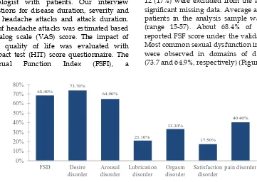


![Table 1. Comparison of risk factors among two groups Risk factors Surgical Clipping [n (%)]](https://thumb-us.123doks.com/thumbv2/123dok_us/8752447.1748956/29.595.44.557.673.768/table-comparison-factors-groups-risk-factors-surgical-clipping.webp)
