Fatimah A. Jasim (Correspondence)
fjasim119@gmail.com
+This article is published under the terms of the Creative Commons Attribution License 4.0 Author(s) retain the copyright of this article. Publication rights with Alkhaer Publications. Published at: http://www.ijsciences.com/pub/issue/2017-03/
DOI: 10.18483/ijSci.1197; Online ISSN: 2305-3925; Print ISSN: 2410-4477
TiO
2
Nanoparticles Induce Lung Fibrosis and
Proteinosis through Influence on Matrix
Metalloproteinase Expression
Fatimah A. Jasim
1
, Dhamia K. Suker
2
, Adnan I. Albadran
3
1PhD. Student, Department of Biology, College of Science, University of Basrah, Basrah , Iraq.
2
Professor, Cell and Biotechnology research unit, College of Science, University of Basrah, Basrah , Iraq.
3
Professor, Department of Biology, College of Science, University of Basrah, Basrah , Iraq.
Abstract:
Background: nanotechnology applications, speared very quickly while very little has been done to measure and assess the hazard of nanoparticles (NPs) to an ecosystem and to the biological systems.Lung exposure to titanium dioxide nanoparticle (TiO2 NP) may induce pulmonary alveolar proteinases and fibrosis through influence on matrix
metalloproteinase expression (MMPs).
Methods: In order to study this, TiO2 NP was instilled into the lung, then, histopathological alteration and MMPs
(MMP-1, MMP-2, Collagen-I) expression using RT-qPCR were assessed at 4 days, a month and 3 months post-instillation. Data were analyzed using ANOVA test and gene expression was normalized to that of housekeeping gene, which was hypoxanthine phosphoribosyltransferase (HPRT).
Results: The results showed that TiO2 NP induces acute inflammation in lung tissue after 4 days post-instillation
with significantly decrease (p<0.05) in MMPs expression. While inducing fibrosis and proteinosis with significantly increase (p<0.05) in MMPs expression after a month post- instillation. Otherwise, after 3 months post-instillation the fibrosis and proteinosis were decreased and the expression significantly increased (p<0.05).
Conclusion: TiO2 NP induces many alterations in lung structure after 4 days and a month from intratracheal instillation this included metaplasia in bronchus epithelial and in alveolar epithelial, Fibrosis, angiogenesis and proteinaceous through affected on MMPs expression which decreased the expression of MMP-1, MMP-2, MMP-12 and Collagen-I in the lung.
Keywords: Lung fibrosis; Pulmonary alveolar proteinosis; TiO2 NPs; MMPs
Introduction
Nanoparticles are microscopic matters have one or more dimensions less than 100 nm and have attracted powerful scientific interest. Distinguishing volume-dependent properties of nanoparticles are mainly due to their relatively large surface area. They are utilized as a targeted transmission system for transport of tiny and major molecules by changing their pharmacokinetics and pharmacodynamics properties. Nanoparticles are old in the environment as their presence had been discovered a long time ago for e.g. air pollutants, but they had been reported and formulated for different beneficial purposes such as medicine delivery, tissue targeting, cancer therapy, diagnostic operator and for imaging purpose (1).
Titanium dioxide nanoparticles (TiO2 NP), which
generally utilized in prettifying, paints, sunscreens, and nutrition, induced emphysema and lung sore in mice (2). TiO2 particles are not immunologically
active and may be an essential supplement in beat normal gut cell hypo-responsiveness to endogenous luminal molecules. They assumed that the TiO2
http://www.ijSciences.com
Volume 6
– March
2017 (03
)
2
arthritis (RA) and damage the joints by the degeneracy of the cartilage and bone, as well as motivating angiogenesis and inflammation (4). The MMP prevent fibrosis or, for that issue, any illness or remedy process by controlled their catalysis. Contrasted with a normal remedy, the action of an effector MMP might be over/or under performed fibrosis, and conversion in overall activity can be created by two mechanisms: First, biosynthesis of the MMP might be varied. Expression of most MMPs managed at the level of transcription. Therefore, over/or downregulation of MMP would appear, the effects on regulated gene expression in a specific cell type are also modified. The second mechanism(s) that MMP activity form is different controls over enzyme efficiency, experience data demonstrating or submitting functional roles, both protective and injurious, for specific MMPs in lung, kidney and liver fibrosis. Although there are many mechanisms for obstetrics fibrosis diffuse among these organs, notably involving the constant activation of myofibroblasts from occupant interstitial cells and common immune characteristics, it is remarkable to note that the roles for specific MMPs are different among organ systems (5). Through the severe inflammatory stage of acute respiratory distress syndrome, irregular releasing of active cytotoxic moderators from penetrating leukocytes, including matrix metalloproteinases (MMPs), nitrogen species, proteolytic enzymes such as elastase and reactive oxygen, effects in injury to pulmonary epithelial and endothelial cells and, if acute, damages the lung scaffold (6).
Methods
Preparation of Nano-TiO2 Solution
Manufactured nano-TiO2 was purchased from Sigma, particles with a size 21 nm. Nano-TiO2 was sterilized in 121 °C for 20 min to decrease the danger of bacterial pollution, then powdered TiO2 was scattered into a suspension with 0.9% (w/w) sodium chloride (NaCl). For adequately scattered particles, solutions containing TiO2 particles were sonicated for 5 min by Ultrasonicator processor and vortexed before it's utilization in treatment (7).
Animals and Experiment Design
A total of 63 male rats (8weeks age) with average body weight of 123.87±22.47 g were used. Animals housed in cages kept in standard conditions in animals’ room, 25˚C temperature with relative humidity at 60% and a 12 hour light/dark cycle, distilled water and sterilized food for rats were available ad libituium. Rats were divided into seven groups. The control group was treated with 0.9% w/w
NaCl solution, and the treated groups with (0.5, 5, 50
mg/kg) (8), 1.5, 15, 150 mg/kg (B.W.) of nano-TiO2
(9,10). All groups underwent repeat exposure (twice a week, for four consecutive weeks) by 0.1 ml/100 g (B.W) intratracheal instillation. Rats anesthetized with 16.5mg/kg xylazine and 112.5mg/kg ketamine until their breathing was slow, then placed in a supine position with the head elevated. A tracheal cannula was inserted through the vocal cord to reach the trachea bronchial and insert nano TiO2 solution into
the lungs (11). After 4 weeks of intra-tracheal instillation, three rats from each group were randomly sacrificed after being anesthetized with xylazine and ketamine in 4 days, 1 month and 3 months, animals and lung were weighted.
Histopathological Examinations
Histopathological examinations performed by using standard laboratory procedures. Tissue samples were removed from experimental groups and rinsed thoroughly for 1 mint in normal saline. Then, the tissue was fixed in a Carnoy's fluid for 60 minutes and transferred to 95% percent or absolute alcohol for 1 hours. Thereafter, processed to paraffin embedding routine. Sections of 5-7 µm were stained with hematoxylin and eosin stain as well as Van Geison stain, then examined under light microscope for histopathological alteration and collagen contents (12).
Gene Expression of Extracellular Metalloproteinase
http://www.ijSciences.com
Volume 6
– March
2017 (03
)
3
equation [1]. The ΔCT was calculated by subtracting target CT from that of HPRT genes while the NO. of copies was calculated according to the standard curve
shown in appendix A (16). The RT-qPCR product was detected by melting curve.
Ratio (reference target ) = 2
Ct(reference)−Ct(target)….[1]
Table (1) Primers sequences used in RT-qPCR Gene
s
Genebank
accession number Sequences (5´- 3´) Size of products (bp)
MM P-1
NM_001134 530.1
GCCAACAGGTGCAACAACAC
GCATCAAGTTTACCTGGCAGATT 186
MM
P-2 RNCOLL
ACAGTGACACCACGTGACAAGC
CATTCCCTGCGAAGAACACAG 130
Coll
agen-I NM_053356
TGCTGCTTGCAGTAACGTCG
TCAACACCATCTCTGCCTCG 116
HPR T
NM_012583 .2
TTGGAAAGGGTGTTTATTCCTCAT
ATCCAGCAGGTCAGCAAAGAA 158
Statistical aAnalysis:
Statistical analysis of all data was carried out using the ANOVA test with differences less than 0.5 (p<0.05) considered to be statistically significant. This calculation was carried out according to the Statistical Package for Social Science (SPSS version 20) and the least significant difference (L.S.D) at a level less than (0.05) was also used.
RESULTS
Coefficients of Tissue
The coefficients of tissue are shown in the table [2]. It was defined as grams (wet weight of tissue/body weight). In 4 days after intratracheal instillation, at low doses, there was significantly decreased in coefficients and increased coefficients at the high dose in lung tissue (p<0.05). After a month and 3 months of instillation, the coefficient was significantly decreased in lung tissues at all doses (p<0.05) compared with that of the control group which depended on the dose concentration.
Table (2) Ended weight and organs weight percentage of rats during the study. TiO2NPs (mg/kg) B.W.
Groups 0 0.5 1.5 5 15 50 150
4days body 126.34±4.54 164.35±0.65 194.33±8.48 164.17±0.05 127.33±9.63 144.20±0.06 126.18±15.77 lung 1.53±0.10 0.74±0.064* 0.85±0.112* 0.88±0.121* 1.12±0.297* 0.99±0.11* 1.99±0.34* month body 171.33± 1.3 324.56±1.99 176.13±7.37 318.63±11.06 184.33±0.25 317.36±11.49 199.33±0.58
lung 1.404±0.07 1.058±0.13* 1.179±0.048* 0.895±0.03* 0.931±0.04* 0.814±0.01* 1.23±0.09* 3months body 171.33±4.56 326.17±4.06 176.13±7.36 318.63±11.05 184.73±6.47 317.36±11.49 199.33±0.58
lung 1.40±0.07 1.05±0.12* 1.18±0.48* 0.9±0.28* 0.93±0.43* 0.81±0.08* 1.26±0.049*
Histopathological Examinations:
The histological observation showed the normal structure of the lung in the control group. Histopathological observation after 4 days of intratracheal instillation, on the treated groups with (0.5, 1.5, 5, 15, 50, 150) mg/kg of TiO2 included
acute inflammatory cells infiltration (polymorph leukocyte and neutrophil) in alveolar septa with many
http://www.ijSciences.com
Volume 6
– March
2017 (03
)
4
with small round deeply basophil nuclei (fig.1), metaplasia occurred in some region of the bronchial epithelium which represented by the changes from pseudostratified ciliated epithelium to cuboidal or simple or stratified squamous epithelium, enlarge lymphatic nodule which extended into the bronchus epithelial Beginning of fibrosis featured by a heavy increase in density of collagen fiber surrounded the bronchus and the bronchioles also around the dilated veins with numerous fibrocytes (fig.2)
After a month from an intra-tracheal instillation, the treated groups with lower doses show some improvement in healing and this included the alveolar sacs return to the normal structure while there was still some alteration noticed such as enlarge lymphatic follicle around the bronchiole, aggregation of inflammatory cells around the dilated blood vessels. Lymphatic follicles frequently appeared in alveolar septa, An increase in density of collagen fiber as bundle and strands. TiO2 NPs deposition was
decreased in all groups compared with the preceding time, it was found in alveolar septa and inside macrophages. Metaplasia occurred in squamous epithelial of alveoli, which transferred to stratified cuboidal epithelial, there was an eosinophil material filled the alveoli and in interalveolar septa probably present a proteinaceous (fig.3). Big foamy macrophages fill the alveoli, TiO2 induce a chronic
granulomatous inflammation (fig.4).After 3 months of intratracheal instillation, the treated groups exhibited a healing improvement in their structure except the lower doses possess a developed of emphysema due to alveolar sacs extending, terminal bronchioles with normal collagen fiber detected, extending of tunica adventitia of pulmonary artery while the proteinaceous was decreased, but the immune response was still presented, including the forming of lymphoid follicles in the alveolar septa and around the blood vessels. The result showed an increase in the smooth muscle and tunica adventitia thickness due to increase collagen fiber(fig.5).
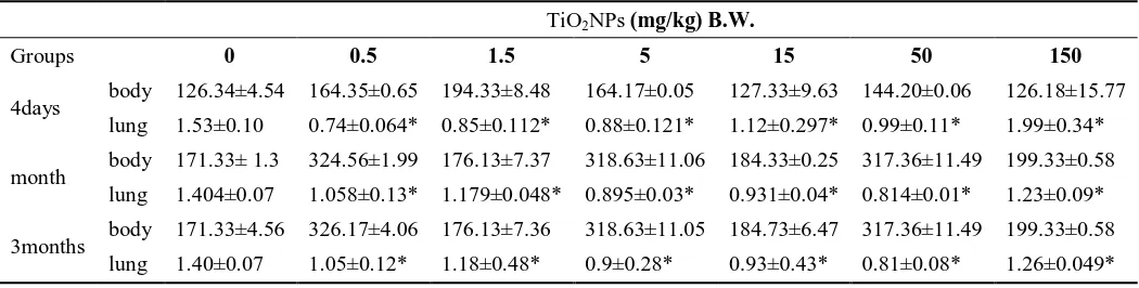
Fig. 1. Histological section of lung from (A) the control group and (B, C, D, F, G) treated group with (0.5, 1.5, 0.5, 5, 15, 50, 150) mg/kg of TiO2 NP respectively, after 4 days from intratracheal instillation, (A) showing the normal
http://www.ijSciences.com
Volume 6
– March
2017 (03
)
5
arrows). (B-G)showing the alteration in lung structure, a heavyinfiltration of lymphocyte in the alveolar space and septa (thick arrows), heavy bleeding in alveolar septa and deposition of TiO2 NP in alveolar septa and macrophage).
H&E stain.
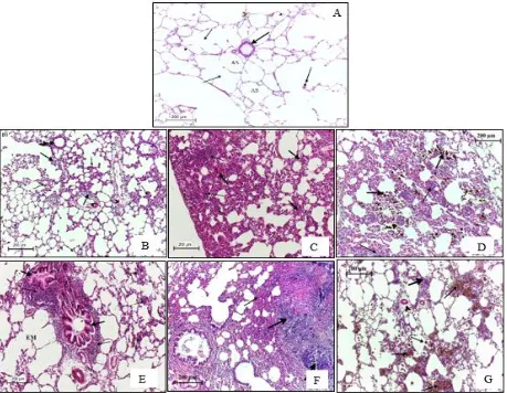
Fig. 2. Section of lung from the control group (A, B) and treated group (C, D, F, G, H) with (0.5, 1.5, 0.5, 5, 15, 50, 150) mg/kg of TiO2 NP respectively, after 4 days post-instillation, (A, B) showing the normal bronchus lining
http://www.ijSciences.com
Volume 6
– March
2017 (03
)
6
Fig. 3. Illustrated the alteration on lung of the treated groups (B, C, D, E, F, G) with (0.5, 1.5, 0.5, 5, 15, 50, 150) mg/kg of TiO2 NP respectively comparing with control group (A) after a month post-instillation, showing
lymphocyte cluster in alveolar (thin arrow), proteinaceous material present in alveoli (thick arrows). H&E stain
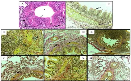
Fig. 4. A cross section o lung of the control group (A, B) and the treated group with (C, D, F, G, H) with (0.5, 1.5, 0.5, 5, 15, 50, 150) mg/kg of TiO2 NP respectively, after a month post-instillation, showing metaplasia in alveolar
http://www.ijSciences.com
Volume 6
– March
2017 (03
)
7
detected in alveoli, collagen bundle greatly increased and a chronic granulomatous inflammation also induced. A, C, D, E, G: Van Geison stain; B, F, H: H&E stain.
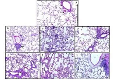
Fig. 5. Histopathological alteration on lung section of the treated groups (B, C, D, E, F, G) with (0.5, 1.5, 0.5, 5, 15, 50, 150) mg/kg of TiO2 NP respectively comparing with control group (A) after 3 months post-instillation, the
section revealed cluster of mature lymphocytes, smooth muscle and tunica adventitia of pulmonary artery increased in thickness, decreased in proteinaceous in the alveoli. H&E stain.
Gene expression of Extracellular Metalloproteinase:
Purification of Total RNA:
Agarose gel electrophoresis was performed to identify the cleansed all out cell’s RNA from rat lung tissue (Fig. 6). The concentration of RNA was 202.43 ng/µl measured on A 260˚ nm by NanoDrop
spectrophotometer.
Amplification of Extracellular Metalloproteinase Gene:
http://www.ijSciences.com
Volume 6
– March
2017 (03
)
8
Fig. 6. 0.8% Gel electrophoresis (at 60 volts for 30 min) analysis of total RNA extract from all lung’s groups; lane 1: ladder (100 bp DNA marker), lane 2 to 8: total RNA extracted from the experiment groups (control and treated groups).
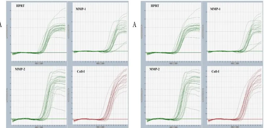
Fig. 7. A: Quantification of mRNA level by real-time quantitative PCR (RT-qPCR) for genes, B: Melting point for all genes.
http://www.ijSciences.com
Volume 6
– March
2017 (03
)
9
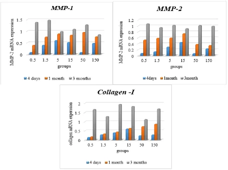
The increase in expression of MMP-1, MMP-2, and Collagen-I was continued after 3 months of intra-tracheal instillation in all experiment groups with no significant different (p>0.05). (fig.8)
Fig. 8. Effect of TiO2 NP on the mRNA expression of MMP-1, MMP-2, and Collagen-I in the lung after 4 days, a
month and 3 months of intra-tracheal instillation.
Table (3) Effect of TiO2 NP on the amplification of extracellular metalloproteinase mRNA by RT-qPCR analysis
after 4days, a month and 3 months of intra-tracheal instillation for consecutive 4 weeks.
Genes TiO2NP (mg/kg BW)
4days 0 0.5 1.5 5 15 50 150
refer-HPRT Ct 23.106 22.701 22.774 23.105 22.997 23.275 23.067
http://www.ijSciences.com
Volume 6
– March
2017 (03
)
10
MMP-1 Ct 23.515 26.514 24.227 24.235 24.877 27.123 24.517
Relative
Copies 32.705 3.669E+3 4.00E+2 1.738E+2 3.854E+3 1.36E+5 7.189E+2 Ratio of
MMP-1/HPRT
0.755±0.049 0.072±0.014a 0.306±0.112a 0.508±0.282 0.393±0.36a 0.07±0.012a 0.367±0.02a
MMP-2 Ct 22.040 25.538 25.144 24.278 23.625 26.254a 25.292a
Relative
Copies 28.1838 4.68E+4 1.395E+4 4.94E+3 5.457E+2 2.519E+5 4.13E+7 Ratio of
MMP-2/HPRT
1.661±0.432 0.145±0.0432a 0.209±0.089a 0.482±0.210a 0.721±0.374a 0.128±0.018a 0.408±0.33a
Coll-I Ct 28.721 31.049 30.17 30.045 29.103 31.0519 30.511
Relative
Copies 7.27E+2 1.179E+5 2.76E+4 1.856E+4 4.872E+3 1.364E+6 7.647E+4 Ratio of
Coll-I/HPRT 0.021±0.007 0.003±0.0006
a
0.006±0.0008a 0.008±0.001a 0.011±.002a 0.005±0.0035a 0.006±0.0001a 1month
refer-HPRT Ct 22.773 22.701 23.441 22.772 22.997 23.734 22.734
Copies 2.85E+7 2.43E+8 4.17E+6 2.43E+8 2.61E+6 5.79E+3 2.62E+6
MMP-1 Ct 21.995 23.333 22.423 22.257 22.106 22.694 22.993
Relative
Copies 1.01E+5 2.696E+5 3.28E+4 1.19E+4 1.49E+4 7.08E+4 1.61E+5 Ratio of
MMP-1/HPRT
1.83±0.75 0.656±0.15a 1.419±0.68 1.461±0.39 1.943±0.75 1.537±0.39 0.822±0.95
MMP-2 Ct 20.005 20.897 20.846 20.764 20.795 22.020 21.587
Relative
Copies 1.56E+3 9.75E+3 1.91E+4 6.28E+3 9.31E+3 1.46E+5 6.74E+4 Ratio of
MMP-2/HPRT
6.844±0.807 3.602±1.045a 3.874±0.92a 4.065±0.74a 4.954±2.3 2.564±1.1a 2.341±1.00a
Coll-I Ct 24.214 26.412 26.355 25.808 26.804 24.858 24.5166
Relative
Copies 3.84E+3 2.24E+5 2.01E+5 1.98E+5 1.35E+7 1.34E+4 1.72E+4 Ratio of
Coll-I/HPRT 0.386±0.386 0.077±0.016
a
0.134±0.02 0.157±0.12 0.168±0.24 0.347±0.11 0.347±0.171 3months
refer-HPRT Ct 22.739 23.275 24.065 23.526 22.976 22.752 23.186
Copies 4.35E+3 2.82E+6 1.39E+5 4.19E+4 1.12E+5 2.19E+6 2.14E+5
MMP-1 Ct 23.303 24.109 24.302 24.129 24.562 24.718 24.224
Relative
Copies 22.005 1.38E+3 5.53E+2 1.36E+2 1.18E+3 5.53E+2 1.23E+3 Ratio of
MMP1/HPRT 0.679±0.062 0.944±0.359 0.969±0.625 0.668±0.133 0.749±0.415 0.927±0.183 0.56±0.319
MMP-2 Ct 21.084 23.453 21.004 22.268 20.214 22.211 21.232
Relative
Copies 3.22E+6 5.59E+6 2.56E+4 5.46E+5 1.39E+4 2.41E+5 2.03E+4 Ratio of
MMP2/HPRT 0.926±0.103 0.984±0.087 0.873±0.036 0.946±0.022 0.843±0.044 0.938±0.051 0.919±0.065
Coll-I Ct 26.464 26.373 27.553 26.301 25.829 26.600 26.494
Relative
Copies 6.51E+4 3.24E+4 4.98E+5 9.01E+4 1.15E+4 5.52E+4 2.18E+5 Ratio of
http://www.ijSciences.com
Volume 6
– March
2017 (03
)
11
aThe mean difference is significant (p < 0.05)
Discussion
Dependent on the variant endpoints, we detected the concerted toxic influence of exposure to TiO2 NP.
Probably TiO2 NPs causes cell damage related to the time and dose. Toxins exposure can happen through skin touch, inhalation, injection, and ingestion. The clusters of TiO2 NPs that deposited in lungs observed in high dose groups and some of them were not phagocytosed by alveolar dust cell that resembles with Li et al.(17) study. TiO2 -NPs treatment decreased the body weight significantly but no significant differences appeared in the coefficients of liver, kidney, and testis but, the coefficient of the brain was significantly higher in TiO2-NPs- exposed rats than the control group (18). In Abu-Dief et al. (11) study, intraperitoneal injection of different doses of TiO2 NPs can significantly decrease body weight and increase coefficients of the liver. Bermudez etal. who explained decrease body weight and shorter lifetime due to retention and overload of TiO2 NPs in
vivo (19). Duan and his associates stated that TiO2
NPs were difficult to clearance in vivo result in its deposition in the liver and hepatic lesion (20). It has been demonstrated that TiO2 excite special
apoptotic pathways (21), Roulet et al. (14) recognized inflammatory histological changes included vascular and bronchial infiltration of inflammatory cells. Angiogenesis occurring in tissues beyond the area of existing blood vessels as the results of reaction between fibroblasts with endothelial cells (22). TiO2 NPs trends to
conglomerate when administered by aerosol, especially at high doses, the size of nanoparticle conglomerates in the physiological environment may have a crucial influence on macrophage phagocytosis, as it represents the real size that could elucidate a pulmonary immune cell response, besides that, the inflammation produced stimulated by an elevate positive zeta potential which may induce NPs to injury the safety of the phagolysosomal membrane inside the phagolysosome under acid conditions (23). Porter et al. (24) reported histopathology scores for alveolitis, phagocytosed NPs, alveolar histiocytosis, and interstitial fibrosis, phagocytosed TiO2 NPs in
alveolar macrophages were significantly higher for mice exposed to nanospheres (7.5 and 30 µg) and short nanobelt (30 µg) at 28 and 112 days post-exposure compared with vehicle exposed controls, there was also a tendency for the long nanobelt to cluster or aggregate within cell cytoplasm of alveolar macrophages compared with the nanospheres, phagocytosis of nanospheres (30 µg dose) was also more evident in tracheobronchial lymph nodes at 112 days exposure compared with 28 days
post-exposure. Research with high-aspect ratio nanoparticles shows blocked rescue from the lungs after inhalation exposure leading to the pathogenesis of diseases such as pulmonary fibrosis and mesothelioma (24). At present, there is evidence that high-aspect ratio TiO2 NPs are relatively more
pathogenic, and may elicit "frustrated phagocytosis" by macrophages, lysosome, and impaired lung clearance. Indeed, long-term studies show a link between particle retention and pulmonary pathology (25). Scarino etal. showed that lung tissue of chicken egg ovalbumin exposure to TiO2 NPs inducing
pulmonary inflammation, as shown by the leukocytes surrounding bronchioles and extent into alveoli (26). Rats also were unique in the development of gradual fibroproliferative lesions and alveolar epithelial metaplasia in response to exposure to a high concentration of p-TiO2 particles for 90 days (27).
Rats and mice, but not hamsters, experienced overload at 10 mg/m2 nano-TiO2. Moreover, only rats had fibroproliferative lesions and alveolar epithelial bronchiolization (28). An inflammatory response is then observed and develops into active chronic inflammation, an increase in collagen deposition (coming from fibroblast proliferation) and epithelial cell proliferation, as well as metaplasia, were observed in rats subjected to a high dose of carbon black (29). The physical contact between the epithelium surface and the NPs may have been a source of injuries and restrain the enough gas exchange, leading to the high mortality rates after TiO2 NP instillation.
Matrix metalloproteinase expression was affected by TiO2 NP which demonstrated in this study. There was
decreased in the expression of (MMP-1, MMP-2,
MMP12 and Collagen I) genes in 4 days
http://www.ijSciences.com
Volume 6
– March
2017 (03
)
12
modulators but also the determination of cell behavior. Crucially, some MMPs seem to encourage a fibrotic response while others appear to play a protection role (31).
In consequence of the degradation of the extracellular matrix by metalloproteinases, healthy cells can also be influenced by proliferation, apoptosis, or pathological morphogenesis. MMPs can also change the activity, they can even modify other proteins expressions (32). Armand et al. (33) demonstrated that five TiO2 NPs stimulated a significant
dose-dependent increase in MMP-1 mRNA expression 48 hours after the initial exposure but not at earlier time points, micrometric TiO2 also stimulated MMP-1
mRNA-protein expression., so MMP-1 expression modulation by TiO2 particles was mirrored by a
significant dose-dependent decrease. Furthermore, Armand and coworkers reported that a dissociation between MMP-1 expression (mRNA and protein) and its activity.
In normal tissues, MMPs are expressed at low levels, and their participation manifests to play an important role in the development of a several of pathological processes including fibrosis. the involvement of MMPs demonstrated in the pathogenesis of pulmonary and hepatic fibrosis. For instance, broncho-alveolar lavage fluids from individuals with sarcoidosis or pulmonary fibrosis contain high levels of collagenase, concept to be neutrophil-derived, a fact that has been proposition to be related to the development of fibrosis in these subjects, the involvement of MMP-2 in extracellular matrix precipitation was also suggested in a model of bleomycin stimulated pulmonary fibrosis in rabbits (34). MMP-2 is a protease have gelatinolytic activity (hence its alternate name, gelatinase A), which is demonstrated to be expressed constitutively in various lungs cell types. This enzyme has a broad spectrum of substrates and is involved in modulating different cellular functions, including angiogenesis, tissue remodeling, and potentiation of the inflammatory response, MMP-2 is thought to contribute to the pathogenesis of a variety of pulmonary disorders, including chronic obstructive pulmonary disease, asthma, lung cancer and pulmonary fibrosis (35). Geraghty etal. (36) recently demonstrated that neutrophil elastase may raise MMP-2 expression from epithelial cells, potentially leading to increased remodeling and inflammatory response in cystic fibrosis. For example, erasure of MMP2 is preventive in allotransplant models because it significantly decreases cellular infiltration and fibrosis (37).
Conclusion
TiO2 NPs are widely used, many applications as the additive, including drugs deliver, a white pigment in paint, a food colorant, in cosmetic creams and sunscreens as well as in the environmental purification of air, water and soil as pesticide destruction products. So the potential health of TiO2 NPs had gained increasing attention and because of the smaller particles of TiO2 has more reactivity, effectively and toxicity; in this study we demonstrated that TiO2 NP affected on MMPs
expression and histopathological alteration on lung including fibrosis, acute inflammation, and proteinaceous.
Acknowledgments
This article was part of a Biostatistics PhD thesis entitled “Histopathological and immunohistochemistry effects of TiO2 NP after repeated intra-tracheal instillation and evaluation of metalloproteinase expression in animal model” in University of Basrah, College of science.
References
1. Garg, A., Visht, S., Sharma, P. K. & Kumar, N. Formulation, Characterization and Application on Nanoparticle: A Review. Der Pharmacia Sinica, 2011; 2(2), 17–26.
2. Ambalavanan, N., Stanishevsky, A., Bulger, A., Halloran, B., Steele, C., Vohra, Y., & Matalon, S. Titanium oxide nanoparticle instillation induces inflammation and inhibits lung development in mice. American Journal of Physiology.
Lung Cellular and Molecular Physiology, 2013; 304(3),
L152-61.
3. Powell, J. J., Harvey, R. S., Ashwood, P., Wolstencroft, R., Gershwin, M. E., & Thompson, R. P. H. Immune potentiation of ultrafine dietary particles in normal subjects and patients with inflammatory bowel disease. Journal of
Autoimmunity, 2000; 14(1), 99–105.
4. Jackson, B. C., Nebert, D. W., & Vasiliou, V. Update of human and mouse matrix metalloproteinase families.
Human Genomics, 2010; 4(3), 194–201.
5. Giannandrea, M., & Parks, W. CDiverse functions of matrix metalloproteinases during fibrosis. Disease Models &
Mechanisms, 2014; 7(2), 193–203.
6. Burnham, E. L., Janssen, W. J., Riches, D. W. H., Moss, M., & Downey, G. P. The fibroproliferative response in acute respiratory distress syndrome: mechanisms and clinical significance. Eur. Respir. J., 2014; 43(1), 276–285. 7. Fu Y., Zhang Y., Chang X., Zhang Y., Ma S., Sui J., Yin L.,
Pu Y.& Liang G. Systemic Immune Effects of Titanium Dioxide Nanoparticles after Repeated Intratracheal Instillation in Rat. Int. J. Mol. Sci., 2014; 15, 6961-6973.
8. Abd Al-abbas, M. J. MLST of S Aureus Isolates Identified
by 16SrRNA Gene Sequencing. Lambert Academic
Publishing. 2012;221pp.
9. Liu R., Yin L., Pu Y., Liang G., Zhang J., Su Y., Xiao Z. & Ye B. Pulmonary toxicity induced by three forms of titanium dioxide nanoparticles via intratracheal instillation in rats. Progress in Natural Science, 2009; 19, 573–579. 10. Shinohara, N., Oshima, Y., Kobayashi, T., Imatanaka, N.,
Nakai, M., Ichinose, T., … Gamo, M. Dose-dependent clearance kinetics of intratracheally administered titanium dioxide nanoparticles in rat lung. Toxicology, 2014; 325. 11. Abu-Dief, E. E., Khalil, K. M., Abdel-Aziz, H. O.,
http://www.ijSciences.com
Volume 6
– March
2017 (03
)
13
Dioxide Nanoparticles in Adult Male Albino Rat Liver and Possible Prophylactic Effects of Milk Thistle Seeds. Life Science Journal, 2015;12(2), 115–123.
12. Baisch, B. L., Corson, N. M., Wade-Mercer, P., Gelein, R., Kennell, A. J., Oberdörster, G., & Elder, A. Equivalent titanium dioxide nanoparticle deposition by intratracheal instillation and whole body inhalation: the effect of dose rate on acute respiratory tract inflammation. Particle and Fibre Toxicology, 2014.;11, 5.
13. Drury, R. A. B., Wallington, E. A. & Carmeron, Sir R. Carleton’s histological technique. 4th
ed. Oxford university Press, London, England. 1967, pp:129-133,166-176. 14. Roulet A., Armand L., Dagouassat M., Rogerieux F.,
Simon-Deckers A., Belade E., Nhieu J. T. V., Lanone S., Pairon J.-C., Lacroix G. & Boczkowski J. Intratracheally administered titanium dioxide or carbon black nanoparticles do not aggravate elastase-induced pulmonary emphysema in rats. BMC Pulmonary Medicine, 2012; 12:38.
15. Sakata, Y., Yamamoto, K., Mano, T., Nishikawa, N., Yoshida, J., Hori, M., Miwa, T., and Masuyama, T. Activation of matrix metalloproteinases precedes left ventricular remodeling in hypertensive heart failure rats: Its inhibition as a primary effect of angiotensin-converting enzyme inhibitor. Circulation. 2004; 109:2143-2149. 16. Ma, L., Zhao, J., Wang, J., Liu, J., Duan, Y., Liu, H., …
Hong, F. The Acute Liver Injury in Mice Caused by Nano-Anatase TiO2. Nanoscale Research Letters, 2009; 4, 1275– 1285.
17. Li, J., Li, Q., Xu, J., Li, J., Cai, X., Liu, R., … Li, W. Comparative study on the acute pulmonary toxicity induced by 3 and 20 nm TiO2 primary particles in mice.
Environmental Toxicology and Pharmacology, 2007; 24(3),
239–244.
18. Amara, S., Khemissi, W., Mrad, I., Rihane, N., Slama, I. B., Mir, M. E., … Sakly, M. Effect of TiO2 nanoparticles on emotional behavior and biochemical parameters in adult Wistar rats. General Physiology and Biophysics, 2013; 32, 229–234.
19. Bermudez, E., Mangum, J. B., Asgharian, B., Wong, B. A., Reverdy, E. E., Janszen, D. B., … Everitt, J. I. Long-term pulmonary responses of three laboratory rodent species to subchronic inhalation of pigmentary titanium dioxide particles. Toxicological Sciences, 2002; 70(1), 86–97. 20. Duan, Y., Liu, J., Ma, L., Li, N., Liu, H., Wang, J., … Hong,
F. Toxicological characteristics of nanoparticulate anatase titanium dioxide in mice. Biomaterials, 2010; 31(5), 894– 899.
21. Hussain, S., Boland, S., Baeza-Squiban, A., Hamel, R., Thomassen, L. C. J., Martens, J. A., … Marano, F.. Oxidative stress and proinflammatory effects of carbon black and titanium dioxide nanoparticles: Role of particle surface area and internalized amount. Toxicology, 2009; 260(1–3), 142–149.
22. Kendall, R. T., & Feghali-Bostwick, C. A. Fibroblasts in fibrosis: Novel roles and mediators. Frontiers in Pharmacology, 2014; 5 MAY(May), 1–13.
23. Cho, W. S., Duffin, R., Thielbeer, F., Bradley, M., Megson, I. L., MacNee, W., … Donaldson, K. Zeta potential and solubility to toxic ions as mechanisms of lung inflammation caused by metal/metal oxide nanoparticles. Toxicological Sciences, 2012; 126(2), 469–477.
24. Porter, D. W., Wu, N., Hubbs, A. F., Mercer, R. R., Funk, K., Meng, F., … Holian, A. Differential mouse pulmonary dose and time course responses to titanium dioxide
nanospheres and nanobelts. Toxicological Sciences, 2013; 131(1), 179–193.
25. Silva, R. M., Teesy, C., Franzi, L., Weir, A., Westerhoff, P., Evans, J. E., & Pinkerton, K. E. Biological response to nano-scale titanium dioxide (TiO2): role of particle dose,
shape, and retention. Journal of Toxicology and Environmental Health. Part A, 2013; 76(16), 953–972. 26. Scarino, A., Noël, A., Renzi, P. M., Cloutier, Y., Vincent,
R., Truchon, G., … & Charbonneau, M. Impact of emerging pollutants on pulmonary inflammation in asthmatic rats: ethanol vapors and agglomerated TiO2 nanoparticles.
InhalationToxicology, 2012; 24(8), 528–538.
27. Bermudez, E., Mangum, J. B., Asgharian, B., Wong, B. A., Reverdy, E. E., Janszen, D. B., … Everitt, J. I. Long-term pulmonary responses of three laboratory rodent species to subchronic inhalation of pigmentary titanium dioxide particles. Toxicological Sciences, 2002; 70(1), 86–97. 28. Davis, J., Wang, a, & Shtakin, J. Nanomaterial Case
Studies: Nanoscale Titanium Dioxide in Water Treatment and in Topical Sunscreen. US EPA: Research Triangle Park, NC, (November) 2010.
29. Ostiguy, C., Soucy, B., Lapointe, G., Woods, C., Menard, L., & Trottier, M. Health Effects of Nanoparticles. Chemical Substances and Biological Agents. Studies and Research Projects. (second ed.), 2008; Montréal.
30. Esa, S. A., Rawy, A. M., El-behissy, M. M., & Kamel, M. H. Study of the level of sputum matrix metalloproteinase-9 (MMP-9) and tissue inhibitor metalloproteinase-1 (TIMP-1) in COPD patients. Egyptian Journal of Chest Diseases and Tuberculosis, 2016; 65, 303–309.
31. Pardo, A., Sandra, C., Maldonado, M., & Moisés, S. Role of matrix metalloproteinases in the pathogenesis of idiopathic pulmonary fibrosis. Respiratory Research, 2016; 17(23), 1– 10.
32. Safranek, J., Pesta, M., Holubec, L., Kulda, V., Dreslerova, J., Vrzalova, J., … Treska, V. Expression of 7, MMP-9, TIMP-1 and TIMP-2 mRNA in lung tissue of patients with non-small cell lung cancer (NSCLC) and benign pulmonary disease. Anticancer Research, 2009; 29(7), 2513–2517.
33. Armand, L., Dagouassat, M., Belade, E., Simon-Deckers, A., Le Gouvello, S., Tharabat, C., … Lanone, S. Titanium dioxide nanoparticles induce matrix metalloprotease 1 in human pulmonary fibroblasts partly via an interleukin-1b-Dependent mechanism. American Journal of Respiratory
Cell and Molecular Biology, 2013; 48(3), 354–363.
34. Robert, S., Gicquel, T., Victoni, T., Valença, S., Barreto, E., Bailly-Maître, B., … Lagente, V. Involvement of matrix metalloproteinases (MMPs) and inflammasome pathway in molecular mechanisms of fibrosis. Bioscience Reports, 2016; 36(4), 1–11.
35. Gaggar, A, Hector, A, Bratcher, P. E., Mall, M. a, Griese, M., & Hartl, D. The role of matrix metalloproteinases in cystic fibrosis lung disease. The European Respiratory Journal, 2011; 38(3), 721–7.
36. Geraghty, P., Rogan, M. P., Greene, C. M., Boxio, R. M. M., Poiriert, T., O’Mahony, M., … McElvaney, N. G. Neutrophil Elastase Up-Regulates Cathepsin B, and Matrix Metalloprotease-2 Expression. The Journal of Immunology, 2007; 178(9), 5871–5878.