*Corresponding author:Ajai Singh ISSN: 0976-3031
Research Article
ANALYSIS OF HEAVY METAL EXPOSURE AS A RISK FACTOR FOR CLINICO-RADIOLOGICAL
SEVERITY OF PERTHE’S DISEASE
Ajai Singh
1*., Sabir Ali
1., Manish Yadav
1., Salma Siddiqui
1and Abbas Ali Mahdi
21
Department of Orthopaedic Surgery, King George's Medical University, Lucknow,
Uttar Pradesh, India - 226 018
2
Biochemistry, King George's Medical University, Lucknow, Uttar Pradesh, India- 226 018
DOI: http://dx.doi.org/10.24327/ijrsr.2017.0810.1037
ARTICLE INFO ABSTRACT
Purpose: To analyzed the heavy metals [lead (Pb) and Cadmimum (Cd)] in the peripheral blood of
the perthes disease patients and to correlate their blood concentration with the clinico-radiological profile of perthes disease.
Materials and Methods: In this cohort study, total 41 cases were enrolled as per strict inclusion-
exclusion parameters. Along withclinico-radiological examination severity of disease was done by then further further classifying them accordingly to Herring classification. The heavy metals [Pb and Cd] estimation was done using Inductively Coupled Plasma-Optical Emission Spectrometer (ICP-OES) and there blood concentration were correlated with severity of disease. Result: Total 41 patients with perthes disease patients were grouped into Group I [(with Hearing B) n=16], Group II [(with Hearing B/C) n=3] and Group III [(with Hearing C) n=22)] as per Herring lateral pillar classification. Total 30 patients (73%) showed abnormal Pb concentration while 11 (27%) patients showed normal Pb concentration. On other hand, 26 patients (63%) showed abnormal Cd concentration while 15 (37%) patients showed normal Cd concentration. Serum Pb level of 42.81±24.33 in group C and serum Cd level of 0.04±0.07 in group B/C observed were maximum concentration. Statistical significant differences were found in Pb level between B and C (radiological parameter) and Abduction and Internal rotation (clinical parameter). Significant positive correlations were found Pb level with the clinico-radiological severity of perthes disease patients.
Conclusion: Heavy metal [Lead (Pb)] blood concentration is positively correlated with
clinico-radiological severity of perthes disease. But to establish its causal role, further research is required.
INTRODUCTION
Perthes disease is the clinical manifestation of defective vascular supply of unknown etiology of femoral capital epiphysis having unknown etiology, primarily affecting children between 4-8 years of age. This happens when the lateral epiphyseal vessel which is the primary source vascular supply of the femoral capital epiphyseal region is affected (Bahmanyar et al., 2008; Singh et al., 2016(a)). The annual incidence of perthes disease in children below 15 years of age ranges from 0.2 to 19.1 per 100,000 (Boccia et al., 2008). Based on incidence study, it was noted that minimum incidence of perthes disease in Dupi Talukon western coastal plain of south India was 4.4/1,00,000 children aged 5-14 yrs, while the prevalence survey suggested that the incidence may be as high as 14.8/1,00,000 children. The some of the known risk factors are: children exposed to maternal smoking, low birth weight
neonates, low socioeconomic groups and children of white ethnicity (Boccia et al., 2008; Catterall A, 1981; Campbell I.G, 2002; Singh et al, 2016(b)). Exact pathogenetic mechanism of perthes disease is still unknown and it may probably be caused by a multifactorial etiology which is still remaining a controversy (De Inocencio J, 2004; Singh et al., 2014). Though few studies have suggested some association of environmental factors with this disease, but their role is not confirmed and causal relationship has not been established (Daniel et al., 2012).
It is a known fact that several hazardous chemicals present in our surrounding environment, (either by natural process or by many anthropogenic activities) causing so many adverse effects on environment as well on human health. Even the household wastes that can be categorized as hazardous waste include disposed old batteries, shoepolish, paint tins, old medicines and
International Journal of
Recent Scientific
Research
International Journal of Recent Scientific Research
Vol. 8, Issue, 10, pp. 21247-21253, October, 2017
Copyright © Ajai Singh et al, 2017, this is an open-access article distributed under the terms of the Creative Commons Attribution License, which permits unrestricted use, distribution and reproduction in any medium, provided the original work is properly cited.
DOI: 10.24327/IJRSR
CODEN: IJRSFP (USA)
Article History: Received 17th July, 2017 Received in revised form 12th
August, 2017
Accepted 04th September, 2017
Published online 28th October, 2017
Key Words:
medicine bottles etc. Hospital waste contaminated by chemicals used in hospitals like formaldehyde, phenols or even mercury used in thermometers is considered as hazardous. India generates around 7 million tonnes of hazardous wastes every year (Rajput et al., 2009). Along with that the combustion of wood and other biomass, which also have qualitatively similar to the burning of tobacco in terms of emissions of particulate matter and gases, causes adverse health effects in humans are the serious problem at present (Balmes et al., 2015; Falfan et al., 2014).
Now a day due to huge urbanization, heavy metals are one of the significant environmental pollutants that are inhaled / ingested by the human beings by mean of air or water pollution (Nagajyoti et al., 2010; Jaishanker et al., 2014). Heavy metals are generally referred to as those metals which possess a specific density of more than 5 g/cm3. Although, these metals in very low concentration are quit essential to maintain various biochemical and physiological functions in living organisms but they become noxious when they exceed certain threshold concentrations (Kim et al., 2012; Singh et al., 2016(c)). Although it is acknowledged that heavy metals have many adverse health effects which last for a long period of time, heavy metal exposure continues in daily life activities and risk is increasing dangerously in many parts of the world including India.
In the present study we planned to analyze the heavy metals [lead (Pb) and Cadmimum (Cd)] in the peripheral blood of the patients with perthes disease and to correlate their blood concentration with the clinico-radiological profile of perthes disease.
Our research question was whether due to huge urbanization and pollution, the heavy metal toxicity which is on alarming phase is associated/correlated with clinico-radiological profile of perthes disease or not? Our research hypothesis was if heavy metals [especially lead (Pb) and Cadmimum (Cd)] are associated with vitals steps of perthes disease pathogenesis (namely vascularization, osteoblastogenesis, mineralization) then their excessive exposure may affect natural history (clinic-radiological profile) of perthes disease.
MATERIAL AND METHOD
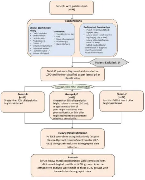
In this cohort study, out of total 55 patients presenting with hip lurch, total 41 cases were enrolled having perthes disease. The 14 patients were excluded because of secondary causes. The whole of the study was carried out in the Department of Orthopaedic Surgery in collaboration with Department of Biochemistry of our institution. The Institutional Review Board and Ethics Committee approved this study which was carried out during January 2012 to May 2016. Before enrolment, each parent/guardian’s written informed consent was obtained.
All children of either sex with age more than 4 years and less than 14 years diagnosed (clinical and radiological) as a case of perthes disease and parent / guardians are willing for inclusion were included in the study. The children of age less than 4 years or more than 14 years, children suggestive of other causes of avascular necrosis such as sickle cell anaemia, steroid use, significant trauma hip (such as fracture & dislocation), DDH, previous hip surgery or multiple epiphyseal dysplasia, children/parents not willing to be included, children on cancer chemotherapy, Immuno-compromised patients, clinical rickets with clinical deformities and children with different skeletal dysplasia were excluded.
Table 1 Patients Demographic Data
Herring Lateral Pillar Classification p-value B
(N=16)
B/C (N=03)
C (N=22)
Age 8.75±2.64 8.33±1.52 9.22±2.59 p=0.7749
Sex
Male 13 (31.7) 2 (4.8) 16 (39.0)
p=0.7768
Female 3 (7.3) 1 (2.4) 6 (14.6)
Socioeconomic Status
Super Rich ≥ 12,00,000 0 0 0
X2 =4.411
p=0.8183
Rich Class 2,40,000- 0 0 0
Upper Class 60,000- 2 (4.8) 0 1 (2.4)
Middle Class 20,000- 2 (4.8) 0 3 (7.3)
Lower Class 12,000- 3 (7.3) 1 (2.4) 4 (9.7)
Poor Class 6,000- 5 (12.1) 2 (4.8) 6 (14.6)
Wretched Class ≤ 6,000 4 (9.7) 0 8 (19.5)
Site
Unilateral L 8 (19.5) 2 (4.8) 8 (19.5) X2 =1.949
p=0.7451
R 6 (14.6) 1 (2.4) 9 (21.9)
Bilateral 2 (4.8) 0 5 (12.1)
Total No of family members
1 to 2 6 (14.6) 2 (4.8) 7 (17.0) X2 =1.391
p=0.4987
3 or more 10 (24.3) 1 (2.4) 15 (36.5)
House
Pukka House
Room with separate kitchen 4 (9.7) 1 (2.4) 4 (9.7) X2 =0.7259
p=0.6956
Room without separate kitchen 2 (4.8) 0 3 (7.3)
Kucha House
Room with separate kitchen 2 (4.8) 1 (2.4) 2 (4.8) X2 =1.595
p=0.4504
Room without separate kitchen 8 (19.5) 1 (2.4) 13 (31.7)
Occupation of guardian
Professional 0 0 1 (2.4)
X2 =7.11
p=0.8502
Semi professional 1 (2.4) 0 3 (7.3)
Clinical/Shop Owner/Farm
Owner 3 (7.3) 0 1 (2.4)
Skilled Worker 4 (9.7) 1 (2.4) 6 (14.6)
Semi Skilled Worker 6 (14.6) 1 (2.4) 5 (12.1)
Unskilled Worker 2 (4.8) 1 (2.4) 4 (9.7)
Unemployed 0 0 2 (4.8)
Occupational Hazardous activity
At Home 4 (9.7) 1 (2.4) 5 (12.1) X2 =0.8296
p=0.6605
After the demographic data recording, the diagnosis of perthes was established as per standard clinico-radiological parameters. The cinical parameters were: onset of groin pain or discomfort with abductor lurch, limitation of hip joint movements (especially abduction and internal rotation). The radiographic signs considered for establishing diagnosis of perthesinclude condensation or fragmentation of the epiphyseal ossification center with or without loss of femoral head height with or without sphericity.
These patients were subjected to uniform institutional standard protocol (Multiple drilling of head with or without derotation osteotomy). The clinico-radiological examination was done and the patients were categorized accordingly based on their X-ray findings (Lateral pillar classification) [Fig-1]. In Clinical examination, parameters such as presence or absence of Trendelenburg sign, range of motion (Abduction, Int. rotation) were done. Venous blood (2ml) was collected in plain vials and estimation of Lead (Pb) and Cadmium (Cd) were done using Inductively Coupled Plasma-Optical Emission Spectrometer (ICP-OES) as per standard protocol. Furthermore, the serum heavy metal (Pb& Cd) concentrations were correlated with clinico-radiological profile of perthes groups classified as per lateral pillar classification. Also the comparative analyses were made to these perthes groups with the exclusive demographic data.
RESULTS
In our study we analysed 41 patients with perthes disease out of which 31 (75.6%) were males and 10 (24.3%) were females. The mean age of all the patients was 8.9± 2.54 range (4-14) years. All patients were grouped into Group I [(with Hearing B) n=16], Group II [(with Hearing B/C) n=3] and Group III [(with Hearing C) n=22)] as per Herring lateral pillar
classification. The mean age of the patients of group B, B/C and C were 8.75±2.64, 8.33±1.52 and 9.22±2.59 respectively.
Demographic parameters shown in table-1 & 2 showing no significant association. However while analyzing socioeconomic status, most of the patients were from the poor class. We found that twenty six patients were having 3 or more family members. Most of the perthes disease patients were from rural areas, residing in Kucha house without any separate kitchen (n=22). Most of the perthes disease patient’s guardians are semi-skilled workers (n=12) and skilled workers (n=11), in which n=10 attendants doing the occupational hazardous activity at home. Also, most of the perthes disease patients were using the non-municipal water (n=25), were exposed to passive smoking (n=19) by any mean and were non-vegetarian (n=23).
However all these demographic characteristics of patients were statistically non-significant [Table 1 &2].
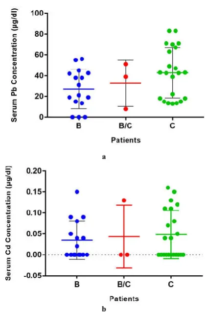
On the basis of heavy metal blood concentration, these patients were divided into two groups (as per heavy metal concentration) in two group-one with normal and other with abnormal concentration group. Total 30 patients (73.1%) showed abnormal Pb concentration while 26 patients (63.4%) showed abnormal Cadmium (Cd) level in serum. Serum Pb level of 42.81±24.33 in group C and serum Cd level of 0.04±0.07 in group B/C observed to be maximum [Table-3 & 4; Fig-2].
Statistical significant differences were found in Pb level between B and C (radiological parameter) and Abduction and Internal rotation (clinical parameter). Significant positive correlations were found Pb level with the clinico-radiological severity of perthes disease patients.
Table 2 Patients general environmental exposure
Herring Lateral Pillar Classification B
(N=16) B/C (N=03)
C (N=22)
Locality
Urban 5 (12.1) 0 7 (17.0) X2 =1.341
p=0.5115
Rural 11 (26.8) 3 (7.3) 15 (36.5)
Water Intake
Municipal 7 (17.0) 1 (2.4) 8 (19.5) X2 =0.2564
p=0.8797
Non Municipal 9 (21.9) 2 (4.8) 14 (34.1)
Smoking
Active 0 0 0
--
Passive 5 (12.1) 1 (2.4) 13 (31.7)
Food Habit
Vegetarian 6 (14.6) 2 (4.8) 10 (24.3) X2 =0.9191
p=0.6316
Non-Vegetarian 10 (24.3) 1 (2.4) 12 (29.2)
Figure 1 Flowchart of Methodology
Table 3 Number of patients with serum Lead and Cadmium levels
Heavy Metals
Range (µg/dl)
B (n=16)
B/C (n=03)
C (n=22)
Pb
Normal (10-20) 04 (25%) 00 07 (39%)
Abnormal 12 (74%) 03 (100%) 15 (61%)
Cd
Normal (0.05-0.2) 05 (31%) 01 (33%) 09 (41%)
Abnormal 11 (69%) 02 (67%) 13 (59%)
Table 4 Serum Lead and Cadmium level in patients.
Herring Lateral Pillar Classification B
(N=16)
B/C (N=03)
C (N=22)
Lead (µg/dl)
27.18±18.88 32.66±22.18 42.81±24.33
Cadmium
However, no statistical significant observations were founded in Cd level between the groups [Table-5, 6 & 7; Fig-3a, &3b]. No patients showed any other skeletal manifestation (like osteoporosis, teeth abnormalities, angular deformities, kyphosis etc.) of heavy metal toxicity.
DISCUSSION
Pollution has reached an alarming stage age worldwide including in India. One can escape from the hazardous pollutant and may inhale or ingested by several different means. Different anthropogenic activities carried out due to various reasons are the major cause of environmental pollution (Jaishankar et al., 2014). As far as humans are concerned, pollution may bethe one of the main reason for serious illnesses. Various pollutants with their different chemical composition, reaction emission, properties, persistence in the environment, ability to be transported in long or short distances and their eventual impacts on human health have been reported. Among these pollutants different gaseous (SO2, NOx, CO etc.) and organic pollutants (dioxins etc.), heavy metals (Pb, Cd, Hg etc.)in air and/or water are the major areas of concern. Industrial activities lead to considerable generation of hazardous waste especially in developing countries like in India.
The determination of whether or not a substance poses a health risk to humans is based on clinical, epidemiological, and/or animal studies which demonstrate that exposure to a substance is associated with health effects. In the context of human health ‘‘risk’’ is the probability that a noxious health effects may occur. In current scenario, due to huge urbanization and pollution, the heavy metal toxicity is on alarming phase. Therefore, author hypothesized that there may be an association/correlation of serum heavy metal concentration with the clinico-radiological profile of perthes disease in children. To prove above hypothesis, in this study author aims
Figure 2 Number of patients with serum Lead and Cadmium levels
Table 5 Difference in serum Lead and Cadmium level in patients categorized as per lateral pillar classification
B vs B/C B/C vs C B vs C ANOVA Lead (µg/dl) p=0.6576
t= 0.4512
p=0.50 t= 0.6827
p=0.03* t= 2.14
p=0.1114 F=2.32
Cadmium (µg/dl)
p=0.7951 t= 0.2638
p=0.8954 t= 00.1329
p=0.4540 t= 0.757
p=0.7631 F= 0.2723
*Significant
Table 6 Difference in serum Lead and Cadmium level in patients categorized as per clinical parameters
Clinical Parameters
Serum Lead (Pb) Concentration
P-value Normal Heavy
Metal Concentration
[n=11]
Abnormal Heavy Metal Concentration
[n=30]
Trendelenburg sign
+ive 7 24
p=0.4132
-ive 4 6
Abduction 18.21±2.35 (11-25) 16.23±2.44 (09-18) t=2.324
p=0.025*
Internal rotation 08.47±1.13 (06-12) 06.25±1.07 (04-10) t=2.143
p=0.038* Serum Cadmium (Cd) Concentration
Normal Heavy Metal Concentration
[n=15]
Abnormal Heavy Metal Concentration
[n=26] Trendelenburg
sign
+ive 6 15
p=0.3408
-ive 9 11
Abduction 19.55±2.61 (9-21) 18.23±2.13 (13-25) p=0.0863 t= 1.760
Internal rotation 07.09±1.36 (06-10) 07.82±1.07 (04-12) t= 1.191
p= 0.2408 *Significant
Table 7 Correlation of serum Lead and Cadmium level with severity patients categorized as per clinico-radiological
parameters.
Pearson r
Radiological
Parameter Clinical Parameters
Pillar Classification
Trendelenburg
sign Abduction Int. rotation
Pb r
2=0.1084;
p=0.0356*
r2=0.1632;
p=0.0930
r2=0.1902;
p=0.0498*
r2=0.1831;
p=0.0590*
Cd r
2=0.0140;
p=0.4603
r2=0.1457;
p=0.1342
r2=0.1377;
p=0.1574
r2=0.1118;
p=0.2515 *Significant
a
b
to analyzed the heavy metals [lead (Pb) and Cadmium (Cd)] in the peripheral blood of the perthes disease patients and to correlate these concentrations with the clinico-radiological profile of perthes disease.
These observations, especially the heavy metal concentration to a toxic level, compel us to think about that by what mean we prone to heavy metal toxicity? Is the serum heavy metal metals concentration was the real culprit to alter the human physiology that leads to various disorders, especially to the bones? Whether our epidemiological life style/status affects our physiology? All these questions must be answered by us as this is really very important.
Many of the previous studies reported that apoptosis of osteoblasts and osteoclasts is a strictly regulated process and plays vital role in physiological bone turnover and in the development of various pathological conditions in skeleton (Lechner et al., 2000), but none of the studies have questioned the association of these mechanism with the etiopathogenesis of perthes disease patients so far. Alteration in osteogenesis/bone remodeling is the one of the most important factor in the pathophysiology of perthes disease, which leads to development of severe deformity in the affected hip and thus affecting the clinical course of the disease (Molloy et al., 1996; Singh et al., 2016(d); Sanja et al., 2015; Tsai et al., 2015). As no study was still conducted on perthes disease patients in relation to heavy metal, the present study was first to correlate the serum heavy metal analysis with the clinico-radiological progression of perthes disease along with epidemiological profiling of the enrolled patients via questionnaire.
Carmouche et al. (2005), experimental animal’s exposed to Pb showed inhibition of fracture healing by showing a significant decrease in osteoprogenitor cell frequency, increased chondrogenesis, and delay in cartilage mineralization. Monir et al. (2010), also pointed that Pb suppressed the Wnt/β-catenin signaling in fracture repair which might cause delay in endochondral ossification and conversation of cartilage tissue to bone. Also Pb effectively reduced the overall size of the mineralized callus. In fact, Pb exposure has shown reduced bone mineral properties and bone strength in mice and rats (Monir et al., 2010). This may affect the regeneration phase of perthes disease.
Several studies have revealed that bone is one of the target organs for Cd toxicity (Aoshima et al., 2003; Wang et al., 2003; James et al., 2013; Sadeghi et al., 2014). It also had been noted that high concentrations of Cd (>10 µg/dl) inhibits the process of vascularization by inhibiting vascular endothelial growth factor (Aoshima et al., 2003). In separate studies by Wang et al (2003); Brzoska et al (2004) and Coonse (2007) it was observed that exposure of Cd leads to various bone pathologies like osteoporosis, osteopenia. Several studies (Wang et al., 2003; Brzoska et al., 2004; Coonse et al., 2007; Chen et al., 2011; Chen et al., 2013; Bhattacharyya MH, 2009; Wittman et al., 2002) revealed that Cd might affect the bone remodeling process by stimulating the differentiation and activity of osteoclasts, but inhibits osteoblast activity and differentiation, which may resulting in decreased bone mineral density and prone to fracture incidence. Study by Chen et al. in 2011and further in 2013 observed that even low-level exposure to Cd disturbs the bone metabolism and furthermore
increased the urinary Ca excretion and subsequent skeletal demineralization, that may leads to major bone loss and increase bone fragility. Jin et al. (2004) and Martynowicz (2004) in a study demonstrated the inhibitory effect of Cd on synthesis and/or release of endothelium derived vasoactive substances (e.g. nitric oxide - NO) that may also leads to increased superoxide anions as a result of Cd-induced oxidative stress. On the other hand, exposure of endothelial cells to Cd significantly increased the secretion of vasoconstrictors (angiotensin-II and endothelin-I) (Bilgen et al., 2003). Cd can directly inhibit endothelial migration and tube formation. These aspects could supposed to contribute vascular (Haversian) canal constriction in the rats exposed to Cd (Bilgen et al., 2003). These observations may be significant as these may affect the necrotic phase as well as regeneration in perthes disease.
As per the above evidences, Pb as well as Cd toxicity may affect the bone remodeling and vascularization process. Thus we may conclude that although there are insufficient evidences that heavy metals contribute in the causation of ischemia of perthes disease but the evidences and also the present study observations do support the hypothesis that the heavy metal toxicity may affect the natural history of perthes disease. However, due to being a single centric study with a small sample size, we are unable to generalized our observation, which was the limitation of the study. The authors favour for more multi-centric studies with large sample sizeto establish the association of heavy metal with the clinic-radiological profile of perthes disease. This can help in opening new horizons in the diagnosis as well as management of perthes disease.
CONCLUSION
Now a day due to huge urbanization, heavy metals are considered as one of the significant environmental pollutants that are inhaled / ingested by the human beings by different means. In the present study, we observed that the epidemiological/environmental factors with special reference to heavy metals may contribute a significant role in natural history ofperthes disease.This is the first study to explore the role of heavy metal with clinico-radiological severity of perthes disease patient. And definitely this will open a new scope of research in the complex etiological process of perthes disease. However to establish the role of heavy metals in causation of perthes is still an area of future research.
References
1. Aoshima K, Fan J, Cai Y, Katoh T, Teranishi H, Kasuya M, (2003). Assessment of bone metabolism in cadmium-induced renal tubular dysfunction by measurements of biochemical markers. Toxicol Lett, 136:183-192. 2. Bahmanyar S, Montgomery SM, Weiss RJ, Ekbom A,
(2008). Maternal smoking during regnancy, other prenatal and perinatal factors, and the risk of Legg-Calve-Perthes disease. Pediatrics. 122:e459- 464. 3. Balmes JR, (2015). Indoor biomass burning and health
consequences. In Air Pollution and Health Effects. Springer London. 381-402.
human exposures. Toxicol Appl Pharmacol. 238:258-265.
5. Bilgen I, Oner G, Edremitlioglu M, Alkan Z, Cirrik S, (2003). Involvement of cholinoceptors in cadmium-induced endothelial dysfunction. J Basic Clin Physiol Pharmacol. 14:55-76.
6. Boccia S, Hung R, Ricciardi G, Gianfagna F, Ebert MP, Fang JY, et al, (2008). Meta- and pooled analyses of the methylenetetrahydrofolatereductase C677T and A1298C polymorphisms and gastric cancer risk: a huge-GSEC review. Am J Epidemiol 167: 505- 516.
7. Brzóska MM, Moniuszko-Jakoniuk J, (2004). Low-level lifetime exposure to cadmium decreases skeletal mineralization and enhances bone loss in aged rats. Bone. 35:1180-1191.
8. Campbell I G, (2002). Methylenetetrahydrofolate reductase polymorphism and susceptibility to breast cancer. Breast Cancer Res. 14: 1-4.
9. Carmouche JJ, Puzas JE, Zhang X, Tiyapatanaputi P, Cory-Slechta DA, Gelein R, et al, (2005). Lead exposure inhibits fracture healing and is associated with increased chondrogenesis, delay in cartilage mineralization, and a decrease in osteoprogenitor frequency. Environmental health perspectives. 113:749-755.
10. Catterall A, (1981). Legg-calve Perthes syndrome. ClinorthopRelat Res. 158:41-52.
11. Chen X, Wang G, Li X, Gan C, Zhu G, Jin T, et al, (2013). Environmental level of cadmium exposure stimulates osteoclasts formation in male rats. Food ChemToxicol. 60:530.
12. Chen X, Zhu G, Gu S, Jin T, Shao C, (2009). Effects of cadmium on osteoblasts and osteoclasts in vitro. Environ ToxicolPharmacol, 28:232-236.
13. Chen X, Zhu G, Shao C, Jin T, Tan M, GuS,et al, (2011). Effects of cadmium on bone microstructure and serum tartrate-resistant acid phosphatase 5b in male rats. ExpBiol Med. 236:1298-1305.
14. Chung SM, (1976). The arterial supply of the developing proximal end of the human femur. J Bone Joint Surg Am.58:961- 970.
15. Coonse KG, Coonts AJ, Morrison EV, Heggland SJ, (2007). Cadmium induces apoptosis in the osteoblast-like cell line, Saos-2. J Toxicol Environ Health A. 70:575-581.
16. Daniel AB, Shah H, Kamath A, Guddettu V, Joseph B, (2012). Environmental tobacco and wood smoke increase the risk of Legg-Calve-Perthes disease. ClinorthopRelat Res. 470(9):2369-75.
17. De Inocencio J, (2004). Epidemiology of musculoskeletal pain in primary care. Arch Dis Child. 89:431- 434.
18. Falfan-Valencia R, Resendiz-Hernandez JM, Ramirez-Venegas A, Sansores R, Camarena A, Vargas-Rojas MI, et al, (2014). SNPs In TNF Promoter Gene Prove Differences In COPD Secondary To Wood Smoke And Tobacco Smoking. InC34. Insights IntoCopd Pathogenesis From Pre-Clinical Studies. American Thoracic Society. A4280-A4280.
19. Jaishankar M, Mathew BB, Shah MS, Gowda KRS, (2014). Biosorption of Few Heavy Metal Ions Using
Agricultural Wastes. Journal of Environment Pollution and Human Health. 2(1): 1-6.
20. James KA, Meliker JR, (2013). Environmental cadmium exposure and osteoporosis: a review. Int J Public Health. 58:737-745.
21. Jin T, Nordberg G, Ye T, Bo M, Wang H, Zhu G, et al, (2004). Osteoporosis and renal dysfunction in a general population exposed to cadmium in China. Environ Res. 96:353-359.
22. Kim J, Lim W, Ko Y, Kwon H, Kim S, Kim O, et al, (2012). The effects of cadmium on VEGF-mediated angiogenesis in HUVECs. J ApplToxicol. 32:342-349. 23. Lechner A, Schutze N, Siggelkow H, Seufert J, Jakob F,
(2000). The immediate early gene product hCYR61 localizes to the secretory pathway in human osteoblasts. Bone. 27(1):53-60.
24. Martynowicz H, Skoczynska A, Wojakowska A, Turczyn B, (2004). Serum vasoactive agents in rats poisoned with cadmium. Int J Occup Med Environ Health. 17:479-485.
25. Molloy MK, MacMahon B, (1966). Incidence of Legg-Perthes disease (osteochondritisdeformans). N Engl J Med. 275:988-90.
26. Monir AU, Gundberg CM, Yagerman SE, van der Meulen MC, Budell WC, Boskey AL, et al, (2010). The effect of lead on bone mineral properties from female adult C57/BL6 mice. Bone. 47:888-894.
27. Nagajyoti PC, Lee KD, Sreekanth TVM, (2010). Heavy metals, occurrence and toxicity for plants: a review. Environ ChemLett. 8(3): 199-216.
28. Rajput R, Prasad G, AK C, (2009). Scenario of solid waste management in present Indian context. Caspian Journal of Environmental Sciences. 7(1):45-53.
29. Sadeghi N, Oveisi MR, Jannat B, Hajimahmoodi M, Behzad M, Behfar A, et al, (2014). The relationship between bone health and plasma zinc, copper lead and cadmium concentration in osteoporotic women. J Environ Health Sci Eng.12:125.
30. SanjaSrzentic, GordanaNikcevic, DuskoSpasovski, ZoranBascarevic, ZoricaZivkovic, ZoricaTerzic-Supic, et al, (2015). Predictive genetic markers of coagulation, inflammation and apoptosis in Perthes disease-Serbian experience. Eur J Pediatr. 174: 1085-1092.
31. Singh A, .Srivastava R N, Shukla P, Pushkar A, Ali S, (2014(b). Management of Late Onset Perthes: Evaluation of Distraction by External Fixator-5-Year Follow-Up. Advances in Orthopedics. Article ID 135236, 6 pages
32. Singh A, Ali S, Yadav M, Mahdi A A, Srivastava R N, Sharma V, (2016(c). Heavy Metals in Perthes Disease: A Review. Journal of Bone and Joint Diseases. 31: 9-14.
33. Singh A, Ali S, Hussain S R, Kumar V, Mahdi A A, Srivastava R N, (2016(a). Analysis of correlation of CYR61 and MTHFR Gene Polymorphism in Legg-Calve-Perthes disease. South American Journal of Clinical Research Special Edition. 1(3), 1-7.
Legg-Calve-Perthes disease. South American Journal of Clinical Research. 1-7.
35. Singh A, Kumar V, Ali S, (2016). Evidence Based Approach to Perthes. BJMMR. 16(7): 1-11.
36. Tsai TL, Pan WH, Chung YT, Wu TN, Tseng YC, Liou SH, et al, (2015). Association between urinary lead and bone health in a general population from Taiwan..J Expo Sci Environ Epidemiol. doi: 10.1038/jes.2015.30.
37. Wang H, Zhu G, Shi Y, Weng S, Jin T, Kong Q, et al, (2003). Influence of environmental cadmium exposure on forearm bone density. J Bone Miner Res. 18:553-560. 38. Wittman R, Hu H, (2002). Cadmium exposure and nephropathy in a 28-year-old female metals worker. Environ Health Perspect. 110:1261-1266.
*******
How to cite this article: