Risk strati
fi
cation for major adverse
cardiac events and ventricular
tachyarrhythmias by cardiac MRI
in patients with cardiac sarcoidosis
Masakazu Yasuda,1Yoshitaka Iwanaga,1Takao Kato,2Toshiaki Izumi,2 Yasutaka Inuzuka,3Takashi Nakamura,1Yuki Miyaji,1Takayuki Kawamura,1 Shigeru Ikeguchi,3Moriaki Inoko,2Takashi Kurita,1Shunichi Miyazaki1
To cite:Yasuda M,
Iwanaga Y, Kato T,et al. Risk
stratification for major adverse cardiac events and ventricular tachyarrhythmias by cardiac MRI in patients with cardiac sarcoidosis. Open Heart2016;3:e000437. doi:10.1136/openhrt-2016-000437
▸Additional material is
available. To view please visit the journal (http://dx.doi.org/ 10.1136/openhrt-2016-000437).
MY and YI both authors contributed equally to this work.
Received 14 March 2016 Revised 28 May 2016 Accepted 30 June 2016
1Division of Cardiology,
Kindai University Faculty of Medicine, Osakasayama, Japan
2Kitano Hospital, Osaka,
Japan
3Shiga Medical Center for
Adults, Moriyama, Japan
Correspondence to
Dr Yoshitaka Iwanaga; yiwanaga@med.kindai.ac.jp
ABSTRACT
Background:The presence of myocardial fibrosis by cardiac MRI has prognostic value in cardiac sarcoidosis, and localisation may be equally relevant to clinical outcomes.
Objective:We aimed to analyse cardiac damage and function in detail and explore the relationship with clinical outcomes in patients with cardiac sarcoidosis using cardiac MRI.
Methods:We included 81 consecutive patients with cardiac sarcoidosis undergoing cardiac MR. Left ventricular mass and fibrosis mass were calculated, and localisation was analysed using a 17-segment model. Participants underwent follow-up through 2015, and the development of major adverse cardiac events including ventricular tachyarrhythmias was recorded.
Results:Increased left ventricular fibrosis mass was associated with increased prevalence of ventricular tachyarrhythmias (p<0.001). When localisation was defined as the sum of late gadolinium enhancement in the left ventricular basal anterior and basal anteroseptal areas, or the right ventricular area, it was associated with ventricular tachyarrhythmias (p<0.001). Kaplan-Meier analysis during a median follow-up of 22.1 months showed that both the mass and localisation groupings for fibrosis were significantly associated with major adverse cardiac events or ventricular tachyarrhythmias and that when combined, the risk stratification was better than for each variable alone (p<0.001, respectively). By Cox-proportional hazard risk analysis, the localisation grouping was an independent predictor for the both.
Conclusions:In patients with cardiac sarcoidosis, both fibrosis mass and its localisation to the basal anterior/ anteroseptal left ventricle, or right ventricle was associated with the development of major adverse cardiac events or ventricular tachyarrhythmias. Cardiac MR with late gadolinium enhancement may be useful for improving risk stratification in patients with cardiac sarcoidosis.
INTRODUCTION
Cardiac involvement in sarcoidosis occurs in 20–30% of patients in autopsy studies,1 2and
has been closely associated with prognosis.3 4 A previous study reported that sudden cardiac death (SCD) occurred in two-thirds of patients with histological evidence of cardiac sarcoidosis (CS).5Ventricular tachyar-rhythmia (VA), resulting from myocardial granulomas and fibrosis that cause electric instability, has been identified as the under-lying mechanism of SCD. The 5-year mortal-ity rate in patients with CS when structural or functional abnormality is present, according to the diagnostic criteria of the Japanese Ministry of Health and Welfare ( JMHW), has been reported at 40%, with the cause of death being the progression of heart failure (HF) or VA.6However, the factors related to the clinical manifestations and outcomes of CS remain unclear and the risk stratification and clinical management are difficult, par-ticularly for VA and SCD.
Late gadolinium-enhancement cardiac MR (LGE-CMR) has been used to detect myocar-dial damage/fibrosis, and recent findings have suggested that it is more sensitive than clinical diagnostic criteria for detecting
KEY QUESTIONS
What is already known about this subject? ▸ The presence of myocardial fibrosis by cardiac
MRI has prognostic value in cardiac sarcoidosis.
What does this study add?
▸ The fibrosis localisation to the basal anterior/ante-roseptal left ventricle, or right ventricle is asso-ciated with the development of major adverse cardiac events or ventricular tachyarrhythmias.
How might this impact on clinical practice? ▸ Cardiac MRI with late gadolinium enhancement
is promising for predicting adverse events including ventricular tachyarrhythmias.
on September 12, 2020 by guest. Protected by copyright.
CS.7 8 A recent study demonstrated that LGE-positive patients had a higher rate of adverse events (a Cox HR of 31.6 for death, aborted SCD, or appropriate ICD dis-charge), compared with LGE-negative patients in a cohort of patients with suspected CS.9Although myocar-dial fibrosis indicated by LGE may predict potentially lethal events, the utility of LGE in patients with sarcoid-osis who present with high-grade cardiovascular symp-toms, such as HF, VA and advanced atrioventricular block (AVB), has not yet been established. In addition, detailed risk stratification in patients with CS using LGE-CMR remains challenging. Therefore, we analysed cardiac damage and function in detail and explored the relationship with clinical outcomes including VA, using CMR in patients with CS. We assumed that not only
fibrosis mass but also its localisation might help to iden-tify the risk of clinical outcomes in patients with CS.
METHODS Study protocol
We enrolled consecutive patients with CS referred for CMR between 2008 and 2015 at three referral centers (Kindai University Hospital, Osakasayama, Japan; Kitano Hospital, Osaka, Japan; and Shiga Medical Center for Adults, Moriyama, Japan). The study was approved by the Institutional Review Boards.
Patients were enrolled if they fulfilled the following criteria: (1) definite CS: CS diagnosed by the 2006 revised JMHW criteria;10 or (2) suspected CS: strongly suspected CS based on a constellation of clinical charac-teristic manifestations and positive findings on echocar-diography, 18F-fluorodeoxyglucose-positron emission tomography (FDG-PET), or CMR, with or without extra-CS, and after the exclusion of other known cardiac diseases.11 The characteristic positive findings of CMR and FDG-PET were based on those detailed in previous reports.7 12
CMR image acquisition
CMR was performed using a 1.5 T scanner (Intera 1.5T; Philips Medical Systems, the Netherlands), according to a standardised protocol. Cine images were acquired with a steady-state free-precession breath-hold sequence in three long-axis planes and contiguous short-axis slices (10 mm, no gap) from the atrioventricular ring to the apex. The visualisation of myocardial oedema was per-formed using a T2-weighted triple inversion recovery breath-hold pulse sequence. LGE images were obtained 10–15 min after intravenous administration of 0.2 mmol/ kg gadolinium-DTPA (Magnevist; Bayer Schering Pharma, Germany) using an inversion recovery gradient echo sequence with 5 mm slice thickness at the same pos-ition as the long-axis and short-axis cines at end-diastole. The inversion time was adjusted per patient to an optimal null signal from a normal myocardium, typically between 250 and 350 ms.
Image analysis
LV volume, LV mass (LVM), and wall thickness were cal-culated with commercially available workstations (Aze Virtual Place; Aze , Japan) as previously described.13 The endocardial and epicardial borders of the LV myo-cardium were manually planimetered on successive short-axis cine images at end-diastole and LVM was derived by the summation-of-the-disks method and multiplication of the myocardial muscle volume by 1.05 g/cm3. To assess LGE, all short-axis slices from the base to the apex were visually inspected to identify areas of normal (completely nulled) myocardium. The mean signal intensity (and SD) was derived, and a threshold of 6 SD above the mean was used to define the areas of LGE.14 Summing the planimetered areas of LGE in all short-axis slices yielded the total volume (LV fibrosis mass; g), which was also expressed as a proportion of total LV myocardium (% LGE).
Local wall thickness, LGE, and T2-weighted images were also analysed based on the American Heart Association’s (AHA) 17-segment model. To evaluate the relationship between localisation of LGE and clinical outcomes, a localisation score (LS) was defined as the summation of LGE enhancement in segments identified by LGE localisation analysis. All analyses were performed by an experienced specialist, and reviewed and
con-firmed by a second expert. Both independent observers were blinded to the patients’ identities and clinical
pro-files. Any discrepancies in analysis were adjudicated by a senior observer.
Clinical follow-up
Long-term clinical follow-up was for 50 months after the CMR testing and accomplished by patient-completed questionnaire, telephone interview or chart review. Major advanced cardiac events (MACE) were defined as the combination of cardiac mortality, admission for worsen-ing HF, advanced AVB and VA. VA was defined as >3 con-secutive ventricular contractions, appropriate implantable cardioverter defibrillator (ICD) discharge or ventricular
fibrillation (VF). Sustained ventricular tachycardia (VT) was defined as a ventricular rhythm faster than 100 bpm lasting at least 30 s. All events were based on clinical diag-nosis but were validated by reviewing the patients’ medical records. Advanced AVB, VA or sustained VT was also confirmed by ECG or ICD records.
Statistical analysis
As part of the univariable analysis for continuous vari-ables, comparisons among groups were performed with student’s t-tests, one way analysis of variance, and Mann-Whitney tests. Pearson χ2 and the Fisher’s exact test were used to assess differences in categorical vari-ables. Cut-off levels of LV fibrosis mass index for VA events and the sensitivities and specificities of the cut-off levels were calculated using a receiver operating characteristics (ROC) curve analysis. Event-free survival curves were analysed using the Kaplan-Meier method
on September 12, 2020 by guest. Protected by copyright.
and comparison between curves was carried out using log-rank tests. The multivariate analysis of clinical out-comes was evaluated with Cox’s proportional hazard model, using JMP V.10.0 (SAS Institute , USA). HRs and 95% CIs were calculated. A p value <0.05 was considered significant. All results are expressed as mean±SD.
RESULTS
Baseline patient characteristics
In total, 81 consecutive patients were enrolled, of which 35 were diagnosed as definite CS using the 2006 revised JMHW criteria and 46 were diagnosed as suspected CS clinically (table 1). Furthermore, 38 (46.9%) patients had extracardiac involvements and 43 (53.1%) patients were diagnosed as having isolated CS, with 31 (38.3%) patients having prior VA events, 20 (24.7%) having docu-mented advanced AV block, and 24 (29.6%) being com-plicated by HF. Plasma B-type natriuretic peptide (<18.4 pg/mL) and ACE inhibitors levels (<21 IU/L) were increased in 84.0% and 32.9% of the patients, respectively. Although just 15 (18.5%) patients had taken corticosteroids at baseline, a further 27 (33.3%) had taken them during follow-up.
CMR findings and VA events
In the CMR examination at baseline, a reduced EF (<50%) was observed in 58.8% and LGE was present in 94.9%, with lesions frequently observed in the basal
septum (Segment 2: 75.9%; Segment 3: 65.8%) and RV (57.0%). The mean % LGE and FM index were 16.6% ±12.8% and 11.3±9.6 g/m2, respectively. During a median follow-up of 22.1 months, 25 (30.7%) patients had VA episodes of which 8 (9.9%) where new onset and the other 17 (21.0%) where recurrences. Any VA event was observed in 39 (48.1%) patients.
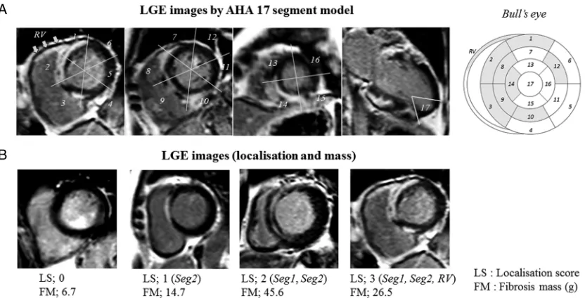
As shown in table 2, all VA events (ie, a history of VA and new-onset VA events) were highly associated with the existence of LGE in the basal anterior (segment 1) and basal anteroseptal (segment 2) LV, as well as the RV ( p<0.01). Among patients with VA, almost all had LGE in segment 2 and LGE was more frequently present in segment 1 and the RV than in patients without VA. Accordingly, we defined the LS as the summation of the LGE in segment 1, segment 2, and the RV, with a poten-tial score of 0 to 3. Figure 1 shows representative LGE images according to the AHA’s 17-segment model and representative cases stratified by the LS. As shown in
figure 2, when patients with all VA events were stratified by LS, LS was significantly associated with all VA events ( p<0.001). Increased LV FM index was also associated with all VA events ( p<0.001), and ROC analysis showed that the area under the ROC curve (AUC) for FM as an indicator of all VA events was 0.760 (95% CI 0.651 to 0.870) (figure 2). The optimal cut-off value of LV FM index was 5.12 g/m2, with a sensitivity of 86% and a spe-cificity of 62%.
Table 1 Baseline clinical characteristics and CMR findings N=81
Age, years 62.9±13.0 Device implantation
Female 49 (60.5) Pacemaker 11 (13.6)
ACE, IU/L 18.1±10.0 ICD/CRT-D 8 (9.9)
BNP, pg/mL 151.3±172.3 CMR parameters
Involvement LV EDV, mL 157.7±61.7
Lung 20 (24.7) LV ESV, mL 91.2±58.7
Skin 12 (14.8) LV EF, % 45.9±14.6
Eye 17 (21.0) LV mass index, g/m2 68.2±19.1
Other 6 (7.4) T2 positive 47 (58.8)
JMHW criteria, positive 35 (43.2) % LGE area,% 16.6±12.9
Medication Fibrosis mass index, g/m2 11.3±9.6
ACEi or ARB 29 (35.8) LV morphological abnormality
β-blocker 25 (30.9) Thinning 21 (25.9)
Diuretic 26 (32.1) Aneurysmal 13 (16.0)
Corticosteroid 15 (18.5)
Antiarrhythmic drug 15 (18.5) Cardiac symptoms
NYHA functional class≥II 24 (29.6) Admission due to HF 16 (19.8)
Syncope 22 (29.7)
Atrial fibrillation 10 (12.3) Advanced AV block 20 (24.7)
VA 31 (42.5)
Values are the mean±SD or number (%).
ACEi, ACE inhibitor; ARB, angiotensin receptor blocker; AV, atrioventricular; BNP, B-type natriuretic peptide; CRT-D, cardiac
resynchronisation therapy-defibrillator; EDV, end-diastolic volume; EF, ejection fraction; ESV, end-systolic volume; HF, heart failure; HT, hypertension; ICD, implantable cardioverter defibrillator; JMHW, Japanese Ministry of Health and Welfare; LGE, late gadolinium enhancement; LV, left ventricular; NYHA, New York Heart Association; VA, ventricular tachyarrhythmia.
on September 12, 2020 by guest. Protected by copyright.
Clinical outcome and risk stratification
During a median follow-up of 22.1 months (lower quar-tile, 7.4 months; upper quartile, 38.1 months), 30 MACEs occurred: 3 patients died due to SCD or HF, 7 were admitted for worsening HF and 4 patients had advanced AVB. Twelve severe ventricular arrhythmias epi-sodes such as sustained VT and VF occurred. In 10 patients, the origin was estimated by 12-lead ECG and 8 patients showed the ECG with inferior axis. They were from left/right ventricular outflow tract or high LV septum, which were associated with LS (the existence of LGE in segment 1, 2 or RV).
For the Kaplan-Meier analysis, patients were divided into two groups according to the optimal cut-off for LV FM index (<5.12 g/m2(n=29) or≥5.12 g/m2(n=45)), or the LS (≤1 (n=32) or≥2 (n=47)). Patients with an LV FM
≥5.12 g/m2or an LS≥2 showed significantly worse prog-noses in terms of both MACE and VA events (figure 3). In sustained VT or VF events, only increased LS was asso-ciated with worse prognosis ( p=0.006). Lower EF (<45%) was mildly associated with the worse prognosis in the MACE ( p=0.045), and extracardiac sarcoidosis involvement was not associated with the both prognoses ( p=0.330 and P=0.930, respectively).
Table 2 Association of VA events with localisation of LGE according to the 17-segment AHA model
AHA 17 segment
LGE (%) Fisher’s test
p Value
VA event (+) VA event (−)
1. Basal anterior 63.2 22.0 0.0002
2. Basal anteroseptal 92.1 61.0 0.0014
RV 76.3 41.5 0.0015
9. Mid inferoseptal 55.3 29.3 0.0238
12. Mid anterolateral 34.2 12.2 0.0306
3. Basal inferoseptal 79.0 53.7 0.0317
5. Basal inferolateral 36.8 14.6 0.0372
4. Basal inferior 44.7 22.0 0.0542
14. Apical septal 31.6 14.6 0.1070
15. Apical inferior 21.1 7.3 0.1071
7. Mid anterior 36.8 19.5 0.1311
10. Mid inferior 42.1 24.4 0.1499
8. Mid anteroseptal1 44.7 23.9 0.1697
17. Apex 10.5 4.9 0.4204
6. Basal anterolateral 29.0 19.5 0.4310
13. Apical anterior 18.4 12.2 0.5374
11. Mid inferolateral 21.1 19.5 1.0000
16. Apical lateral 13.2 14.6 1.0000
AHA, American Heart Association; LGE, late gadolinium enhancement; LV, left ventricle; RV, right ventricle; VA, ventricular tachyarrhythmias.
Figure 1 Representative LGE images using the AHA 17-segment model (A) and representative cases stratified by the LS (B). AHA, American Heart Association; FM, fibrosis mass; LGE, late gadolinium enhancement; LS, localisation score.
on September 12, 2020 by guest. Protected by copyright.
In multivariate Cox proportional hazard analysis with age, gender and FM index, the LS was independently associated with MACE and VA events, as shown intable 3. When the FM index as a continuous variable and the LS as throughout its range were entered into the analysis, the LS was still an independent predictor for MACE and VA (P<0.001 and P=0.002, respectively). Furthermore, as shown infigure 4, the combined analysis for the higher FM index and LS groups showed that the combination was associated with the highest risk of MACE and VA events. Notably, all patients with admission for worsening HF or advanced AVB also belonged to this group. In contrast, patients with a FM index <5.12 g/m2and an LS≤1 had no MACEs, or no VA events.
Comparison between definite and suspected CS
In comparison of baseline clinical characteristics between patients with definite and suspected CS,
frequent extracardiac involvement and increased plasma ACE level were observed in definite CS ( p<0.01, respect-ively) (see online supplementary table S1). Also, cortico-steroid therapy was frequently performed ( p=0.003). In CMR analysis, no significant differences were observed between them (see online supplementary table S2). In Kaplan-Meier analysis, each group also showed that the higher LS group (≥2) had a worse prognosis in both MACE and VA events (figure 5).
DISCUSSION
The major findings of this study are as follows. First, VA was the most frequent cardiac event in CS, and VA events, including a prior history of VA, occurred in 48.1% of patients. Second, an LV FM index ≥5.12 g/m2 was shown to predict MACE and VA. Third, a developed LS, defined as the summation of the LGE in the basal anterior LV, basal anteroseptal LV and RV, was able to
Figure 2 Relationship between LV fibrosis mass index or localisation score and VA events. Stratification of patients with VA events by localisation score (A). Receiver operating characteristic (ROC) curves for LV fibrosis mass index as an indicator of all VA events (B). AUC, area under the ROC curve; VA, ventricular tachyarrhythmia.
Figure 3 Kaplan-Meier analysis for the MACE-free (A and B) and VA-free survival (C and D) in patients stratified by baseline LV fibrosis mass index (A and C) and localisation score (B and D). Lower group, solid line; higher group, dotted line. MACE, major adverse cardiac event; VA, ventricular tachyarrhythmia.
on September 12, 2020 by guest. Protected by copyright.
predict VA when ≥2. Fourth, the combination of both FM and LS improved the risk stratification for predicting clinical outcomes, including VA.
Prognosis and VA in patients with CS
In a retrospective autopsy series of SCD in sarcoidosis, 25 of 41 patients had evidence of cardiac involvement and 10 of 25 patients had no evidence of sarcoid in any other organ.15 Fleming et al16 reported that SCD occurred in 48 of 197 (24%) patients with CS in their cohort. In a retrospective study of Japanese patients with CS, sustained VT was shown to be an independent risk factor for mortality, suggesting that VA is very important for prognosis of CS.6 In the present study, only two patients had SCD caused by a VA, or 12 patients had severe ventricular arrhythmias. We therefore tried to identify a better risk stratification tool for VA in CS that was more closely associated with SCD.
In an autopsy series of SCD in sarcoidosis, the ven-tricular septum had the largest percentage of involve-ment and ∼50% of the cases were involved in the RV apex and septum.15 Kumar et al17 reported that among 21 patients with CS and VT, 16 of 18 patients had an RV scar and 14 of 15 patients had an LV scar, with LV scar-ring frequently present in the septal (11/15) and anter-ior (7/15) LV. Similarly, our findings indicated LGE in basal septum at a high frequency among the patients with VAs (segment 2, 92.1%; segment 3, 79.0%),
followed by the RV (73.7%) and basal anterior LV (segment 1, 60.5%). In a recent cohort study of CS by Crawfordet al,18the presence of LGE in the RV was asso-ciated with adverse events, including VT/VF. Consistent with their studies, our results suggest that the presence of LGE in the RV may be important for the origin of VT. For further risk stratification, we utilised a three-segment model (three-segment 1, 2 and RV), which showed highly significant differences in the presence of LGE between patients with and without VA (table 2). These were then used to define the LS, which was itself highly associated with not only all VA events (figure 2) but also follow-up VA events (P<0.001, data not shown). The reason the LS was useful for risk stratification in VA is unclear. However, myocardial scar has been known to be an origin and a specific substrate of VA and it is pro-posed that the scars in the His-Purkinje system, LV outflow-tract in the basal septum and the anterior wall may participate in VA generation or SCD processes in CS using voltage cardiac mapping.17 Indeed, in the present study, the origin of sustained VT was estimated in 10 patients and the 80% was from left or right ven-tricular outflow tract or high LV septum, suggesting the close relationship between VT origin and LS (ie, the existence of LGE in segment 1, 2 or RV). With regard to the importance of RV involvement, Schuller et al19 reported that RV dysfunction, in particular, was asso-ciated with appropriate ICD therapy in patients with CS.
Table 3 Multivariate Cox proportional hazard analysis
Variables HR 95% CI p Value
MACE <0.001
Age 0.958 0.921 to 0.999 0.043
Gender; male/female 1.235 0.545 to 2.722 0.604
LV fibrosis mass; high/low 2.198 0.669 to 10.327 0.210
Localisation score; high/low 20.487 3.504 to 398.919 <0.001
VA events <0.001
Age 0.987 0.946 to 1.035 0.580
Gender; male/female 1.525 0.620 to 3.685 0.350
LV fibrosis mass; high/low 1.308 0.395 to 6.098 0.685
Localisation score; high/low 14.546 2.579 to 276.708 <0.001 MACE, major adverse cardiac events; VA, ventricular tachyarrhythmias.
Figure 4 Risk stratification of clinical outcomes by the combined analysis of LV fibrosis mass and localisation score: MACE (A), VA events (B) and all VA events (a history of VA and new-onset VA events) (C). MACE, major adverse cardiac event; VA, ventricular tachyarrhythmia.
on September 12, 2020 by guest. Protected by copyright.
Further mechanistic studies for VA generation will clarify and improve the risk stratification in CS.
LGE and VA in cardiomyopathies
In studies of LGE in patients with VA, including both ischaemic and non-ischaemic cardiomyopathies, the pres-ence offibrosis was an independent predictor of adverse outcomes.20 In a meta-analysis by Kuruvilla et al21, the presence and extent of LGE were associated with increased risk of all-cause mortality, HF hospitalisation, and SCD in patients with overall non-ischaemic cardiomy-opathy. However, given that the basic pathophysiology is different for each cardiomyopathy, the relationship between LGE and each event may also diverge, suggest-ing that LGE quantification cannot be uniformly adapted to all cardiomyopathies. In a cohort study of 1293 patients with hypertrophic cardiomyopathy, the extent of LGE was associated with an increased risk of SCD, with the adjusted HR reported as 1.46 per 10% increase in LGE.22 In a study of dilated cardiomyopathy, mid-wallfibrosis was present in 142 (30%) patients, and its assessment with LGE-CMR imaging provided independent prognostic information.23
Similarly, the presence of LGE among patients with suspected CS yielded a Cox HR of 31.6 for death, aborted SCD and appropriate ICD discharge, and of 33.9 for events including death and VT during a 3.4-year follow-up in a recent prospective study.9 However, they found no relationship between cardiac events including VA and LGE extent, the studied cohort consisted of a broad range of patients with systemic sarcoidosis, and the % LGE in all patients was lower than that in our study cohort (4.4% vs 16.6%, respectively). In the present study, our cohort may be representative of more advanced CS, resulting in the increased LV FM by
quantification being significantly associated with MACE or VA events by Kaplan-Meier analysis, and the ROC curve analysis showing an optimal cut-off value of 5.12 g/ m2 in LV FM index for VA events. A recent study in 43 Japanese patients with CS taking steroid therapy, where % LGE was 19%±10% (5-SD method), showed that large-extent LGE (ie, % LGE ≥20%) was independently associated with combined adverse outcomes.24Crawford
et al18also reported that LGE involving≥9 segments in a
combined left and right ventricular segmental analysis resulted in a 92% sensitivity and 88% specificity for differ-entiating patients with VT/VF from those without VT/VF (AUC, 0.90). In addition, the results of the present study suggest an incremental effect of the LS on LGE extent (LV FM) by LGE assessment, and that a combined ana-lysis of LS and LV FM may be better for the risk stratifi ca-tion of clinical outcomes in CS.
Study limitations
Several limitations should be considered when interpret-ing our results. First, the study population was relatively small and could have resulted in low statistical power. However, the size of our cohort was average relative to that of other studies of CMR in CS.17 18 Also, the retro-spective nature of the study may suggest some impact of the missing or insufficient data on the present results.
Second, about half of the patients did not fulfil the JMHW diagnostic criteria (suspected CS), most of whom had isolated CS. Recently, several reports have suggested that there are similar clinical characteristics and clinical outcomes for suspected CS that does not fulfil the JMHW diagnostic criteria but that satisfies only the guidelines for clinical cardiac diagnosis.25 To test this, we analysed and compared the data between the patients who did (definite CS) and did not fulfil the
Figure 5 Kaplan-Meier analysis for the MACE-free or VA-free survival in patients with definite CS (A and C) or suspected CS (B and D) stratified by localisation score. Lower group, solid line; higher group, dotted line. CS, cardiac sarcoidosis; MACE, major adverse cardiac event; VA, ventricular tachyarrhythmia.
on September 12, 2020 by guest. Protected by copyright.
JMHW diagnostic criteria (suspected CS), and found that the clinical characteristics were indeed similar and that the higher LS group (≥2) had a worse prognosis in each group (figure 5).
Third, the study population consisted of patients with CS who could undergo CMR. In particular, most patients with previously implanted cardiac devices and those with severe renal failure were not included in the present study since they could not tolerate the procedure. Therefore, the event rate and clinical characteristics might be underestimated in the present analysis.
Contributors MY and YI are the primary investigators. TK, TI, YI, TN, TK, YM are the secondary investigator (data sampling/analysis). SI, M I, TK, SM are consultants and supervisors.
Competing interests None.
Ethics approval Kindai University Faculty of Medicine.
Provenance and peer review Not commissioned; externally peer reviewed.
Open Access This is an Open Access article distributed in accordance with the Creative Commons Attribution Non Commercial (CC BY-NC 4.0) license, which permits others to distribute, remix, adapt, build upon this work non-commercially, and license their derivative works on different terms, provided the original work is properly cited and the use is non-commercial. See: http:// creativecommons.org/licenses/by-nc/4.0/
REFERENCES
1. Silverman KJ, Hutchins GM, Bulkley BH. Cardiac sarcoid: a clinicopathologic study of 84 unselected patients with systemic sarcoidosis.Circulation1978;58:1204–11.
2. Longcope WT, Freiman DG. A study of sarcoidosis; based on a combined investigation of 160 cases including 30 autopsies from The Johns Hopkins Hospital and Massachusetts General Hospital. Medicine (Baltimore)1952;31:1–132.
3. Kim JS, Judson MA, Donnino R,et al. Cardiac sarcoidosis. Am Heart J2009;157:9–21.
4. Doughan AR, Williams BR. Cardiac sarcoidosis.Heart 2006;92:282–8.
5. Roberts WC, McAllister HA Jr, Ferrans VJ. Sarcoidosis of the heart. A clinicopathologic study of 35 necropsy patients (group 1) and review of 78 previously described necropsy patients (group 11). Am J Med1977;63:86–108.
6. Yazaki Y, Isobe M, Hiroe M,et al. Prognostic determinants of long-term survival in Japanese patients with cardiac sarcoidosis treated with prednisone.Am J Cardiol2001;88:1006–10.
7. Smedema JP, Snoep G, van Kroonenburgh MP,et al. Evaluation of the accuracy of gadolinium-enhanced cardiovascular magnetic resonance in the diagnosis of cardiac sarcoidosis.J Am Coll Cardiol 2005;45:1683–90.
8. Ohira H, Tsujino I, Ishimaru S,et al. Myocardial imaging with 18-F-fluoro-2-deoxyglucose positron emission tomography and
magnetic resonance imaging in sarcoidosis.Eur J Nucl Med Mol Imaging2008;35:933–41.
9. Greulich S, Deluigi CC, Gloekler S,et al. CMR imaging predicts death and other adverse events in suspected cardiac sarcoidosis. JACC Cardiovasc Imaging2013;6:501–11.
10. Tahara N, Tahara A, Nitta Y,et al. Heterogeneous myocardial FDG uptake and the disease activity in cardiac sarcoidosis.JACC Cardiovasc Imaging2010;3:1219–28.
11. Youssef G, Beanlands RS, Birnie DH,et al. Cardiac sarcoidosis: applications of imaging in diagnosis and directing treatment.Heart 2011;97:2078–87.
12. Tadamura E, Yamamuro M, Kubo S,et al. Effectiveness of delayed enhanced MRI for identification of cardiac sarcoidosis: comparison with radionuclide imaging.AJR Am J Roentgenol2005;185:110–5. 13. Nakamura T, Iwanaga Y, Yasuda M,et al. Relationship of
basal-septal fibrosis with LV outflow tract obstruction in hypertrophic cardiomyopathy: insights from cardiac magnetic resonance analysis. Int J Cardiovasc Imaging2016;32:613–20.
14. Flett AS, Hasleton J, Cook C,et al. Evaluation of techniques for the quantification of myocardial scar of differing etiology using cardiac magnetic resonance.JACC Cardiovasc Imaging2011;4:150–6. 15. Tavora F, Cresswell N, Li L,et al. Comparison of necropsy findings
in patients with sarcoidosis dying suddenly from cardiac sarcoidosis versus dying suddenly from other causes.Am J Cardiol
2009;104:571–7.
16. Fleming HA, Bailey SM. Sarcoid heart disease.J R Coll Physicians Lond1981;15:245–6, 249–53.
17. Kumar S, Barbhaiya C, Nagashima K,et al. Ventricular tachycardia in cardiac sarcoidosis: characterization of ventricular substrate and outcomes of catheter ablation.Circ Arrhythm Electrophysiol 2015;8:87–93.
18. Crawford T, Mueller G, Sarsam S,et al. Magnetic resonance imaging for identifying patients with cardiac sarcoidosis and preserved or mildly reduced left ventricular function at risk of ventricular arrhythmias.Circ Arrhythm Electrophysiol 2014;7:1109–15.
19. Schuller JL, Zipse M, Crawford T,et al. Implantable cardioverter defibrillator therapy in patients with cardiac sarcoidosis. J Cardiovasc Electrophysiol2012;23:925–9.
20. Dawson DK, Hawlisch K, Prescott G,et al. Prognostic role of CMR in patients presenting with ventricular arrhythmias.JACC Cardiovasc Imaging2013;6:335–4.
21. Kuruvilla S, Adenaw N, Katwal AB,et al. Late gadolinium enhancement on cardiac magnetic resonance predicts adverse cardiovascular outcomes in nonischemic cardiomyopathy: a systematic review and meta-analysis.Circ Cardiovasc Imaging 2014;7:250–8.
22. Chan RH, Maron BJ, Olivotto I,et al. Prognostic value of quantitative contrast-enhanced cardiovascular magnetic resonance for the evaluation of sudden death risk in patients with hypertrophic cardiomyopathy.Circulation2014;130:484–95.
23. Gulati A, Jabbour A, Ismail TF,et al. Association of fibrosis with mortality and sudden cardiac death in patients with nonischemic dilated cardiomyopathy.JAMA2013;309:896–908.
24. Ise T, Hasegawa T, Morita Y,et al. Extensive late gadolinium enhancement on cardiovascular magnetic resonance predicts adverse outcomes and lack of improvement in LV function after steroid therapy in cardiac sarcoidosis.Heart2014;100:1165–72. 25. Takaya Y, Kusano KF, Nakamura K,et al. Comparison of outcomes
in patients with probable versus definite cardiac sarcoidosis.Am J Cardiol2015;115:1293–7.
on September 12, 2020 by guest. Protected by copyright.