Open Access
Research
clc
is co-expressed with
clf
or
cntfr
in developing mouse muscles
Béatrice de Bovis
1, Damien Derouet
2, Jean-François Gauchat
3, Greg Elson
4,
Hugues Gascan
2and Odile deLapeyrière*
1Address: 1INSERM UMR 623, Developmental Biology Institute of Marseille (CNRS – INSERM – Univ. Méditerranée), Campus de Luminy, Case 907, 13288 Marseille Cedex 09, France, 2INSERM U564, CHU d'Angers, 4 rue Larrey, 49033 Angers Cedex, France, 3Département de
Pharmacologie, Université de Montréal, 2900 Édouard-Montpetit, Montréal QC H3T 1J4, Canada and 4NovImmune SA, 1211 Geneva, Switzerland
Email: Béatrice de Bovis - [email protected]; Damien Derouet - [email protected];
Jean-François Gauchat - [email protected]; Greg Elson - [email protected]; Hugues Gascan - [email protected]; Odile deLapeyrière* - [email protected]
* Corresponding author
Abstract
Background: The ciliary neurotrophic factor (CNTF) receptor is composed of two signalling receptor chains, gp130 and the leukaemia inhibitory factor receptor, associated with a non-signalling CNTF binding receptor α component (CNTFR). This tripartite receptor has been shown to play important roles in the development of motor neurons, but the identity of the relevant ligand(s) is still not clearly established. Recently, we have identified two new ligands for the CNTF receptor complex. These are heterodimeric cytokines composed of cardiotrophin-like cytokine (CLC) associated either with the soluble receptor subunit cytokine-like factor-1 (CLF) or the soluble form of the binding receptor itself (sCNTFR).
Results: Here we show that, during development, clc is expressed in lung, kidney, vibrissae, tooth, epithelia and muscles during the period of development corresponding to when motoneuron loss is observed in mice lacking a functional CNTF receptor. In addition, we demonstrate that it is co-expressed at the single cell level with clf and cntfr, supporting the idea that CLC might be co-secreted with either CLF or sCNTFR.
Conclusion: This expression pattern is in favor of CLC, associated either with CLF or sCNTFR, being an important player in the signal triggered by the CNTF receptor being required for motoneuron development.
Background
CLC (cardiotrophin-like cytokine) shares homology with CNTF (ciliary neurotrophic factor) and CT-1 (cardio-trophin-1) and requires co-expression with either CLF (cytokine-like factor-1) or the soluble form of the CNTFR to be secreted [1,2]. The CLC-CLF heterodimer displays activities only on cells expressing a functional CNTF receptor [1] and therefore CLC is likely to be part of the
developmentally important second ligand for CNTFR. The existence of such a second ligand has been suggested by the phenotype of mice lacking any of the three receptor subunits comprising the functional CNTF receptor com-plex (LIFRβ, gp130 and CNTFR) which exhibit significant reductions in motoneuron number [3-5] whereas CNTF-deficient mice have no motoneuron loss during develop-ment [6]. There are however two prerequisites for CLC to Published: 31 January 2005
Cell Communication and Signaling 2005, 3:1 doi:10.1186/1478-811X-3-1
Received: 21 July 2004 Accepted: 31 January 2005
This article is available from: http://www.biosignaling.com/content/3/1/1
© 2005 de Bovis et al; licensee BioMed Central Ltd.
Cell Communication and Signaling 2005, 3:1 http://www.biosignaling.com/content/3/1/1
play a major role in motoneuron development: 1) CLC must be expressed in the environment of motoneurons during development. 2) As it cannot be secreted alone, it must be co-expressed with either CLF or sCNTFR, in the same cell.
Results and Discussion
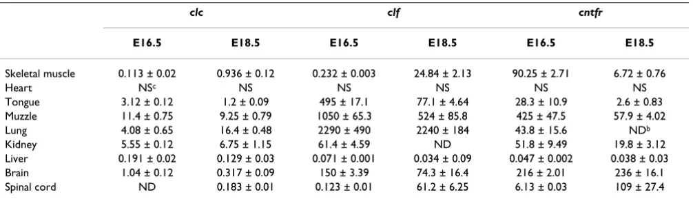
Developmental expression of clcSince the expression of clc has only been studied in adult mouse tissues [7], we first examined the expression of genes encoding CLC or its co-secreted proteins, CLF and CNTFR in various embryonic tissues using reverse tran-scription and quantitative real-time polymerase chain reaction (RT-PCR). In all tissues tested from E16.5 and E18.5 (Table 1), the level of expression of clc is very low when compared with that of clf or cntfr. The highest level of clc expression was observed in the muzzle, a very heter-ogeneous region containing different positive tissues, as described below. Clc expression is also observed in lung, kidney, brain and skeletal muscles such as the tongue or limb muscles.
To further assess the potential involvement of CLC in the
development of motoneurons, we performed in situ
hybridization experiments to determine the pattern of expression of clc in the environment of developing motoneurons and compare it with the expression of both
clf and cntfr. Motoneuron death occurs between E14.5 and E18.5 in mice lacking in the ability to produce a func-tional CNTF receptor complex [5], suggesting that expres-sion of CNTFR and its relevant ligands is critical between these timepoints. We therefore studied clc mRNA expres-sion levels at E16.5. Clc is expressed in muscles along the whole rostro-caudal axis, at the brachial level (Fig. 1A) as well as at the lumbar level (Fig. 1E and [8]. It is also
expressed in the tongue (Fig. 1C) like clf (Fig. 1D). The identity of muscle cells (Fig. 1G) was confirmed by double staining performed on transgenic mice with the nlacZ
reporter gene under the control of the muscle-specific MLC promoter [9]. All clc-positive muscle fibers also stained positive for clf (Fig. 1E, 1F, 1G, 1H and [8]). clc
expression was not detected in certain clf-positive muscles however, such those around the vibrissae (Fig. 1I and 1J). Since the level of clc expression is generally low, this could reflect the limited sensitivity of the in situ hybridization technique used. To determine the onset of clc and clf
expression in the muscles, the motoneuron targets, we performed in situ hybridizations at different stages. Clc
and clf are expressed, although at low levels, as soon as the muscles develop and are clearly observed at E14.5 (Fig. 1K and 1L).
Clc is also expressed in several organs in which reciprocal epithelial-mesenchymal interactions are essential, such as the developing vibrissae (Fig. 1I and 2I), tooth, kidney, and lung. In the kidney, clc is expressed in the comma-shaped body (Fig. 2A). Strikingly, CLF and CNTFR are expressed in different structures, clf being synthesized in the tips of the ureteric (Fig. 2B) and cntfr being synthe-sized by mesenchyma cells surrounding these structures (Fig. 2C). In the lung, both clc and cnftr are expressed faintly in distal airway epithelium whereas clf is strongly expressed in distal and proximal epithelia (Fig. 2D, 2E and 2F). Sections through molar tooth germs (Fig. 2G and 2H) show that clf is expressed in both the mesenchyma surrounding the dental follicle which gives rise to alveolar bone and the inner enamel epithelium whereas clc is expressed only in the former. Clc and clf are also co-expressed in the epithelium bordering the mandibles and the lips although clf is also expressed in mesenchyma (Fig.
Table 1: RT-PCR analysis of clc, clf and cntfr expressiona
clc clf cntfr
E16.5 E18.5 E16.5 E18.5 E16.5 E18.5
Skeletal muscle 0.113 ± 0.02 0.936 ± 0.12 0.232 ± 0.003 24.84 ± 2.13 90.25 ± 2.71 6.72 ± 0.76
Heart NSc NS NS NS NS NS
Tongue 3.12 ± 0.12 1.2 ± 0.09 495 ± 17.1 77.1 ± 4.64 28.3 ± 10.9 2.6 ± 0.83 Muzzle 11.4 ± 0.75 9.25 ± 0.79 1050 ± 65.3 524 ± 85.8 425 ± 47.5 57.9 ± 4.02 Lung 4.08 ± 0.65 16.4 ± 0.48 2290 ± 490 2240 ± 184 43.8 ± 15.6 NDb
Kidney 5.55 ± 0.12 6.75 ± 1.15 61.4 ± 4.59 ND 51.8 ± 9.49 19.8 ± 3.12 Liver 0.191 ± 0.02 0.129 ± 0.03 0.071 ± 0.001 0.034 ± 0.09 0.047 ± 0.002 0.038 ± 0.03 Brain 1.04 ± 0.12 0.317 ± 0.09 150 ± 3.39 74.3 ± 16.4 216 ± 2.01 236 ± 16.1 Spinal cord ND 0.183 ± 0.01 0.123 ± 0.01 61.2 ± 6.25 6.13 ± 0.03 109 ± 27.4
aExpression of clc, clf and cntfr was determined using reverse transcription and quantitative real-time PCR as detailed in Experimental Procedures
and expressed as fM of cDNA/µg total RNA.
2I and 2J). Together these results are in agreement with the expression pattern described for both clf [10] and cntfr
[11].
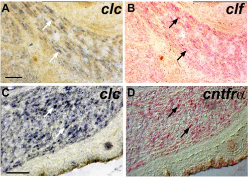
Co-expression of clc, clf and cntfr in the developing muscle
In transfected cells CLC requires either CLF or sCNTFR to be secreted [1,2]. This cooperative effect requires the expression of genes for both factors in the same cell. To ascertain whether a single muscle cell can express at least CLC and CLF or CLC and sCNTFR, we studied co-expres-sion on hind-limb muscle sections. We performed double
in situ hybridization of clc and clf and of clc and cntfr. Most muscle cells expressed both clc, (revealed using NBT/
BCIP; Fig. 3A, C) and clf or cntfr (Fig. 3B, D; revealed using Fast Red). Co-expression was observed at the single cell level demonstrating that in vivo CLC could be co-secreted either with CLF or sCNTFR.
Conclusions
Clc is expressed in developing muscles during the period of motoneuron loss in mice lacking a functional CNTF receptor and it is co-expressed with both CLF and CNTFR. This expression pattern is in favor of the hypothesis that CLC is an important player in the signal triggered by the CNTF receptor and that is required for motoneuron development. In addition, our results show that in the kidney, clc is expressed in cells neighboring those
express-In E16.5 mouse embryos clc is expressed in muscles Figure 1
Cell Communication and Signaling 2005, 3:1 http://www.biosignaling.com/content/3/1/1
Clc is expressed in epithelia Figure 2
ing clf or cntfr but it is not co-expressed with these genes suggesting either the possible existence of an additional protein capable of inducing secretion of CLC or that CLC is not secreted in these cells and therefore not functional. Because genetic deletion of cntf fails to perturb neuronal development before birth, we can hypothesize some func-tional redundancies in vivo that will require the analysis of double or triple knockout mice for CNTFR ligands to clar-ify their respective involvement in mouse neural development.
Methods
RT and real time PCR
Total RNA was extracted using Trizol reagent (Invitrogen) from E16.5 or E18.5 mouse tissues according to the
man-ufacturer's instructions. Complementary cDNA was syn-thesised from 2 µg of RNA by random hexamer priming using MMLV reverse transcriptase (Promega). Quantita-tive PCR was performed using a capillary real-time LightCycler (Roche Diagnostics), and the data analysed using "Fit Point Method" (Roche Diagnostics). For com-parison of gene expression levels, all quantifications were normalized to endogenous gapdh to account for variabil-ity in the initial concentration of RNA and for differences in the efficiency of the reverse transcription reactions. The following primers were designed to amplify mouse clc: 5'-GCTACCTGGAGCATCAACT-3',
5'-GGTGACTG-TACGCCTCATAG-3'; clf:
5'-CAGTCAGGAGACAATCT-GGT-3', 5'-ACGTGAGATCCTTCATGTTC-3'; cntfr:
5'-CTACATCCCCAATACCTACA-3',
5'-GTGAATTCGT-Double-labeling detects co-expression of clc and clf or clc and cntfr in individual muscle cells Figure 3
Cell Communication and Signaling 2005, 3:1 http://www.biosignaling.com/content/3/1/1
CAAAGGTGAT-3'; gapdh:
5'-TGCGACTTCAACAG-CAACTC-3', 5'-CTTGCTCAGTGTCCTTGCTG-3'. Results are expressed in fmole of cDNA/µgRNA.
Probes
Plasmid cDNA clones were linearized and transcribed with T7 or T3 polymerase using digoxigenin (Dig) or flu-orescein (Fluo)labeling reagents (Roche Diagnostics). Probes were used at a concentration of 500 ng/ml. The
cntfr clone was as previously described [12] and the mouse clf [13] and clc probes corresponded to the isolated cDNAs.
In situ hybridization
In situ hybridization was performed as described previ-ously [14] on 20 µm-thick frozen transverse cryostat sec-tions prepared from mouse embryos fixed with 4% paraformaldehyde in PBS, and cryopreserved in 15% sucrose in PBS before embedding in OCT compound (Miles). Alternatively, 100 µm-thick vibratome sections were prepared from fixed embryos embedded in glutaral-dehyde/gelatin. After hybridization overnight at 70°C with Dig-labeled riboprobes, the slides were washed twice in 1X SSC, 50% formamide at 70°C for 30 min and blocked in the presence of 4% blocking reagent (Roche Diagnostics) and 20% inactivated sheep serum. The slides were then incubated with anti-Dig-alkaline-phosphatase (AP)-conjugated antibody (1/5000, Roche Diagnostics), washed and revealed by NBT/BCIP staining.
In order to confirm that muscle fibers, per se, express clc
and clf, double in situ hybridization / immunohistochem-istry was carried out as described [15] on sections from E16.5 MLCnlacZ mice, which express the nlacZ reporter gene under the control of a muscle-specific myosin light chain promoter. After in situ hybridization, slides were rinsed in PBT (PBS, 0.1% Triton), and sections were suc-cessively incubated for 1 h with blocking solution con-taining 2% BSA, 2% heat-inactivated donkey serum in PBT and then overnight at 4°C with rabbit anti-β -galac-tosidasel (1/1000, Cappel). After three washes in PBT, slides were incubated 1 h at RT with a biotin donkey anti-mouse secondary antibody. Slides were then washed in PBS, and TBS (50 mM Tris-HCl, 0.15 M NaCl, pH 7.6), and incubated for 30 min at RT in ABC streptavidin/HRP in TBS. Staining was revealed with DAB (D4293, Sigma) in the presence of H2O2.
Double in situ hybridization was performed as described previously [14]. Briefly, Dig- and Fluo-labeled probes were mixed in hybridization buffer and applied to sec-tions. After hybridization at 70°C overnight and washing at 65°C, the first probe was revealed using a 1:2000 dilu-tion of anti-Fluo-alkaline phosphatase (AP)- conjugate (Roche Diagnostics) and Fast Red (Sigma) as a substrate.
Sections were photographed at this stage. After AP inacti-vation with 0.1 M glycine, pH 2.2, the second probe was revealed using a 1:5000 dilution of anti-Dig-AP and NBT/ BCIP staining. Fast Red precipitates were then removed by incubating the slides in increasing concentrations of etha-nol culminating in two final incubations in 100% ethaetha-nol for 10 min before cleaning with Histoclear and mounting with Eukitt (VWR, Strasbourg, France). Photomicrographs of the NBT/BCIP results were then taken for comparison with those showing the Fast Red results on the same sections.
Competing interests
The author(s) declare that they have no competing interests.
Authors' contributions
BB performed in situ hybridizations whereas DD and HG performed RT-PCR analyses. GE and JFG provided the clc
and clf probes before publication. OL participated in the experimental design and coordination of the research. All authors read and approved the final manuscript.
Acknowledgements
We thank members of INSERM U.623 and U.564 for many helpful discus-sions and encouraging support. This work was funded by INSERM, CNRS, the Association Française contre les Myopathies (AFM), the post-genome program from Région Pays-de-la-Loire, the Canadian Institutes of Health Research (IRSC) and the Multiple Sclerosis Scientific Research Foundation (SP). Damien Derouet was supported by INSERM and the Région Pays-de-la-Loire.
References
1. Elson GC, Lelievre E, Guillet C, Chevalier S, Plun-Favreau H, Froger J, Suard I, de Coignac AB, Delneste Y, Bonnefoy JY, et al.: CLF asso-ciates with CLC to form a functional heteromeric ligand for the CNTF receptor complex.Nat Neurosci 2000, 3:867-72. 2. Plun-Favreau H, Elson G, Chabbert M, Froger J, deLapeyriere O,
Lelievre E, Guillet C, Hermann J, Gauchat JF, Gascan H, et al.: The cil-iary neurotrophic factor receptor alpha component induces the secretion of and is required for functional responses to cardiotrophin-like cytokine.Embo J 2001, 20:1692-1703. 3. DeChiara TM, Vejsada R, Poueymirou WT, Acheson A, Suri C,
Cono-ver JC, Friedman B, McClain J, Pan L, Stahl N, et al.: Mice lacking the CNTF receptor, unlike mice lacking CNTF, exhibit profound motor neuron deficits at birth.Cell 1995, 83:313-322.
4. Li M, Sendtner M, Smith A: Essential function of LIF receptor in motor neurons.Nature 1995, 378:724-727.
5. Nakashima K, Wiese S, Yanagisawa M, Arakawa H, Kimura N, Hisat-sune T, Yoshida K, Kishimoto T, Sendtner M, Taga T: Developmen-tal requirement of gp130 signaling in neuronal survival and astrocyte differentiation.J Neurosci 1999, 19:5429-34.
6. Masu Y, Wolf E, Holtmann B, Sendtner M, Brem G, Thoenen H: Dis-ruption of the CNTF gene results in motor neuron degeneration.Nature 1993, 365:27-32.
7. Senaldi G, Varnum BC, Sarmiento U, Starnes C, Lile J, Scully S, Guo J, Elliott G, McNinch J, Shaklee CL, et al.: Novel neurotrophin-1/B cell-stimulating factor-3: a cytokine of the IL-6 family.Proc Natl Acad Sci U S A 1999, 96:11458-63.
8. Forger NG, Prevette D, deLapeyriere O, de Bovis B, Wang S, Bartlett P, Oppenheim RW: Cardiotrophin-like cytokine/cytokine-like factor 1 is an essential trophic factor for lumbar and facial motoneurons in vivo.J Neurosci 2003, 23:8854-8.
Publish with BioMed Central and every scientist can read your work free of charge
"BioMed Central will be the most significant development for disseminating the results of biomedical researc h in our lifetime."
Sir Paul Nurse, Cancer Research UK
Your research papers will be:
available free of charge to the entire biomedical community
peer reviewed and published immediately upon acceptance
cited in PubMed and archived on PubMed Central
yours — you keep the copyright
Submit your manuscript here:
http://www.biomedcentral.com/info/publishing_adv.asp
BioMedcentral
separate enhancers at the MLC1F/3F locus. Dev Biol 1997,
187:183-99.
10. Alexander WS, Rakar S, Robb L, Farley A, Willson TA, Zhang JG, Hartley L, Kikuchi Y, Kojima T, Nomura H, et al.: Suckling defect in mice lacking the soluble haemopoietin receptor NR6.Curr Biol 1999, 9:605-8.
11. Ip NY, McClain J, Barrezueta NX, Aldrich TH, Pan L, Li Y, Wiegand SJ, Friedman B, Davis S, Yancopoulos GD: The alpha component of the CNTF receptor is required for signaling and defines potential CNTF targets in the adult and during development.Neuron 1993, 10:89-102.
12. Arce V, Pollock RA, Philippe JM, Pennica D, Henderson CE, deLapey-rière O: Synergistic effects of Schwann- and muscle-derived factors on motoneuron survival involve GDNF and cardio-trophin-1 (CT-1).J Neurosci 1998, 18:1440-1448.
13. Elson GC, Graber P, Losberger C, Herren S, Gretener D, Menoud LN, Wells TN, Kosco-Vilbois MH, Gauchat JF: Cytokine-like fac-tor-1, a novel soluble protein, shares homology with mem-bers of the cytokine type I receptor family.J Immunol 1998,
161:1371-9.
14. Garcès A, Livet J, Grillet N, Henderson CE, Delapeyrière O: Respon-siveness to neurturin of subpopulations of embryonic rat spi-nal motoneuron does not correlate with expression of GFR alpha 1 or GFR alpha 2.Dev Dyn 2001, 220:189-97.
15. Carroll P, Gayet O, Feuillet C, Kallenbach S, de Bovis B, Dudley K, Alonso S: Juxtaposition of CNR protocadherins and reelin expression in the developing spinal cord.Mol Cell Neurosci 2001,