Plasminogen and angiostatin levels
in female benign breast lesions
A. A. TykhomyroV1, I. L. VoVchUk2, T. V. GrINeNko1
1Palladin Institute of Biochemistry, National Academy of Sciences of Ukraine, kyiv; 2odessa I. I. mechnikov National University, Ukraine;
e-mail: artem_tykhomyrov@ukr.net
It is known that benign breast tissue exhibit relatively low angiogenic capacity. Activation of an-giogenesis in mammary pre-malignant lesions could be associated with disease progression and high risk of transformation into the breast cancer. however, insight into the underlying molecular mechanisms in-volved in angiogenesis regulation in non-cancerous breast pathologies is still poorly defined. The purpose of the present study was to determine levels of plasminogen and its proteolytic fragments (angiostatins) in mammary dysplasia (mastopathy and breast cyst) and benign neoplasms (fibroadenomas). Plasminogen and angiostatins were analyzed using immunoblotting and quantified by densitometric scanning. The significant increase in plasminogen levels was found in fibrocystic, cysts, and non-proliferatious fibroadenoma masses (4.7-, 3.7-, and 3.5-fold, respectively) compared to healthy breast tissues (control). In the same benign lesions, 6.7-, 4-, and 3.7-fold increase in plasminogen 50 kDa fragment (angiostatin) levels as compared with control were also observed. Activation of matrix metalloproteinase-9, which was detected using gelatine zymography, could be responsible for plasminogen cleavage and abundance of angiostatin in fibrocystic and cyst masses. In contrast, dramatic decrease of both plasminogen and angiostatin levels (3.8- and 5.3-folds, respectively) was shown in tissues of proliferatious form of fibroadenoma in comparison with that of the dormant type of this neoplasm. Based on the obtained results, we concluded that angiostatin, a potent vessel growth inhibitor and anti-inflammatory molecule, can play a crucial role in pathophysiology of non-cancerous breast dis -eases. Further studies are needed to evaluate potential diagnostic and clinical implications of these proteins for prediction and therapy of benign breast pathologies.
k e y w o r d s: plasminogen, angiostatin, matrix metalloproteinases, angiogenesis, benign breast diseases, mastopathy, breast cyst, fibroadenoma.
T
he term “benign breast diseases” (BBDs) en-compasses a heterogeneous group of lesions, including developmental abnormalities,in-flammatory lesions, epithelial and stromal prolife rations, and neoplasms. Mastopathy (fibrocystic
disease), breast cysts, and fibroadenoma are recog
-nized to be the most prevalent types of BBDs [1]. According to the definition published by the World Health Organization (WHO) in 1984, mastopathy is the fibrocystic disease of breast, characterized by
a disbalance between epithelial and connective tis-sues growth with high proliferative and regressive
changes of the breast tissue [2]. Cysts are nonpro
-liferative benign breast conditions, associated with hormone-dependent transformation of apocrine
epi-thelial cells [3]. Fibroadenoma is the most common benign neoplasm of the breast. It is usually a disease
of early reproductive life – the peak incidence is be
-tween the ages of 15 and 35 years. BBDs are among prevalent strong risk factors for breast cancer. Today,
it has been admitted that breast cancer is located 3-5 times more often in women with benign lesions of
breast and 3040 times more often if the women are suffering from nodulose form of mastopathy with signs of epithelial proliferation [4].
It is known that abnormal blood vessel out
-growth plays an important role in the genesis and
development of various tumors, and growth of neo-plastic masses is associated with formation of their own vessels due to pathological neovascularization
[5]. J. Folkman [6] has shown that neoplastic tis
-sues have a peculiar capacity to induce angiogene sis in the surrounding tissues, and lack of vessel
formation can exclude neoplastic growth. It should
be emphasized that angiogenesis is regulated by a
Angiogenesis in invasive breast cancer is well
docu-mented [7], but relatively few studies have addressed
the role of angiogenesis in pre-malignant disease or benign neoplasms where the switch of angiogenesis
balance may also occurs during their development.
“Angiogenic switch” is a crucial step for the
progres-sion of tumor from the benign to malignant state [8].
Though the benign states and breast cancers share
many similar pathological processes, their angio
-genic abilities are rather different. The angio-genic
capacities are rare in fibrous or adipose tissue and in tissues from fibroadenomas, fibrocystic disease,
and normal lobules, but it is pronounced in
carcino-mas and intraductal papillocarcino-mas [9]. The molecular
mecha nisms responsible for maintenance of low an-giogenic potential in benign lesions are still uncove-red.
Limited proteolysis of extracellular proteins by
matrix metalloproteinases (MMPs) and some serine proteases is an important step in the regulation of
angiogenesis. Among the proteolytic degradation
products derived from extracellular matrix proteins and hemostasis factors, a number of fragments have
potent angiogenesis inhibiting properties [10]. One
of them, angiostatin (AS), is an internal proteolyti
-cally derived fragment of plasminogen (Plg), span
-ning various numbers of its kringle domains. AS is
considered to be one of the most powerful endoge-nous inhibitors of neovascularization. AS suppresses
angiogenesis by inhibiting endothelial cell prolifera
-tion, migration and can even promote endothelial
apoptosis [1113]. As a rule, AS circulates in abun
-dance in plasma of patients with different cancers
and inflammatory states, however, the changes in
blood concentrations of this molecule are not
al-ways consistent with the alterations of their tissue levels [14, 15]. To our knowledge, no reports have
examined content of Plg/AS in the breast benign
neoplasms. In order to fill this gap, here we aimed to
determine amounts of these proteins in benign
non-cancerous lesions of mammary gland. To check the hypothesis that local levels of AS and its precursor molecule could be related to abnormally increased
rate of breast cell proliferation, we paid special
at-tention on Plg/AS content in fibroadenomas with
different proliferating potential. Evaluation of
activi-ties of the collagenolytic enzymes, which could be potentially responsible for Plg degradation and AS
release in tissues of breast benign formations, was
also among the tasks of this study.
materials and methods
Patients and tissue specimens. Breast tissue specimens were obtained from women with various
BBDs (fibroadenoma, fibrocystic breast disease,
breast cyst, n = 9 for each type of disease) by sur
-gical resection performed in certified Laboratory of Pathomorphology of Odessa Regional Oncology
Center. The diagnoses were established
histological-ly by experienced breast pathologists. Fibroadenoma
nodules were additionally screened for ductal epithe
-lial proliferative changes and subclassified as non
proliferatious and proliferatious forms (n = 5 and 4,
respectively), based on the criteria as described by Rosen et al. [16]. The size of surgical specimens was
at least 0.5 cm3. The study protocol was prepared in accordance with international and local human re-search ethical standards. Informed consent was ob-tained from all patients before the beginning of the
study. The samples of the surrounding healthy tis
-sues were used as controls (n = 6). Tissue specimens
were kept at – 80 °C until they were transported on ice and frozen at – 20 °C when received.
reagents. Secondary antirabbit horseradish
peroxidise (HRP)conjugated IgG, bovine serum
albumin (BSA), protease inhibitors, ethylenediami
-netetraacetic acid (EDTA), Triton X-100, Tween-80,
ammonium persulfate, tetrahydrochloride hydrate,
tetramethylethylenediamine (TEMED), 3,3′di
-aminobenzidine (DAB), nonfat dry milk, gelatine, Coomassie Briliant Blue R250 were purchased from Sigma (USA); tris(hydroxymethyl)aminomethane,
glycine, acrilamide, bisacrilamide, sodium phos
-phates, sodium chloride were obtained from
Heli-con (Russian Federation); sodium dodecyl sulphate (SDS) were purchased from AppliChem (Germany); molecular weight markers (PageRuler Prestained Protein Ladder) were obtained from Fermentas (Germany). All other chemicals were of analytical
reagent grade.
Protein sample preparation. Protein samples of normal and pathological breast tissues for further AS
detection were prepared by grinding and homogeni
-zation of tissue specimens (weighted 100-150 mg)
in icecold 50 mM TrisHCl buffer (pH 7.4), ad
-ditionally containing 150 mM NaCl, 1% SDS, 2.5 mM EDTA, 6.5 μM aprotinin, 1.5 μM pepstatin A,
23 μM leupeptin, 1 mM phenylmethylsulfonyl fluo
-ride, 5 μg/ml soybean trypsin inhibitor. Tissue:buffer ratio was taken equal 1:5 (m/v). For MMP analysis,
homog-enization procedures using of 50 mм TrisHCl (рн
7.4), which contained 150 mм NaCl and was supple
-mented with serine proteinase inhibitors mentioned above (tissue:buffer = 1:3, m/v). After
homogenisa-tion steps, samples were sonicated for approximately
30 sec with using of ultrasonic disintegrator
Sarto-rius (Labsonic® M, Göttingen, Germany) and cen
-trifuged at 16000 g for 45 min at 4 °C. All super
-natants were carefully removed and transferred into
the clean Eppendorf tubes. The total protein concen-tration in each supernatant was determined
spec-trophotometrically by Stoscheck method measuring absorbance at 260, 280, and 320 nm as described elsewhere [17]. The samples were diluted 1:1 with
non-reducing Laemmli Sample Buffer, frozen and
stored at 20 °C before analysis.
Immunoblotting. Protein samples (50 μg/track)
were run in 518% denaturing polyacrilamide gels
(PAAG) and transferred onto nitrocellulose mem
-brane (GE Healthcare, Amersham Bioscience) with
0.45 μm pore diameter by means of electroblot. Af
-ter transferring, membranes were blocked in 5% m/v nonfat dry milk for 90 min at 37 °C and probed with primary antibody, which were obtained as described previously [15, 18]. Briefly, isolated and purified product of Plg limited digesting by porcine elastase, which comprises the first three kringle domains
(K1-3), was used as an antigen for rabbit
immuniza-tion. Polyclonal antibodies, purified on immunoaf
-fine sorbent K13sepharose, appeared to recognize ASlike proteolytic fragments of Plg as well as the parent molecule. AntiAS/Plg antibody was diluted in phosphate buffer saline (PBS) and used in final concentration 5 μg/ml. After overnight incubation at 4 °C, membranes were washed five times with PBS, containing 0.05% Triton X100 (PBST), and then
incubated for 60 min with the HRPconjugated sec
-ondary antibody diluted in PBST 1/3000. Then, un
-bound antibodies were removed by 7times washes in PBST for 5 min each. Specific immunostaining was developed by incubation of membranes with 0.05% DAB in 50 mM TrisHCl buffer (pH 7.4), containing 0.01% H2O2. Relative intensities of bands, correspondent to Plg and AS, were measured using
densitometry software TotalLab TL120 (Nonlinear Inc, USA). Each trace was corrected for background by subtracting a tracing of nonreactive area on the
blot. Antigens of various molecular weights were
identified by extrapolation of plots of relative mo
-bilities of prestained transblot proteins with known molecular weight (PageRuler Prestained Protein
Gelatin zymography. It is generally accepted
that zymographic tests of MMP activity have some advantages over immunologic assays due to lower
costs, more rapid time of execution and the
possi-bility of detection simultaneously multiple forms of MMPs [19]. For detection of MMP activities in the
samples obtained from breast tissues, gelatine zy
-mography was performed using SDSPAAG electro
-phoresis with 7.5% separating and 4% stacking gels
in the absence of reducing agents. The separating
gel was copolymerized with heatdenatured gelatine (5 mg/ml) as enzyme substrate. Tissue extracts in the volumes, containing 50 μg total protein, were loaded onto gelatinePAAG slabs. After running, gels were washed twice with 2.5% Triton X100 in aqueous solution and then rinsed five times with icecold bi
distilled water and incubated in 50 mM Tris-HCl
de-veloping buffer (pH 7,6), containing 20 mM NaCl,
5 mM CaCl2, 1 mM ZnCl2, 0.05% Triton X100, and
0.02% Tween80, at 37 °C for 16 h. Zymograms were stained with Coomassie Briliant Blue R250 and destained with 30% methanol and 10% acetic acid in distilled water. The final gel had a uniform blue background except in those regions to which MMPs
had migrated and cleaved the substrate, so bands of gelatine degradation could be seen as transparent
areas against a blue background. Molecular weights
were determined using standard pre-stained
molecu-lar weight markers.
Statistical analysis. The statistics were
per-formed using nonparametric MannWhitney U-test.
Plg and AS contents, evaluated by immunoblots, are expressed in arbitrary units (a.u.) and presented in histograms as medians. Statistical significance was defined as P less than 0.05.
results and discussion
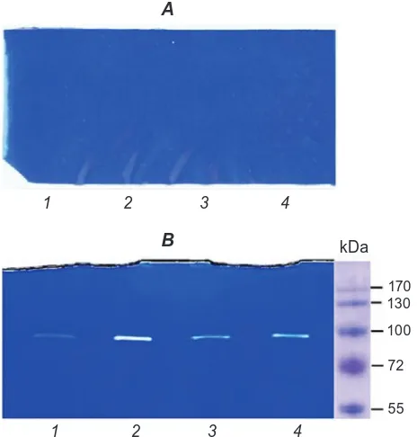
Polyclonal antibodies used for immunob
-lot analysis reacted with bands, the approximate molecu lar weights of which were 90 and 50 kDa
(Fig. 1). Mr values of these bands correspond to
na-tive plasminogen (Mr 9293 kDa) and ASlike frag -ment (Mr 4550 kDa), consisting of first four kringle
domains (K14 or K14.5). Only trace amounts of immunoreactivity were observed in protein samples obtained from histologically normal breast tissues, used as control in the present study. In contrast,
abundance of two major immunoreactive polypep
-tide bands is found in the samples prepared from
benign tissue lesions. Western blot of samples pro
sities of Plg/AS band immunostaining depend on the
proliferation potential of neoplasm. In fibroadeno
-mas with increased proliferative rate, weak specific
immunoreactivity is observed, while nonprolifera
-tious neoplasms represent more intense Plg/AS im-munostaining. This observation can be of peculiar importance because levels of these proteins could
re-flect development of regressive changes in breast tis
-sues or transformation of cells, composing a tumor.
In order to quantify results of immunoblotting, densitometric analysis was performed and intensities
of immunoreactivity of Plg and AS bands as medi
-ans of arbitrary units are presented in Fig. 2. It was
shown that levels of AS isoform were correlated with Plg content in both normal and pathological tissues.
However, in mastopathy, Plg and AS levels were found to be 4.7 and 6.7fold higher than those for unchanged tissues respectively (P < 0.05). Signifi -cant increase in both Plg and AS contents was also
Fig. 1. Immunoblots of protein samples prepared from mammary tissues of female patients with various breast benign diseases and healthy control tissues
Breast cyst Fibroadenoma
non-proliferatious form proliferatious form
100
72
55
45
33
kDa Control Fibrocystic breast disease
Plg
AS
found in cysts as compared with healthy tissues (3.7 and 4folds respectively, P < 0.05). In fibroadenoma (benign neoplasm condition), Plg and AS abundance was 3.5- and 3.7-fold higher in the case of non-pro-liferative form as compared with control (P < 0.05). In order to relate our data with disease features, we measured Plg/AS levels in samples prepared from
fibroadenoma nodules with signs of hyperprolifera
-tion. It is of interest that averaged levels of Plg and
its 50 kDa proteolytic fragment in fibroadenoma
with evident proliferative changes appeared to be
respectively 3.8 and 5.3fold lower than that of non
proliferatious form of this neoplasm.
Gelatine zymography of the samples of healthy mammary tissues and breast benign masses was performed to assess activity of MMPs (gelatinases).
It is important to note that serine protease inhibi-tors were added into the homogenization buffer in
Fig. 2. results of densitometry analysis of plasminogen (A) and angiostatin (B) detected by immunoblotting (data are expressed as medians): 1 – control (n = 6), 2 – fibrocystic disease (n = 9), 3 – breast cyst (n = 9), 4 – fibroadenoma (non-proliferatious form, n = 5), 5 – fibroadenoma (proliferatious form, n = 4). * P < 0.05 as compared to control (Mann-Whitney U-test); # P < 0.05 as compared to non-proliferatious form of fibroadenoma (Mann-Whitney U-test)
100
1 2 3 4 5 50
80
60
A
rb
it
rar
y
u
n
it
s
A
rb
it
rar
y
u
n
it
s
200 250
150
0
40
20
0 100 120
A B
1 2 3 4 5
gelatine digesting, thus eliminating possible
appear-ance of falsepositive results. As shown in Fig. 3, A,
there was no collagenolytic activity in homogenates
isolated from normal breast tissues. In contrast, in most samples prepared from breast benign lesions,
the single abundant proteolytic activity was detected
as band with approximate Mr 90 kDa, which is cor
-responds to MMP9. Though it has been previously
established that MMP2 and 9 are major gelatino
-lytic protease expressing in breast tumors and circu
-lating in blood of cancer patients [19, 20], we were
failed to reveal active MMP2 (Mr 72 kDa) isoform
in benign mammary masses.
The application of immunochemical methods
(ELISA, Western blot) for AS determination in pa
-tient’s blood plasma or serum is a commonly used procedure. However, at present, little is known about
the localization of AS in human tissues. Also, there
is lack of information concerning local levels and physiological significance of AS in BBDs. Here, we for the first time have assessed contents of AS and
its parent molecule (Plg) in breast tissues of women
with fibrocystic disease, cysts, and fibroadenomas. It is known that regulation of angiogenesis is governed by a fine equilibrium between proangio genic factors (vascular endothelial growth factor –
vEGF, fibroblast growth factor – FGF, plateletde
-rived growth factor – PDGF, transforming growth factor – TGF) and antiangiogenic factors such as
thrombospondin, pigment epithelium-derived
fac-tor – PEDF, endostatin, tumstatin. In adults, blood vessel formation is tightly controlled by “angiogenic
Fig. 3. Gelatin zymography of protein samples ob-tained from normal mammary gland tissues (A, 1-4 numbers of samples) and benign breast masses (B: 1 and 2 – fibrocystic disease, 3 and 4 – breast cysts)
B
1 2 3 4
kDa
170 130
100
72
55
A
1 2 3 4
essential for solid tumors, including breast cancers,
to grow [21]. At present, a whole range of angio
-genesis inhibiting fragments of matrix proteins and coagulation related proteins have been recognized, such as angiostatin, endostatin, tumstatin,
alphasta-tin, thrombospondin fragments and many others
[10]. Plg, catalytically nonactive proenzyme, con
faced Janus. On the one hand, Plg can be converted
into active proteinase plasmin, and when bound to
specific cellsurface and extracellular matrix recep
-tors, plasmin participates in degradation of extracel-lular matrices during cell migration, tissue
remodel-ling, tumor cell invasion, and inflammation. On the
other hand, Plg is also a precursor for a group of
antiangiogenic molecules, AS [22]. They represents a family of proteolytic fragments of Plg, consisting of different number of kringle domains as well as single kringle domains (K13, K14, K14.5, K15, K23, K5 are definitely described) [23]. It has been
reported that AS isoforms are generated from Plg
by limited proteolysisrelated enzymes, such as plas
-min, cathepsins, prostatespecific antigen, pancreatic and neutrophil elastase [24]. Among the proteases
that are involved both in tumor angiogenesis and AS
generation, the members of MMP family (MMP1, 2, 3, 7, 9, 12, 14, and 19) has been extensively
studied [25]. AS blocks proliferation, migration, dif
-ferentiation and tube formation of endothelial cells, therefore suppressing microvessel formation, tumor
progression, and metastasis [26]. It is known that
human AS inhibits the growth of transplanted
hu-man and murine primary tumors in mice and causes human primary carcinomas to regress to a dormant state by a net balance of tumor cell proliferation and apoptosis. For example, the full kringles of
Plg (K1-5) caused total regression of human
MDA-MB231 breast tumor xenografted in mice, which
was correlated with a drastic decrease of functional neovascularization into the tumors and inhibition
of metastatic dissemination [27]. Pronounced anti
inflammatory effects of AS are documented and ex
-tensively studied as well [28, 29].
Results of Western blots demonstrate simulta
-neous elevation of Plg and its proteolytic fragment levels in breast benign lesions (Fig. 1). The interpre
-tation of this result may be that cells of benign tis
-sue lumps can expose a number of molecules, which participate in Plg/AS binding with cellular surface.
A variety of proteins, including actin, annexin II, S100A10, cytokeratin 8, tetranectin and αenolase have been identified as potential Plg receptors on the surface of certain types of human breast cancer cells [30]. AS appeared to effectively interact with some Plg receptors. For example, Dudani et al. [31] have shown that surfaceassociated βactin can bind both Plg and AS K14 (kd ~ 140 nmol/L). It is thought that
AS, competing for binding sites with proenzyme,
can inhibit Plg conversion into active proteinase and
thus modulate plasmin-dependent processes such as cell migration/movement, extracellular matrix re-modelling.
In biological fluids or tissues of patients with
different pathologies, a heterogeneous mixture of Plg fragments is often observed, with patterns that
also varied between patients [32]. In our study, AS like fragment 50 kDa appears to be the principal Plg fragment, which is solely produced in breast tissues.
Therefore, it is most likely that certain type of pro
-teinases contributes to Plg processing in mammary
gland. A number of papers highlights that Plg
frag-mentation in breast tumors is usually associated with MMPs, and MMP2 and 9 are the most important
ones [19, 20]. Although neither normal nor pathologi
-cal breast tissues expressed any MMP2 enzymatic activity, presence of activated MMP9 is obviously demonstrated in the samples of fibrocystic and cyst masses (Fig. 3). Our data suggesting the absence of MMP2 activity in mastopathy or cyst masses are in agreement with the previous reports [33], indicating
MMP2 to be constituent proteinase of rather ma
-lignant tumors responsible for cancer cell invasion
and metastasis. In our study, we were not focused on measuring gelatinase activities in fibroadenoma,
nevertheless, a number of earlier researches
indi-cates that active MMP2 and MMP9 infrequently
occur in low levels in benign tumor tissue extracts
[19, 34]. However, active MMP2 occurs more fre
-quently and at higher levels in malignant carcinoma tissue as compared with benign fibroadenoma tissue. Dramatically elevated levels of the both gelatinases
are found in blood of breast cancer patients
com-pared to benign mammary pathologies and healthy controls [35]. MMPs can be released by different
cells, like neutrophils, macrophages or other inflam
-matory cells that need to digest ECM to access or
-gan parenchymas. Since macrophages are the major population of infiltrating cells in tumor stroma, so it is possible that they may have a role in regulating of
angiogenesis through MMP secretion and Plg
con-version into AS [36, 37].
Hyperplasia is recognized to be a benign breast
condition where some breast cells begin to divide
more quickly than normal, however staying differen
-tiated and saving noncancerous phenotype. Patho
-physiological impact and clinical significance of ele vated activity of MMP9 and Plg/AS expression in
breast benign lesions are uncertain. Earlier, Chung et
al. [37] have shown that upregulation of MMP2 and
vascu-lar stiffening, impaired angiogenesis, and
endothe-lial dysfunction in kidneys. In the same way, activa
-tion of MMP-9 and eleva-tion of Plg-derived protein
levels in fibrocystic lesions and breast cyst tissues can be at least partially responsible for angiogenesis impairments and vascular abnormalities typical for breast benign lesions. It remains still unknown if
excessive amounts of AS contribute to the develop-ment of connective tissue sclerosis, disturbances of
local circulation, and progressive atrophy, which oc
-cur in fibrous cysts [38]. Here, it is assumed that AS can play a potential role in regulating proliferation of epithelial or mesenchimal elements in fibroade
noma. Decreased levels of Plg/AS discovered in breast tissues of patients with proliferatious form of
fibroadenoma may be related to proliferative capaci
-ties of the cells. However, it remains to be elucidated if changed AS levels could be considered either as
consequences or as causative factors, which are nec
-essary for benign tumor cells to acquire and sustain high proliferative potential. Further work is needed
to examine the precise mechanisms and molecular
orchestration involved in AS formation in mammary gland tissues and to establish whether AS has a key
independent role in the development of proliferative and regressive changes within breast tissues under benign conditions.
acknowledgments. The support of Odessa Re
-gional Oncology Center is gratefully acknowledged.
Вміст плазміногену та ангіостатиніВ у тканинах доброякісних утВорень молочної залози жінок
А. О. Тихомиров1, І. Л. Вовчук2, Т. В. Гриненко1
1інститут біохімії ім. о. В. палладіна
нан України, київ;
2одеський національний університет
ім. і. і. мечникова, Україна; email: artem_tykhomyrov@ukr.net
Відомо, що доброякісні утворення молочної залози характеризуються відносно низьким рівнем ангіогенезу. активація ангіогенезу за гіперпроліферативних захворювань молочної
залози може бути пов’язана з подальшим розвит
-ком патології та підвищеним ризи-ком злоякісної трансформації. однак молекулярні механізми, що лежать в основі регуляції ангіогенезу в доброякісних новоутвореннях молочної залози, залишаються недостатньо вивченими. метою роботи було визначити вміст плазміногену та його протеолітичних фрагментів (ангіостатинів) у тканинах молочної залози за мастопатії та кістозної хвороби, а також у доброякісних новоутвореннях (фіброаденомах). Детекцію
плазміногену та ангіостатинів проводили за до
-помогою імуноблотингу з подальшим кількісним денситометричним аналізом. показано, що
рівень плазміногену в тканинах молочної зало
-зи за мастопатії, кісти та непроліферуючої фор
-ми фіброадено-ми у 4,7, 3,7 і 3,5 раза відповідно
перевищує цей показник у нормальних ткани
-нах (контроль). Вміст протеолітичного фрагмен
-та плазміногену з молекулярною масою 50 кДа (ангіостатину) в цих доброякісних утвореннях виявився відповідно у 6,7, 4,0 і 3,7 раза вищим за контрольний рівень. Зростання концентрації
ангіостатину може відбуватися внаслідок роз
-щеплення плазміногену тканинною матрич
-ною металопротеїназою9, активну форму якої виявлено методом желатинової зимографії за фібрознокістозної хвороби та кісти. натомість, у тканинах проліферативної форми фіброаденоми рівень плазміногену та ангіостатину виявився відповідно у 3,8 і 5,3 раза нижчим порівняно з непрогресуючою неоплазією. наведені результати дозволяють припустити, що ангіостатини як потужні інгібітори ангіогенезу та протизапальні агенти можуть відігравати важливу роль у патогенезі дисплазій молочної залози. подальші дослідження необхідні для
оцінки діагностичного та клінічного значен
-ня цих протеїнів для прогнозуван-ня та терапії доброякісних захворювань молочної залози.
содержание плазминогена и ангиостатиноВ В тканях доброкачестВенных
образоВаний молочной железы женщин
А. А. Тихомиров1, И. Л. Вовчук2, Т. В. Гриненко1
1институт биохимии им. а. В. палладина
нан Украины, киев;
2одесский национальный университет
им. и. и. мечникова, Украина; email: artem_tykhomyrov@ukr.net
известно, что доброкачественные образо
-вания молочной железы характеризуются от
-носительно низким уровнем ангиогенеза. акти
-вация ангиогенеза при гиперпролиферативных заболеваниях молочной железы может быть связана с дальнейшим развитием патологии и
повышенным риском злокачественной транс
-формации. однако молекулярные механизмы, лежащие в основе регуляции ангиогенеза в доб рокачественных новообразованиях молочной железы, остаются недостаточно изученными.
Целью данной работы было определить содер
-жание плазминогена и его протеолитических фрагментов (ангиостатинов) в тканях молочной железы при мастопатиях и кистозной болезни,
а также в доброкачественных новообразовани
-ях (фиброаденомах). Детекцию плазминогена
и ангиостатинов проводили с помощью имму
-ноблоттинга с дальнейшим количественным денситометрическим анализом. показано, что
уровень плазминогена в тканях молочной желе
-зы при мастопатии, кисте и непролиферирую щей форме фиброаденомы в 4,7, 3,7 и 3,5 раза соответственно превышал этот показатель в нормальных тканях (контроль). содержание протеолитического фрагмента плазминогена с молекулярной массой 50 кДа (ангиостатина) в
этих доброкачественных образованиях оказа
-лось соответственно в 6,7, 4 и 3,7 раза выше по
сравнению с контрольным уровнем. Возраста
-ние концентрации ангиостатина может проис
-ходить вследствие расщепления плазминогена
тканевой матричной металлопротеиназой9, ак
-тивную форму которой выявлено методом же
-латиновой зимографии при фибрознокистозной
болезни и кисте. В то же время в тканях про
-лиферативной формы фиброаденомы уровень
плазминогена и ангиостатина оказался соответ
-ственно в 3,8 и 5,3 раза ниже по сравнению с не
-прогрессирующей неоплазией. представленные
результаты позволяют предположить, что анги
-остатины, будучи мощными ингибиторами ан
-гиогенеза и противовоспалительными агентами,
могут играть важную роль в патогенезе диспла
-зий молочной железы. Дальнейшие исследова
-ния необходимы для оценки диагностического и клинического значения этих протеинов при прогнозировании и терапии доб рокачественных заболеваний молочной железы.
к л ю ч е в ы е с л о в а: плазминоген, ан
-гиостатин, матричные металлопротеиназы, ангиогенез, доброкачественные заболевания молочной железы, мастопатия, киста молочной железы, фиброаденома.
references
1. Mannello F., Tonti G.A. Benign breast diseases: classification, diagnosis, and management.
oncologist. 2006;11(10):11321134.
2. Norwoord S. L. Fibrocystic breast disease. An
update and review. J. obstet. Gynecol. Neonatal.
Nurs. 1990;19(2):116121.
3. Houssami N., Irwig L., Ung O. Review of complex breast cysts: implications for cancer
detection and clinical practice. ANZ J. Surg.
2005;75(12):10801085.
4. ElWakeel H., Umpleby H.C. Systematic review of fibroadenoma as a risk factor for breast cancer.
Breast. 2003;12(5):302307.
5. Kerbel R. S. Tumor angiogenesis. N. engl. J.
med. 2008;358(19):203949.
6. Folkman J. Tumor angiogenesis: therapeutic
implications. N. engl. J. med. 1971;285(21):1182 1186.
7. Bos R., van Diest P. J., de Jong J. S., van der Groep P., van der valk P., van der Wall E. Hypoxiainducible factor1alpha is associated with angiogenesis, and expression of bFGF, PDGFBB, and EGFR in invasive breast cancer.
histopathology. 2005;46(1):3136.
8. Bluff J. E., Menakuru S. R., Cross S. S.,
Higham S. E., Balasubramanian S. P.,
Brown N. J., Reed M. W., Staton C. A.
Angiogenesis is associated with the onset of
hyperplasia in human ductal breast disease. Br.
J. cancer. 2009;101(4):666672.
9. Azzopardi J. G. Benign and malignant proliferative
10. Nyberg P., Xie L., Kalluri R. Endogenous
inhibitors of angiogenesis. cancer res.
2005;65(10):396779.
11. O’Reilly M. S., Holmgren L., Shing y., Chen C., Rosenthal R. A., Cao y., Moses M., Lane W. S., Sage E. H., Folkman J. Angiostatin: a circulating
endothelial cell inhibitor that suppresses angiogenesis and tumor growth. cold Spring harb. Symp. Quant. Biol. 1994;59:471482.
12. Wahl M. L., Kenan D. J., GonzalezGronow M., Pizzo S. v. Angiostatin’s molecular mechanism: aspects of specificity and regulation elucidated.
J. cell Biochem. 2005;96(2):242261.
13. Cho C.F., Chen P. K., Chang P. C., Wu H. L., Shi G. y. Human plasminogen kringle 15 inhibits
angiogenesis and induces thrombomodulin
degradation in a protein kinase Adependent
manner. J. mol. cell. cardiol. 2013;63:7988.
14. Szarvas T., Jäger T., Laszlo v., Kramer G., Klingler H. C., vom Dorp F., Romics I., Ergün S., Rübben H. Circulating angiostatin, bFGF, and Tie2/TEK levels and their prognostic impact in
bladder cancer. Urology. 2012;80(3):1318.
15. Tykhomyrov A. A., Nedzvetsky v. S., Bardachenko N. I., Grinenko T. v., Kuryata O. v.
Statin treatment decreases serum angiostatin levels in patients with ischemic heart disease. Life Sci. 2015;134:2229.
16. Rosen P. P. Pathological examination of breast specimens. In: Rosen PP, ed. Breast Pathology. Philadelphia, PA: LippincottRaven; 1997:837 872.
17. Stoscheck C. M. Quantitation of protein. methods
enzymol. 1990;182:5068.
18. Tykhomyrov A. A., yusova E. I., Diordieva S. I., Corsa v. v., Grinenko T. v. Production and
characteristics of antibodies against K1-3 fragment of human plasminogen. Biotechnologia Acta. 2013;6(1):8696.
19. La Rocca G., PucciMinafra I., Marrazzo A., Taormina P., Minafra S. Zymographic detection and clinical correlations of MMP2 and
MMP-9 in breast cancer sera. Br. J. cancer.
2004;90(7):14141421.
20. Hanemaaijer R., verheijen J. H., Maguire T. M., visser H., Toet K., McDermott E., O’Higgins N., Duffy M. J. Increased gelatinaseA and
gelatinase-B activities in malignant vs. benign breast tumors. Int. J. cancer. 2000;86(2):204
207.
21. Tonini T., Rossi F., Claudio P.P. Molecular
basis of angiogenesis and cancer. Oncogene.
2003;22(42):65496556.
22. Zhernosekov D. D., Iusova E. I., Grinenko T. v. Role of plasminogen/plasmin in functional
activity of blood cells. Ukr. Biokhim. Zhurn.
2012;84(4):519.
23. Doll J. A., Soff G. A. Angiostatin. cancer Treat.
res. 2005;126:175204.
24. Gately S., Twardowski P., Stack M. S., Cundiff D. L., Grella D., Castellino F. J., Enghild J., Kwaan H. C., Lee F., Kramer R. A., volpert O., Bouck N., Soff G. A. The mechanism
of cancer-mediated conversion of plasminogen to the angiogenesis inhibitor angiostatin. Proc. Natl. Acad. Sci. USA. 1997;94(20):1086810872.
25. Heissig B., Hattori K., Friedrich M., Rafii S., Werb Z. Angiogenesis: vascular remodeling of the
extracellular matrix involves metalloproteinases. curr. opin. hematol. 2003;10(2):136141.
26. Xu R., Sun X., Tse L. y., Li H., Chan P. C., Xu S., Xiao W., Kung H.F., Krissansen G. W., Fan S. T.
Long-term expression of angiostatin suppresses metastatic liver cancer in mice. hepatology.
2003;37(6):14511460.
27. Galaup A., Magnon C., Rouffiac v., Opolon P., Opolon D., Lassau N., Tursz T., Perricaudet M., Griscelli F. Full kringles of plasminogen (aa
1-566) mediate complete regression of human
MDAMB231 breast tumor xenografted in nude
mice. Gene Ther. 2005;12(10):831842.
28. Mu W., Long D. A., Ouyang X., Agarwal A., Cruz P. E., Roncal C. A., Nakagawa T., yu X., Hauswirth W. W., Johnson R. J. Angiostatin
overexpression is associated with an
improvement in chronic kidney injury by an
antiinflammatory mechanism. Am. J. Physiol.
renal Physiol. 2009;296(1):145152.
29. Perri S. R., Annabi B., Galipeau J. Angiostatin inhibits monocyte/macrophage migration via
disruption of actin cytoskeleton. FASeB J.
2007;21(14):39283936.
30. Stillfried G. E., Saunders D. N., Ranson M.
Plasminogen binding and activation at the breast
cancer cell surface: the integral role of urokinase activity. Breast Cancer Res. 2007;9(1):111.
31. Dudani A. K., Ben-Tchavtchavadze M., Porter S.,
Tackaberry E. Angiostatin and plasminogen
32. Migita T., Oda y., Naito S., Morikawa W., Kuwano M., Tsuneyoshi M. The accumulation of angiostatinlike fragments in human prostate
carcinoma. clin. cancer res. 2001;7(9):2750
2756.
33. Jezierska A., Motyl T. Matrix metalloproteinase2
involvement in breast cancer progression: a mini-review. med. Sci. monit. 2009;15(2):3240.
34. Shah F. D., Shukla S. N., Shah P. M., Shukla H. K., Patel P. S. Clinical significance of matrix metalloproteinase 2 and 9 in breast
cancer. Indian J. cancer. 2009;46(3):194202.
35. Ranuncolo S. M., Armanasco E., Cresta C., Bal De Kier Joffe E., Puricelli L. Plasma MMP9 (92 kDaMMP) activity is useful in the follow
up and in the assessment of prognosis in breast cancer patients. Int. J. cancer. 2003;106(5):745 751.
36. Xu Z., Shi H., Li Q., Mei Q., Bao J., Shen y., Xu J.
Mouse macrophage metalloelastase generates angiostatin from plasminogen and suppresses tumor angiogenesis in murine colon cancer. oncol. rep. 2008;20(1):8188.
37. Chung A. W., yang H. H., Sigrist M. K., Brin G., Chum E., Gourlay W. A., Levin A. Matrix metalloproteinase2 and 9 exacerbate
arterial stiffening and angiogenesis in diabetes
and chronic kidney disease. cardiovasc. res.
2009;84(3):494504.
38. Mnihovich M. v., Ternov M. M., Miglyas v. G.
Precancer and breast cancer: light and electron assessment of extracellular matrix, angiogenesis and the microenvironment cell. Pathologia.
2011;8(1):3641.