UDC 543.635.24:616.15:616-008.846.1-074/-076
“Three sources and Three componenT parTs”
of free oligosaccharides
I. U. PISmeNetSkaya1, t. D. BUtterS2 1SI Dnepropetrovsk medical academy, Ukraine; 2Oxford Glycobiology Institute, University of Oxford, Uk;
e-mail: [email protected]
metabolism of glycoproteins and glycolipids is accompanied by the appearance of unbound struc-tural analogues of the carbohydrate portion of glycoconjugates or so called free oligosaccharides. there are their several sources inside the cell: 1) multistep pathways of N-glycosylation, 2) the cell quality control and er-associated degradation of misglycosylated and /or misfolded glycoproteins, 3) lysosomal degradation of mature glycoconjugates. In this review the information about the ways of free oligosaccharides appearance in different cell compartments and details of their structures depending on the source is summarized. In addi-tion, extracellular free oligosaccharides, their structures and changes under normal and pathological condi-tions are discussed.
k e y w o r d s: free oligosaccharides, dolichol phosphate cycle, er-associated protein degradation, lysoso-mal degradation, blood plasma, urine.
G
lycosylation is one of the most widespreadpost- and co-translational modifications of
proteins and lipids. All the functional roles of the enormous diversity of glycan structures are not known but there is no doubt about their crucial importance in essential molecular processes main-taining development, maturation and continuing health of living organisms. An aberrant glycosyla-tion in different disorders is a proof of that. Me-tabolism of glycoconjugates is accompanied by the appearance of unbound structural analogues of the carbohydrate portion of glycoconjugates or free oli-gosaccharides (FOS).
Free oligosaccharides are the combined results of three main processes (multistep pathways of
N-linked glycosylation itself, ER-associated degrada-tion (ERAD) of misglycosylated and/or misfolded glycoproteins and lysosomal degradation of mature glycoconjugates) [1–3] and can indicate normal and aberrant pathways of previous and current glyco-sylation. Some of these FOS are the earliest indica-tors of potential glycosylation alterations that would be revealed in the course of the cell quality control and ER-associated degradation. The connection be-tween FOS detection and human disorders has been demon strated and glucosylated FOS have been
re-vealed as quantifiable biomarkers of ER-glucosidase
inhibition in vivo and quite informative in lysosomal
storage disorders [4]. Certain enzyme deficiencies
in the case of congenital disorders of glycosylation
are sufficiently reflected in the FOS structures [5].
Oncotransformations can dramatically change FOS
profiles in urine of patients with hepatocellular car -cinoma [6] and plasma of patients with blood pro-liferative diseases [7]. Thus, free oligosaccharides
have to reflect a general glycostatus of a cell or an
organism.
In this review we will analyse the information
known so far about the ways of FOS appearance, their structures, intracellular compartmentalization and extracellular distribution.
dolichol phosphate cycle at the early stage of n-glycosylation gives rise mostly to negatively charged phosphorylated free oligosaccharides
Dolichol (Dol) is a polyisoprenoid alcohol used as a sugar carrier for N-glycosylation both in the
en-doplasmic reticulum and the Golgi. In ER it takes
part at the early stage of N-glycosylation step-by-step assembling the precursor glycan Glc3Man9GlcNAc2 . To become the carrier in an eukaryotic cell it must be phosphorylated by dolichol kinase (EC 2.7.1.108) to form dolichol phosphate (Dol-P) [8].
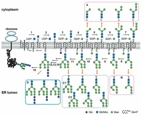
Dolichol phosphate is biosynthesized on the cytoplasmic face of the endoplasmic reticulum and the precursor glycan assembling begins on the same side (Fig. 1).
Sugar residues from nucleotide-activated sugar donors are transferred onto a dolichol phosphate by
specific cytoplasmically oriented glycosyltransferas
-es. In the first reaction, GlcNAc-P is moved from
uridine diphosphate N-acetylglucosamine (UDP-GlcNAc) onto Dol-P giving rise to GlcNAc-PP-Dol with the aid of N-acetylglucosamine transferase (EC
2.7.8.15). In the second reaction, another GlcNAc residue is added to the structure. In the following metabolic steps, specific mannosyltransferases (EC
2.4.1.142; EC 2.4.1.132; EC 2.4.1.257; EC 2.4.1.131)
add five Man from guanosine diphosphate mannoses
(GDP-Man) to the growing saccharide till generation of the heptasaccharide Man5GlcNAc2-PP-Dol. At this point the synthesis of the glycans-precursors on the
cytoplasmic side of ER membrane is finished and the heptasaccharide is flipped across the ER membrane
to the luminal face.
Inside the lumen mannosyltransferas
-es (EC 2.4.1.258; EC 2.4.1.259; EC 2.4.1.261; EC 2.4.1.260) transfer Man from Dol-P-Man to the oligosaccharide elongating the glycan till Man9 Glc-NAc2-PP-Dol. The formation of the
glycan-pre-cursor ends by addition of three glucose residues from Dol-P-Glc with the aid of glucosyltrans-ferases (EC 2.4.1.267; EC 2.4.1.265; EC 2.4.1.266). Dol-P-Man and Dol-P-Glc are synthesized on the
cytosolic side of ER membrane and then flipped into
the lumen.
When the precursor oligosaccharide is trans-ferred from Glc3Man9GlcNAc2-P-P-Dol to appropria-te Asn-X-Ser/Thr sequences in nascent polypep-tides in the lumen of the endoplasmic reticulum, the glycosyl carrier lipid is released as dolichol pyrophosphate (Dol-P-P). A Dol-P-P phosphatase (EC 3.6.1.43) converts Dol-P-P to Dol-P [9]. Dol-P returns to the cytoplasmic side of ER where it can be reutilized for lipid intermediate biosynthesis and additional rounds of N-glycosylation [10].
Dol-P is a rate-limiting intermediate during protein N-glycosylation [11]. Because de novo doli-chol synthesis is slow, Dol-P availability from the dolichol cycle being of particular importance may demand recycling of mature [12] and even imma-ture [13] Dol-P-oligosaccharide intermediates that leads to appearance of free oligosaccharides as byproducts . There are two principal ways of their formation: by cleaving the bond between two resi-dues of phosphoric acid or by breaking the linkage between the second residue of the acid and GlcNAc.
The first way leads to releasing of free glycans with
a negative charge due to one residue of phosphoric acid at the reducing end of the sugars (Fig. 1, a, B). A realization of the different possibility gives rise to neutral free oligosaccharides with N-acetylglu-cosamine at the reducing termini (Fig. 1, C, D).
Interestin gly, some Dol-P-oligosaccharide interme
-diates are a source of only the first group of FOS,
others form the second, and there are some interme-diates that may give the both.
The first evidence that the dolichol cycle is
the source of free phosphooligosaccharides was ob-tained by Hsu et al. during the elucidation of the role
of Man-P-Dol in the protein glycosylation. In mouse
of free neutral oligosaccharides or were utilised in protein glycosylation [18].
When the genes coding the proteins involved in the dolichol cycle are defective and the cycle is interrupted, hypoglycosylation of glycoproteins leads to development of congenital disorders of
gly-cosylation type I (CDG I), rare multisystem genetic
diseases affected nearly all organs and tissues [19].
In these cases the concentration of truncated
Dol-P-oligosaccharides, substrates of limiting reactions and corresponding free oligosaccharides increase
dramatically [20]. Investigations of Epstein-Barr
virus-transformed lymphoblasts derived from a healthy subject and patients with different types of
CDG-I showed low levels of the FOS in normal cells and elevated accumulation in CDG-derived cells. In CDG-Ig cells (with a mutation in the α1,2 mannosyl
transferase – EC 2.4.1.259, EC 2.4.1.261– adding the Fig. 1. the dolichol phosphate cycle as a source of free oligosaccharides. UDP – uridine diphosphate, GDP – guanosine diphosphate, P – phosphate, PP – pyrophosphate, Glc – glucose, GlcNac – N-acetylglucosamine, Dol-P – dolichol phosphate; 1 – GlcNac: dolichol phosphate GlcNac-1-phosphate transferase; 2 – UDP-GlcNac: dolichol pyrophosphate-GlcNac N-acetylglucosaminyltransferase; 3 – chitobiosyldiphosphodolichol β-mannosyltransferase ; 4,5 – α-1,3 mannosyltransferase; 6,7 – α-1,2-mannosyltransferase; 8 – Dol-P-man:
man5GlcNac2-PP-Dol mannosyltransferase; 9,11 – Dol-P-man: man6(8)GlcNac2-PP-Dol mannosyltrans-ferase; 10 – Dol-P-man: man7GlcNac2PP-Dol mannosyltransferase; 12 – α-1,3-glucosyltransferase I; 13 –
α-1,3-glucosyltransferase II; 14 – α-1,2-glucosyltransferase; 15 – oligosaccharyltransferase; A – cytosolic
seventh mannose residue to oligosaccharide precur-sor) negatively charged components increased 3 fold
with respect to the control. Interestingly, this study
revealed only the species with 7 and less mannose residues among phosphorylated oligosaccharides. Moreover, the analysis of ER luminal and cytosolic phosphorylated FOS detected the appearance of spe-cies with 7 mannose residues exclusively in the
cyto-sol. This finding prompted the proposal of an
ER-to-cytosol translocation way for Man6-7GlcNAc2-P [11]. Phosphorylated oligosaccharides can be re-leased from dolichol phosphate by the action of pyrophosphatase (EC 3.6.1.44) at the cytosolic face of the endoplasmic reticulum [18]. Removal of the phosphate from the phosphorylated glycans converts them into oligomannose structures with two GlcNAc residues.
The formation of neutral free oligosaccharides from dolichol pyrophosphate oligosaccharides was shown in the beginning in pig liver [21], hen ovi-duct [22] and calf thyroid microsomes [12]. The oligosaccharides had the glucose-containing oli-gomannose structure with two GlcNAc residues at the reducing end – Glc3Man9GlcNAc2. The investi-gations led to the idea that the oligosaccharyl trans-ferase (EC 2.4.1.119) could carry out both the hydro-lytic and transfer functions. The authors speculated that when acceptor polypeptides were limiting, the enzyme could transfer the mature glycan precursor from the lipid carrier onto water molecules genera-ting neutral FOS. For a long time no direct experi-mental evidence supporting or rejecting the hypo-thesis had been presented. Only recently [23], using mutant strains of Saccharomyces cerevisiae, the hydrolytic activity of the enzyme has been proved. To great surprise it turned to be connected with the presence of a hydrolytic active centre in addition to the transferase centre rather than the acceptor
limi-tation. It means that the oligosaccharyl transferase
catalyses both reactions in-parallel. More truncated glycans, Man7-9GlcNAc2, were also found in the ER lumen [24, 25]. The neutral free oligosaccharides are translocated from ER into the cytosol. However, their contribution to the dolichol cycle-derived FOS as well as to the total pool of free neutral oligosac-charides originating from the ER to the cytosol is not
significant, since the major source is from misfolded/
misassembled glycoproteins, the subjects of ERAD [4, 26, 27].
Thus, in fact there are two cycles of dolichol
phosphate. The first gives rise to phosphorylated free
oligosaccharides with 2-7 mannose residues, the sec-ond – to neutral free oligosaccharides with 7-9 man-nose residues as well as glucosylated oligomanman-nose structures.
er-associated protein degradation (erad) releases glucosylated and non-glucosylated oligomannose free oligosaccharides
Following transfer of the glycan from
lipid-pre-cursor to a nascent polypeptide, α-glycosidases I and II (EC 3.2.1.106; EC 3.2.1.84) remove two glucose
residues and the oligomannose glycans with one glu-cose residue connects to calnexin or calreticulin al-lowing the glycoprotein to enter the calnexin/calreti-culin cycle to be completely folded and assembled with the help of the chaperone system BiP/GRP78 or Hsp70, BiP cochaperones (DnaJ/Hsp40 fami ly members – ERdj1–7), protein disulphide isomerases
(PDI, ERp57, ERp59, ERp72, ERp44 – EC 5.3.4.1)
and some substrate-specific chaperones [28].
α-Glucosidase II (EC 3.2.1.84) cuts the last residue
of glucose and a correctly folded glycoprotein leaves the cycle moving to the Golgi for further process-ing. An aberrant glycoprotein can be retained for a
while in ER due to tandem actions of α-glycosidase II (α-GII) and UDP-glucose:glycoprotein glucosyl -transferase (UGGT, EC 3.6.5.3), which transfers one glucose residue again to the glycan and allows the glycoprotein to return to calnexin/calreticulin cycle, or can be sent to the ER – associated degradation
due to action of ER α-mannosidase I (EC 3.2.1.113),
EDEM (EC 3.2.1.24; EC 3.2.1.113) or Golgi-resident MAN1B1 (3.2.1.113), which remove one mannose residue from Man9GlcNAc2 and forms B isomer of Man8GlcNAc2 on the aberrant glycoprotein [29, 30] or trim the glycans more extensively giving rise to Man7GlcNAc2, Man6GlcNAc2 and Man5GlcNAc2 [31].
α-Glucosidase II is a unique enzyme with a
dual biochemical function. On the one hand, it pro-vides transfer of glycoproteins to and from the cal-nexin/calreticulin cycle, on the other hand, it can send them further either to the Golgi or to the
cyto-sol for degradation. α-GII is a luminal glycoprotein and consists of a catalytic α and a regulatory β subu -nits. The latter is responsible for the endoplasmic reticulum localization of the heterodimer and for the
enhancement of N-glycan trimming. It is not clear in
detail what forces GlcMan9GlcNAc2, the product of
the first reaction of the enzyme, to leave the active
of the first reaction, Glcα1-3Glc cleavage, in the early
investigations seemed to be higher than that of the
second, Glcα1-3Man hydrolysis. The later findings,
however, showed that the second reaction essentially enhanced at the high protein concentrations that in fact take place in ER. Nevertheless, a high competi-tion with calnexin/calreticulin for the substrate and strong suppression of the enzyme’s second reaction by calreticulin have been revealed. The glucosidase has a surprisingly wide range of substrates that in addition to Glc2Man9GlcNAc2 and GlcMan9GlcNAc2 includes GlcMan8GlcNAc2 and GlcMan7GlcNAc2. The enzyme can be inhibited by its nonglucosylated end products – Man9GlcNAc2, Man8GlcNAc2 and Man7GlcNAc2. The latter is the strongest inhibitor and its effect can be even comparable with those of such powerful inhibitors of glucosidases as castano-spermine and deoxynojirimycin. By preventing the formation of monoglucosylated glycoproteins the end products are supposed to regu late the entry of newly synthesized glycoproteins into the calnexin/ calreticulin cycle [32, 33].
Complex relationship of α-glucosidase II with
the calnexin/calreticulin cycle is supplemented by
no less dramatic interactions with UGGT. It is well
documented that UGGT is the folding sensor due to combination of a chaperone and a glycosyltransferase activities. UGGT reactivity is strongly connected with exposed hydrophobic patches of misfolded
gly-coproteins on the advanced folding stages. Its gly
-can specificity appeared to be the highest towards
Man9GlcNAc2, but critically declined with reducing of the mannose residues in the oligosaccharides on the glycoproteins from 8 to 7 by 50 and 85%,
re-spectively. UGGT has a strong affinity for the core
of N-glycans - Man3GlcNAc2 and cannot recognize the oligosaccharides without core GlcNAc2. Only the simultaneous recognition of a misfolded part of a protein and an appropriate high-mannose struc-ture of a glycan leads to conformational changes of the enzyme and an addition of a glucose residue to A arm of the oligosaccharide allowing aberrant molecules to become the subjects of the calnexin/ calreticulin cycle again, but proper folded and/or as-sembled glycoproteins to escape the UGGT activity
and proceed to their final destinations through the second reaction of α-GII, α-mannosidase I activity
and the Golgi processing [33, 34].
Confocal immunofluorescence and high-resolu
-tion immunoelectron microscopy allowed specifica
-tion of intracellular localiza-tion of α-GII, calnexin/
calreticulin and UGGT. They were found not only in “the classical places of expectations“, the rough ER including the nuclear envelope and the transitional ER, but also in the smooth ER and in the pre-Golgi
intermediates. It means that quality control check -points are not restricted to the rough ER, but exist along the secretory pathways [35].
The next member of the quality control system,
α(1,2)-mannosidase (endomannosidase I, MAN1B1),
catalyzes the removal of the terminal mannose resi-due from the middle branch of Man9GlcNAc2 and generates isomer B Man8GlcNAc2, that is the signal for ERAD. The localization of the enzyme in the pe-ripheral and Golgi-associated pre-Golgi intermedi-ates as well as cis and medial cisternae of the Golgi apparatus extends the system of protection against aberrant proteins at least up to the middle of the Golgi. Golgi-resident endomannosidase provides a backup mechanism for deglucosylation of glycopro-teins escaping the ER [26, 27, 36, 37].
By this way the system of cell quality control can discriminate between native and nonnative pro-teins, leads to degrading of aberrant species through ER-associated degradation and guarantees the high
fidelity of secreted and plasma membrane molecu -lar structures [16, 38]. Until the cell can cope with alterations, any aberrant glycoproteins will not ap-pear in functional zones (on the surface of cell mem-branes or outside the cell), though the products of their degradation may have already existed. So there
must be a lag period between the first attempts to
synthesize aberrant glycoproteins in endoplasmic re-ticulum and/or in Golgi apparatus and their embed-ding into the plasma membrane or secretion outside the cell. Therefore, some distinctive products of this non-native glycoprotein degradation could testify the beginning of a disorder much earlier than any cel-lular or tissue dysfunctions will appear.
Misfolded N-glycoproteins, identified during
quality control analysis in the endoplasmic reticulum [39], can be exported into the cytosol via a channel containing Sec61 [40], the same complex that trans-locates nascent polypeptide chains into the endo-plasmic reticulum, or with the help of some different proteins – Derlin family or ubiquitin protein ligases E3 (EC 6.3.2.19) [41].
In cytosol oligosaccharides are removed from
glyco-protein can be deglycosylated by an enzyme with PNGase -like activity in the ER lumen and then the free glycan and polypeptide are translocated into cytosol [42, 2, 16, 27]. A neutral chitobiase
[43] or endo-β-N-acetylglucosaminidase (ENGase,
EC 3.2.1.96) [44, 45] removes one N-acetylglucosa-mine residue from free oligosaccharides Man5-8 Glc-NAc2. The mannose residues are then cleaved by the
neutral cytosolic α-mannosidase (NAM, MAN2C1, EC 3.2.1.24) to produce a specific limit digestion
product , the linier isomer Man5GlcNAc [46]. The smaller glycans – Man2-4GlcNAc – were also found
in the cytosol [47].The final step in the cytosolic
pathway is the transport of the limit digestion oli-gosaccharide into lysosomes by the ATP-dependent,
high-affinity oligosaccharide transporter [18, 3].
Aberrant glycoproteins can be a source of a small amount of cytosolic glucosylated oligoman-nose free glycans [48] that cannot be transported into the lysosome [49] and secreted by the cell [3].
Some findings, such as the increasing rate of ERAD when cytosolic α-mannosidase is overex -pressed, for example, point at a high probability of a feedback mechanism of ERAD regulation through the cytosolic FOS degradation pathway [50].
lysosomes can produce both neutral and negatively charged free oligosaccharides
The mature glycoproteins containing high-mannose, hybrid and complex glycans are turned over in lysosomes [51]. Other glycoconjugates (glycolipids, proteoglycans and glycosylphosphati-dylinositol anchors) share the same fate. They are delivered to lysosomes either by endocytosis from outside the cell or by autophagy within the cell [52] and can give rise to free glycans different from the oligomannose type.
The lysosomal pathways for N-linked glycoteins breakdown (Fig. 2) are bidirectional with pro-teolysis of polypeptide at one side and the sequential release of monosaccharides by exoglycosidases from the nonreducing glycan end [53].
Complete or extensive proteolysis takes place before the N-linked glycan is released from the poly-peptide. The next step in the breakdown of complex or hybrid N-glycans is the removal of any fucose
residues, which is catalyzed by lysosomal
α-L-fucosidase (EC 3.2.1.51). Fucose can be linked by
α1-6 bond to the core N-acetylglucosamine, α1-2, α1-3, or α1-4 bonds to N-acetylglucosamine or α1-2
bond to galactose residues at the nonreducing ends
of the glycans [53]. The peripheral α1-3 linked fucose residues are removed before the core α1-6 linked
resi dues [54]. Removal of fucose allows the hydroly-sis of the asparagine-N-acetylglucosamine amide linkage by glycosylasparaginase (EC 3.5.1.26). This enzyme undergoes dimerization and autoactivation by cleavage into two subunits in the Golgi [55].
Release of the N-acetylglucosamine resi-due at the reducing end of the chitobiose core
can be achieved in two ways by either the
β-N-acetylglucosaminidase (EC 3.2.1.30) or the di-N-acetyl-chitobiase (EC 3.2.1.52).
Some of the glycosidases – for example,
α-neuraminidase (EC 3.2.1.18), β-galactosidase (EC 3.2.1.23), and N-acetyl-β-D-hexosaminidase
(EC 3.2.1.52) – can catalyze the same linkages in other glycoconjugates, such as glycolipids or
gly-cosaminoglycans, whereas α-mannosidase and β-mannosidase (EC 3.2.1.25) appear to be exclusive
to the glycoprotein [52].
N-glycans of lysosomal hydrolases have
mannose-6-phosphate modification, the lysosome
destination recognition marker, catalyzed by two enzymes: UDP-N-acetylglucosamine-1-phospho-transferase (EC 2.7.8.17) and
N-acetylglucosa-mine-1-phosphodiester α-N-acetylglucosaminidase (EC 3.1.4.45). The first enzyme transfers
GlcNAc-1-phosphate to mannose residues on high mannose
glycans in the α-1,6 and/or α-1,3 branch of the oligo -saccharide chains [56], the second one removes the GlcNAc residues exposing Man-6-P group. That is why the second enzyme is called “uncovering
en-zyme”. It is synthesized as an inactive proenzyme
and activated by endoprotease furin (EC 3.4.21.75) in the trans-Golgi network where active “uncovering enzyme” helps to deliver soluble hydrolases to the endosomal/lysosomal compartment allowing their binding to the Man-6-P receptors [57].
Not only lysosomal hydrolases but also seve-ral nonlysosomal proteins have been shown to bear mannose-6-phosphate residues. Among them there
are latent transforming growth factor β1, leukemia
inhibitory factor, proliferin, renin precursor, and
T-cell activation antigen CD26. In this case the modification can facilitate interactions of secreted
glycoproteins with plasma membrane receptors like
for latent transforming growth factor-β1 [58] or regu -late extracellular secretion of proteins directing the
modi fied molecules to the lysosome degradation like
Given GlcNAc-1-phosphotransferase is locali-zed earlier within the Golgi than Golgi mannosidase
I [60] and that the latter can direct the glycoproteins
escaped ER quality control from the Golgi to ER to ERAD [26, 27], among ERAD-generated FOS there might be free glycans with GlcNAc-1-P-6-Man structures.
extracellular free oligosaccharides
Free oligosaccharides have been investigated mostly in cultured cells. The information about their existence in culture medium is very poor. Even less is known about their presence and distribution in
tissues and biological fluids with the exceptions for
urine and milk.
For a long time the idea about FOS com-plete degradation inside the cell in norm and their appearan ce in urine only under disease conditions prevailed and was based on the early investigations of urinary excretions of the degradation products in the cases of so called oligosaccharidoses or lysoso-mal storage disorders with thin-layer chromatogra-phy [61, 62].
The development of more sensitive analyti-cal procedures (high performance liquid chro-matography and mass-spectrometry) allowed de-Fig. 2. Degradation of complex N-glycoprotein and appearance of FOS in lysosome: asn – asparagine; Neu5ac – neuraminic acid ; Gal – galactose; GlcNac – N-acetylglucosamine; man – mannose; Fuc – fu-cose; 1 – proteases; 2,3 – α-fucosidase; 4 – aspartyl-N-acetyl-β-D-glucosaminidase; 5 –
endo-β-N-acetylglucosaminidase; 6,7 – sialidase; 8,9 – β-galactosidase; 10,11 – β-N-acetylhexosaminidase; 12,13 –
tecting the bulk of urine free oligosaccharides and revealing unique patterns for
glycoproteina-ses (α-mannosidosis, sialidosis, galactosialidosis, α-fucosidosis, aspartylglucosaminuria), glycolipi -doses (GM1 and GM2 gangliosidosis, Gaucher
disea se, Sandhoff disease mucolipidosis type II/III)
and glycogen storage disorders (Pompe diseases) [4, 63, 64]. For example, the excess excretion of one-, two- and three-antennary high mannose-type
N-gly-cans is a defining attribute of α-mannosidosis, but
the two-, three- and four-antennary complex-type sugar chains with polylactosamine structures at
non-reducing termini are specific for GM1-gangliosidosis
[65]. The only N-acetylglucosamine residue at the reducing end of these FOS points at their cytosolic origin.
The first evidences of FOS presence in cell me -dium were obtained on human hepatocellular carci-noma HepG2 cells only after inhibition of lysosomal degradation of FOS and glycoproteins with a
spe-cific inhibitor concanamycin A that blocks lysoso
-mal acidification through selective inhibition of the
V-type ATPase [66].
Inhibition of FOS translocation from ER to cy -tosol leads to FOS moving into the cell medium by
the classical secretion pathway through the Golgi. In
this case the FOS have two residues of GlcNAc and may convert into complex and sialic acid-containing types due to processing in the Golgi [67].
Recently more detailed investigations of the HepG2 cell line after treatment with concanamy-cin A [68] revealed that about 7% of total
intra-cellular FOS flowed from the treated cells into the
medium by a way different from the classical se-cretion pathway through the Golgi. Without such a treatment only trace amounts of these components were detected in the culture medium. A compari-son of FOS structures in different compartments of the cell with extracellular free glycans showed that all of them were of oligomannose type and about 90% of medium FOS were the components less than Man7GlcNAc . In the membrane bound compart -ments (intact lysosomes, Golgi apparatus, and ER) these structures comprise about 83%, in cytosol – about 70%. The authors have come to the conclusion that either FOS are degraded in the medium or the secretion is selective towards smaller components and that concanamycin A courses the perturbations in lysosomal/endosomal transport allowing the con-tent of lysosomes to be secreted without the cell damage. So the authors consider the medium FOS as products of lysosomes.
Although the statement that these cellular orga-nelles are the exclusive origin of the medium FOS is in contradiction with the results of other studies, but the idea about the perturbations of lysosomes and their partial contribution to the extracellular pool of FOS through the cytosol is supported by diverse experimental evidence. Dramatic alteration of lyso-some membrane permeabilization, its contribution to relocation of lysosomal content into the cytosol were revealed during the studies of lysosome visua-lization, its injury by various agents, processes of apoptosis and intracellular distribution of lysosomal enzymes in response to various stimuli [69, 70].
Indirect evidence of the lysosomal membrane
permeability for oligosaccharides was the discovery of a disialobiantennary structure among free oligo-saccharides glycans in a cytosolic fraction of mouse liver. The lysosome could be the only source of gly-cans with such a structure [47].
Targeted studies of the membrane
permeabili-ty using FITC-labelled dextrans in activated human
T-lymphocytes revealed size-dependant penetration of the substance to the cytosol in control cells and its substantial changing in the cells in the early stage
of apoptosis caused by staurosporine. In the control FITC-labelled 10 kDa dextrans were found in the cy -tosol of 9% of the cells. The dextrans of 40, 70 and 250 kDa penetrated the membrane in 3-4% of the cells. Under exposition of staurosporine the 10, 40 and 70 kDa molecules were relocated to the cytosol in 62, 41, and 6% of the cells, respectively. The dex-trans of 250 kDa did not change their location [71].
Investigations of intracellular FOS in a bank
of cancer-derived cell lines revealed a striking dif-ference in quantity and structures of cytosolic FOS
among the cells. In the cytosol of stomach cancer MkN7 and MkN45 cells unusually large amount of mono-, di-, tri- and tetra-sialoglycans −
94-96% of the total free oligosaccharides were found. Biochemi cal analysis defined their structures,
showed that NeuAcα2-6Galβ1-4GlcNAcβ1-2Manα1-3Manβ1-4GlcNAc was the most abundant of them. In addition to the sialoglycans, bisecting GlcNAc,
a sialo-biantennary and fucose-containing struc-tures, all of which cannot be derived from ERAD or dolichol-phosphate pathways, were detected. Co-localization of lysosomal enzymes in the same cy-tosolic fractions indicated that FOS were from lyso-somes, the membranes of which were leaking. The researchers came to the conclusion that the cytosolic
-nosis of specific tumours [72]. So, lysosome-derived
cytosolic FOS might represent changes in the orga-nelle integrity.
Intensive improvement of the protocols for
analy sis, using ion-exchange chromatography
fol-lowed by fluorescent labelling with 2-AA (2-ami -nobenzoic acid), purification by lectin-affinity chromatography before separation of 2-AA-labelled FOS by HPLC, combination of enzyme digests
with MALDI-TOF MS and concentration of
2-AA-labelled FOS on different stages of investigations, showed the parallels of ERAD-derived FOS beha-viour in cultured cells, murine and human tissues. A treatment of mice with the imino sugar
N-butyl-deoxynojirimycin, an inhibitor of α-glucosidases I and II that was used in substrate reduction thera
-py for Gaucher type I, Niemann–Pick type C and
Sandhoff diseases (miglustat, Zavesca®), showed a dose-dependent increase of FOS concentrations and
radical changes of HPLC-profiles in all tissues and fluids. [4].
An addition of hydrophilic interaction and anion-exchange HPLC, affinity chromatography with a series of lectins to normal-phase HPLC has expanded potentialities of the methods and allowed the screening of polygenic diseases – hepatocellular carcinoma and colorectal cancer [6]. A purposeful study of human plasma FOS revealed their presence both in normal and pathological conditions (blood oncotransformation and cardiovascular diseases). They were represented by uncharged oligomannose
and negatively charged species. The first group com -prised the glucosylated or non-glucosylated compo-nents Man3-9GlcNAc(2) with domination of species Man4-5GlcNAc(2). The second group consisted of sialic acid-containing types as well as the species with the negative charge based on some different acid residues. Plasma FOS of healthy volunteers
pos-sessed featured reproducible profiles. Pathological processes changed either mostly the first group pro
-files (cardiovascular diseases) or the second (chronic
blood cancer) or both (acute blood cancer) [73, 74].
Investigations of free oligosaccharides in bio
-logical fluids are on the earliest stage of develop -ment. The inherent complexity of oligosaccharides and the existence of various linkage isoforms make the decoding of oligosaccharide structures,
repre-senting each species in FOS profiles, the most chal -lengeable task. An analysis of extracellular FOS in different groups of healthy people could help to
un-derstand dependence of FOS profile changes on ag
-ing and genetics. FOS comparison in a wide range of
illnesses would favour the selection of more specific
or even unique biomarkers in each case and
guaran-tee high fidelity of prediction.
Extracellular free oligosaccharides reflecting
metabolic status, health alterations and treatment responses of the patients might be promising mole-cules in the search for new biomarkers of diseases and therapeutic monitoring. Detailed investigations of these oligosaccharides can suitably enrich the family of glycoconjugates studied intensively for the same purposes by opening an absolutely new per-spective of early prediction of misglycosylation. Due to the system of cell quality control there must be a
lag period between the first attempts to synthesize
aberrant glycoproteins in endoplasmic reticulum and/or in Golgi apparatus and their embedding into the plasma membrane or secretion outside of the cell.
It is among these molecules must be really the earli -est biomarkers so eagerly needed for timely detec-tion and successful treatment of numerous diseases.
acknowledgments. the authors thank the euro-pean molecular Biology Organization, the Interna-tional Union against Cancer and Oxford Glycobiolo-gy Institute (University of Oxford, Oxford, Uk) for the support to P.I.U.
«Три джерела і Три
складові часТини» вільних олігосахаридів
І. Ю. Письменецька1, Т. Д. Баттерс2
1дУ «дніпропетровська медична академія», Україна; 2Інститут глікобіології оксфордського
університету, Велика Британія; e-mail: [email protected]
Метаболізм глікопротеїнів і гліколіпідів
супроводжується появою незв’язаних
структурних аналогів вуглеводної части
-ни глікокон’югатов або так зва-них віль-них олігосахаридів. Усередині клітини існує декілька їх джерел: 1) багатоступеневий шлях
N-глікозилювання, 2) механізми контролю фол
-дингу протеїнів і асоційована з ЕПР деградація
неправильно глікозильованих та/або непра
-вильно згорнутих глікопротеїнів, 3) деградація зрілих глікокон’югатів у лізосомах. В огляді підсумовується відома інформація щодо шляхів появи вільних олігосахаридів у різних ком
залежно від джерела виникнення. Крім того, обговорюються дані про позаклітинні вільні олігосахариди, їх структуру та зміни за нор
-мальних і патологічних станів.
К л ю ч о в і с л о в а: вільні олігосахариди, доліхолфосфатний цикл, асоційована з ЕПР деградація протеїнів, лізосомна деградація, плазма крові, сеча.
«Три исТочника и Три
сосТавные часТи» свободных олигосахаридов
И. Ю. Письменецкая1, Т. Д. Баттерс2
1гУ «днепропетровская медицинская
академия», Украина;
2институт гликобиологии оксфордского
университета, Великобритания; e-mail: [email protected]
Метаболизм гликопротеинов и гликолипи
-дов сопровождается появлением несвязанных структурных аналогов углеводной части гли
-коконъюгатов или так называемых свободных
олигосахаридов. Внутри клетки существует не
-сколько их источников: 1) многоступенчатый путь N-гликозилирования, 2) механизмы кон
-троля фолдинга протеинов и ассоциированная с
ЭПР деградация неправильно гликозилирован
-ных и/или неправильно свернутых гликопро
-теинов, 3) деградация зрелых гликоконъюгатов в лизосомах. В обзоре собрана информация о путях появления свободных олигосахаридов в различных компартментах клетки и детали
их структуры в зависимости от источника воз
-никновения. Кроме того, обсуждаются данные о внеклеточных свободных олигосахаридах, их
структуре и изменениях при нормальных и па
-тологических состояниях.
К л ю ч е в ы е с л о в а: свободные олиго
-сахариды, долихолфосфатный цикл, ассоцииро
-ванная с ЭПР деградация протеинов, лизосом
-ная деградация, плазма крови, моча.
references
1. Varki A. (ed.) Essentials of Glycobiology. Cold Spring Harbor: Cold Spring Harbor Laboratory Press, 2009. 784 p.
2. Donoso G., Herzog V., Schmitz A. Misfolded BiP is degraded by a proteasome-independent
endoplasmic-reticulum-associated degradation pathway. Biochem. J. 2005;387:8970-903.
3. Moore S. H. E. Oligosaccharide transport: pumping waste from the ER into lysosomes. trends Cell Biol. 1999;9:441-446.
4. Alonzi D. S., Neville D. C., Lachman R. H., Dwek R. A., Butters T. D. Glucosylated free oligosaccharides are biomarkers of endoplasmic reticulum alpha-glucosidase inhibition. Biochem. J. 2008;409(2):571-580.
5. Vleugels W., keldermans L., Jaeken J.,
Butters T. D., Michalski J.-C., Matthijs G., Foulquier F. Quality control of glycoproteins bearing truncated glycans in an ALG9-defective
(CDG-IL) patient. Glycobiology. 2009;19(8):910-917.
6. Alonzi D. S., Su y.-H., Butters T. D. Urinary
glycan markers for disease. Biochem. Soc. trans. 2011;39(1):393-398.
7. Pismenetskaya I. U., Butters T. D. Chromatographic profile changes of plasma
free oligosaccharides in subleukemic myelosis. “Scientific Notes of Taurida V. Vernadsky National University”. Series: Biology, Chemistry. 2013;26(65), N 1:153-160. (in Russian).
8. Jones M. B., Julian N. Rosenberg J. N.,
Betenbaugh M. J., krag S. S. Structure
and synthesis of polyisoprenoids used in N-glycosylation across the three domains of life. Biochim. Biophys. acta. 2009;1790:485-494. 9. Rush J. S., Gao N., Lehrman M. A., Waechter C. J.
Recycling of dolichyl monophosphate to the
cytoplasmic leaflet of the endoplasmic reticulum
after the cleavage of dolichyl pyrophosphate on the lumenal monolayer. J. Biol. Chem. 2008;283:4087-4093.
10. Schenk B., Fernandez F., Waechter C. J. The ins(ide) and out(side) of dolichyl phosphate biosynthesis and recycling in the endoplasmic reticulum. Glycobiology. 2001;11:61R-70R. 11. Peric D., Durrant A. C., Delenda C., Dupré T., De
Lonlay P., de Baulny H. O., Pelatan C.,
Bader-Meunier B., Danos O., Chantret I., Moore S. E.
The compartmentalisation of phosphorylated free oligosaccharides in cells from a CDG
Ig patient reveals a novel ER-to-cytosol
translocation process. PLoS One. 2010;5;e11675.
12. Anumula k. R., Spiro R. G. Release of
thyroid microsomal enzymes and slices. J. Biol. Chem. 1983;258:15274-15282.
13. Rosenwald A. G., Stoll J., krag S. S. Regulation
of glycosylation. Three enzymes compete for a common pool of dolichyl phosphate in vivo. J. Biol. Chem. 1990;265:14544-14553.
14. Hsu A. F., Baynes J. W., Heath E. C. The role of a dolichol-oligosaccharide as an intermediate in glycoprotein biosynthesis. Proc. Natl. acad. Sci. USa. 1974;71:2391-2495.
15. Hoflack B., Cacan R., Verbert A. Dolichol
pathway in lymphocytes from rat spleen.
Influence of the glucosylation on the cleavage
of dolichyl diphosphate oligosaccharides into phosphooligosaccharides. eur. J. Biochem. 1981;117:285-290.
16. Chantret I., Moore S. E. H. Free oligosaccharide
regulation during mammalian protein N-glycosylation. Glycobiology. 2008;18:210-224.
17. Belard M., Cacan R., Verbert A. Characterization of an oligosaccharide-pyrophosphodolichol pyrophosphatase activity in yeast. Biochem. J. 1988;255:235-242.
18. Cacan R., Villers C., Belard M., kaiden A., krag S. S., Verbert A. Different fates of the
oligosaccharide moieties of lipid intermediates. Glycobiology. 1992;2:127-136.
19. Grunewald S., Matthijs G., Jaeken J. Congenital disorders of glycosylation: a review. Pediatr. res. 2002;52:618-624.
20. Vleugels W., Duvet S., Peanne R., Mir A. M. Cacan R., Michalski J. C., Matthijs G.,
Foulquier F. Identification of phosphorylated
oligosaccharides in cells of patients with a
congenital disorders of glycosylation (CDG-I).
Biochimie. 2011;93:823-833.
21. Oliver G. J., Harrison J., Hemming F. W. The mannosylation of dolichol diphosphate oligosaccharides and the formation of oligosaccharides and glycoproteins in pig liver microsomal preparations. Biochem. Soc. trans. 1975;3:666-668.
22. Hanover J. A., Lennarz W. J. Transmembrane assembly of membrane and secretory glycoproteins. arch. Biochem. Biophys. 1981;211:1-19.
23. Harada y., Buser R., Ngwa E. M., Hirayama H..
Aebi M., Suzuki T. Eukaryotic oligosaccharyl transferase generates free oligosaccharides during N-glycosylation. J. Biol. Chem. 2013;288(45):32673-32684.
24. Spiro M. J., Spiro R. G. Potential regulation of N-glycosylation precursor through oligo-saccharide-lipid hydrolase action and glucosyltransferase-glucosidase shuttle. J. Biol. Chem. 1991;266:5311-5317.
25. Gao N., Shang J., Lehrman M. A. Analysis
of glycosylation in CDG-Ia fibroblasts
by fluorophore-assisted carbohydrate
electrophoresis: implications for extracellular glucose and intracellular mannose 6-phosphate. J. Biol. Chem. 2005;280:17901-17909.
26. kukushkin N. V., Alonzi D. S., Dwek R. A.,
Butters T. D. Demonstration that endoplasmic reticulum-associated degradation of glyco-proteins can occur downstream of processing by endomannosidase. Biochem. J. 2011;438(1):133-142.
27. Alonzi D. S., kukushkin N. V., Allman S. A.,
Hakki Z., Williams S. J., Pierce L., Dwek R. A., Butters T. D. Glycoprotein misfolding in the
endoplasmic reticulum: identification of released
oligosaccharides reveals a second ER-associated degradation pathway for Golgi-retrieved proteins. Cell mol. Life Sci. 2013;70(15):2799-2814.
28. Araki k., Nagata k. Protein Folding and Quality
Control in the ER. Cold Spring. Harb. Perspect. Biol. 2011;3(11):a007526.
29. Hoseki J., Ushioda R., Nagata k. Mechanism and
components of endoplasmic reticulum associated degradation. J. Biochem. 2010;147(1):19-25.
30. Olzmann J. A., kopito R. R., Christianson J. C.
The mammalian endoplasmic reticulum-associated degradation system. Cold Spring. Harb. Perspect. Biol. 2013;5(9):a013185.
31. Avezov E., Frenkel Z., Ehrlich M., Herscovics A., Lederkremer G. Z. Endoplasmic reticulum
(ER) mannosidase I is compartmentalized
and required for N-Glycan trimming to Man5–6GlcNAc2 in glycoprotein ER-associated degradation. mol. Biol. Cell. 2008;19:216-225.
32. Bosis E., Nachliel E., Cohen T., Takeda y., Ito y.,
Bar-Nun S., Gutman M. Endoplasmic reticulum
glucosidase II is inhibited by its end products.
Biochemistry. 2008;47(41):10970−10980.
33. Ito y., Takeda y. Deciphering the Roles of Glycan
Processing in Glycoprotein Quality Control through Organic Synthesis. Biosci. Biotechnol. Biochem. 2013;77(12):2331-2338.
34. D’Alessio C., Caramelo J. J., Parodi A. J. UDP-GlC:glycoprotein
control. Semin. Cell Dev. Biol. 2010;21(5):491-499.
35. Roth J., yam G. H.-F., Fan J., Hirano k., Gaplovska-kysela k., Le Fourn V., Guhl B.,
Santimaria R., Torossi T., Ziak M., Zuber Ch. Protein quality control: the who’s who, the where’s and therapeutic escapes. Histochem. Cell Biol. 2008;129:163-177.
36. Pan S., Cheng X., Sifers R.N. Golgi-situated
endoplasmic reticulum α-1, 2-mannosidase
contributes to the retrieval of ERAD substrates
through a direct interaction with γ-COP. mol. Biol. Cell. 2013;24(8):1111-1121.
37. Rymen D., Peanne R., Millón M.B., Race V., Sturiale L., Garozzo D., Mills P., Clayton P., Asteggiano C.G., Quelhas D., Cansu A., Martins E., Nassogne M.C., Gonçalves-Rocha M., Topaloglu H., Jaeken J., Foulquier F.,
Matthijs G. MAN1B1 Deficiency: An Unexpected CDG-II. PLoS Genet. 2013;9(12):e1003989. 38. Anelli T., Sitia R. Protein quality control in the
pathway early secretory. emBO J. 2008;27:315-327.
39. Spiro R. G. Role of N-linked polymannose oligosaccharides in targeting glycoproteins for endoplasmic reticulum-associated degradation. Cell mol. Life Sci. 2004;61:1025-1041.
40. Cacan R., Duvet S., Lablau O., Verbert A.,
krag S. S. Mono-glucosylated oligomannosides
are released during the degradation process of newly synthesized glycoproteins. J. Biol. Chem. 2001;276:22307-22312.
41. Hebert D. N., Bernasconi R., Molinari M. Erad substrates: which way out? Semin. Cell Dev. Biol. 2010;21:526-532.
42. Verbert A., Cacan R. Trafficking of
oligo-mannosides released during N-glycosylation: a clearing mechanism of the rough endoplasmic reticulum. Biochim. Biophys. acta. 1999;1473:137-146.
43. Cacan R., Dengremont C., Labiau O.,
kmiecik D., Mir A. M., Verbert A. Occurrence
of a cytosolic neutral chitobiase activity involved in oligomannoside degradation: a study with
Madin-Darby bovine kidney (MDBk) cells.
Biochem. J. 1996;313:597-602.
44. kato T., Hatanaka k., Mega T., Hase S. Purification and characterization of
endo-β-N-acetylglucosaminidase from hen oviduct. J. Biochem. 1997;122:1167-1173.
45. Suzuki T., yano k., Sugimoto S., kitajima k., Lennarz W. J., Inoue S., Inoue y., Emori y. Endo-β-N-acetylglucosaminidase, an enzyme
involved in processing of free oligosaccharides in the cytosol. Proc. Natl. acad. Sci. USa. 2002;99:9691-9696.
46. Hirsch C., Blom D., Ploegh H. L. A role for N-glycanase in the cytosolic turnover of glycoproteins. emBO J. 2003;22:1036-1046.
47. Ohashi S., Iwai k., Mega T., Hase S.
Quantitation and isomeric structure analysis of free oligosaccharides present in the cytosol fraction of mouse liver: detection of a free disialobiantennary oligosaccharide and glucosylated oligomannosides. J. Biochem. 1999;126:852-858.
48. Mellor H. R., Neville D. C., Harvey D. J., Platt F. M., Dwek, R. A., Butters T. D. Cellular effects of deoxynojirimycin analogues: inhibition of N-linked oligosaccharide processing and generation of free glucosylated oligosaccharides. Biochem. J. 2004;381:867-875.
49. Saint-Pol A., Codogno P., Moore S. E. Cytosol-to-lysosome transport of free
polymannose-type oligosaccharides. kinetic and specificity
studies using rat liver lysosomes. J. Biol. Chem. 1999;274:13547-13555.
50. Bernon C., Carré y., kuokkanen E., Slo-mianny M. C., Mir A. M., krzewinski F.,
Cacan R., Heikinheimo P., Morelle W., Michalski J. C., Foulquier F., Duvet S. Overexpression of Man2C1 leads to protein underglycosylation and upregulation of endoplasmic reticulum-associated degradation pathway. Glycobiology. 2011;21(3):363-375.
51. yamashita k., Tachibana y., Mihara k., Okada S., yabuuchi H., kobata A. Urinary
oligosaccharides of mannosidosis. J. Biol. Chem. 1980;255:5126-5133.
52. Winchester B. Lysosomal metabolism of glycoproteins. Glycobiology. 2005;15(6):1R-15R. 53. Johnson S. W., Alhadeff J. A. Mammalian
β-L-fucosidases. Comp. Biochem. Physiol.
1991;99B:479-488.
54. Barker C., Dell A., Rogers M., Alhadeff J. A.,
Winchester B. Canine α -L-fucosidase in relation
to the enzymic defect and storage products in canine fucosidosis. Biochem. J. 1988;254:861-868.
pathogenesis of a disease: structural consequences of aspartyglucosaminuria mutations. Hum. molec. Genet. 2001;10:983-995.
56. Goldberg D. E., kornfeld S. The phosphorylation
of beta-glucuronidase oligosaccharides in mouse P388D1 cells. J. Biol. Chem. 1981;256:13060-13067.
57. Do H., Lee W. S., Ghosh P., Hollowell T.,
Canfield W., kornfeld S. Human mannose
6-phosphate-uncovering enzyme is synthe-sized as a proenzyme that is activated by the endoprotease furin. J. Biol. Chem. 2002;277(33):29737-29744.
58. yang L., Tredget E. E., Ghahary A. Activation
of latent transforming growth factor-beta1 is induced by mannose 6-phosphate/insulin-like
growth factor-II receptor. Wound repair regen. 2000;8(6):538-548.
59. Barnes J., Lim J. M., Godard A., Blanchard F., Wells L., Steet R. Extensive mannose phosphorylation on leukemia inhibitory factor
(LIF) controls its extracellular levels by multiple
mechanisms. J. Biol. Chem. 2011;286(28):24855-24864.
60. Lazzarino D. A., Gabel C. A. Mannose processing is an important determinant in the assembly of phosphorylated high mannose-type oligosaccharides. J. Biol. Chem. 1989;264(9):5015-5023.
61. Norden N. E., Lundblad A., Svensson S., Ockerman P. A., Autio S. A mannose-containing trisaccharide isolated from urines of 3 patients with mannosidosis. J. Biol. Chem. 1973;248:6210-6215.
62. Peelen G. O., de Jong J. G., Wevers R. A. HPLC analysis of oligosaccharides in urine from oligosaccharidosis patients. Clin. Chem. 1994;40(6):914-921.
63. Sowell J., Wood T. Towards a selected reaction
monitoring mass spectrometry fingerprint
approach for the screening of oligosaccharidosis. anal. Chim. acta. 2011;686(1−2):102-106. 64. Xia B., Asif G., Arthur L., Pervaiz M. A., Li X.,
Liu R., Cummings R. D., He M. Oligosaccharide analysis in urine by maldi-tof mass spectrometry for the diagnosis of lysosomal storage diseases. Clin. Chem. 2013;59(9):1357-1368.
65. kobata A. Exo- and endoglycosidases revisited.
Proc. Jpn. acad. Ser. B Phys. Biol. Sci. 2013;89(3):97-117.
66. Saint-Pol A., Bauvy C., Codogno P., Moore S. E. Transfer of free polymannose-type oligosaccharides from the cytosol to lysosomes in cultured human hepatocellular carcinoma HepG2 cells. J. Cell Biol. 1997;136:45-59.
67. Durrant C., Moore S. E. H. Perturbation of free
oligosaccharide trafficking in endoplasmic
reticulum glucosidase I-deficient and
castanospermine-treated cells. Biochem. J. 2002;365:239-247.
68. Chantret I., Fasseu M., Zaoui k., Le Bizec C., Sadou y. H., Dupre T., Moore S. H. E. Identification
of Roles for Peptide: N-Glycanase and Endo-b-NAcetylglucosaminidase (Engase1p) during Protein N-Glycosylation in Human HepG2 Cells. PLoS ONe. 2010;5(7):e11734.
69. Thibodeau M. S., Giardina C., knecht D. A., Helble J., Hubbard A. k. Silica-induced
apoptosis in mouse alveolar macrophages is initiated by lysosomal enzyme activity. toxicol. Sci. 2004;80(1):34-48.
70. Pupyshev A. B. Lysosomal membrane perme-abilization as apoptogenic factor. tsitologiia. 2011;53(4):313-324. (in Russian).
71. Bidère N., Lorenzo H. k., Carmona S.,
Laforge M., Harper F., Dumont C., Senik A. Cathepsin D triggers Bax activation, resulting
in selective apoptosis-inducing factor (AIF)
relocation in T lymphocytes entering the early commitment phase to apoptosis. J. Biol. Chem. 2003;78(33):31401-31411.
72. Ishizuka A., Hashimto y., Naka R., kinoshita M., kakehi k., Seino J., Funakoshi y., Suzuki T., kameyama A., Narimatsu H. Accumulation of free complex-type N-glycans in MkN7 and
MkN45 stomach cancer cells. Biochem. J.
2008;413(2):227-237.
73. Pismenetskaya I. U. Spectra detailing of
blood plasma free oligosaccharides of healthy volunteers.“Visnyk of the Lviv University” Series Biology. 2012;60:105-110. (in Ukrainian).
74. Pismenetskaya I. U., Butters T. D. Free
oligosaccharides of human plasma as a new
hunting field for disease biomarkers. Abstract
book of 7th Lviv-Lublin conference of Experimental and Clinical Biochemistry. Lviv, Ukraine, 2013. P. 152.