Bazhenova N. State of plasma hemostasis in patients with hypertension in combination with non-alcoholic fatty liver disease. Journal of Education, Health and Sport. 2018;8(10):294-303. eISNN 2391-8306. DOI http://dx.doi.org/10.5281/zenodo.1481210
http://ojs.ukw.edu.pl/index.php/johs/article/view/6289
The journal has had 7 points in Ministry of Science and Higher Education parametric evaluation. Part b item 1223 (26/01/2017). 1223 Journal of Education, Health and Sport eISSN 2391-8306 7
© The Author(s) 2018;
This article is published with open access at Licensee Open Journal Systems of Kazimierz Wielki University in Bydgoszcz, Poland
Open Access. This article is distributed under the terms of the Creative Commons Attribution Noncommercial License which permits any noncommercial use, distribution, and reproduction in any medium, provided the original author (s) and source are credited. This is an open access article licensed under the terms of the Creative Commons Attribution Non commercial license
(http://creativecommons.org/licenses/by-nc/4.0/) which permits unrestricted, non commercial use, distribution and reproduction in any medium, provided the work is properly cited. This is an open access article licensed under the terms of the Creative Commons Attribution Non commercial License (http://creativecommons.org/licenses/by-nc/4.0/) which permits unrestricted, non commercial
use, distribution and reproduction in any medium, provided the work is properly cited. The authors declare that there is no conflict of interests regarding the publication of this paper.
Received: 02.10.2018. Revised: 19.10.2018. Accepted: 31.10.2018.
UDC 616.151.5:616.12-008.331.1:616.36-003.826
State of plasma hemostasis in patients with hypertension in combination with
non-alcoholic fatty liver disease
N. Bazhenova
Bogomolets National Medical University, Kyiv, Ukraine
https://orcid.org/0000-0002-5640-4317
Abstract
Background. Among liver diseases, non-alcoholic fatty liver disease (NAFLD) is the
most common. NAFLD is an independent risk factor for the development and progression of
cardiovascular diseases. Objective. To determine the state of the anticoagulant, fibrinolytic
and coagulation hemostasis in patients with hypertension and concomitant non-alcoholic fatty
liver disease in the presence of obesity. Materials and methods. 150 patients (64 men and 86
women) were examined. Groups of patients: I - 50 patients with HT stage 2, II - 48 patients
with NAFLD without HT, group III - 52 patients who had HT stage 2 with concomitant
NAFLD. Results. PT value decrease in the NAFLD group by 12.2% (p<0.01). The level of
TT is reduced by 16.2% (p<0.001) in the group with NAFLD. APTT decreases in patients
with NAFLD by 13.3% (p<0.001) and when combined with NAFLD with hypertension by
13.6% (p<0.05). Fibrinogen increases in the HT group by 31.5% (p<0.001), and in the
NAFLD group combined with HT - by 39.8% (p<0.001). SFMK levels significantly increase
times (p<0.001), in the NAFLD+HT group - 5.3 times (p<0.001). There is a decrease in AT
III by 16.4% in both the HT group (p<0.01) and the NAFLD group (p<0.01), combined
pathology leads to more significant inhibition AT III - by 20.3% (p<0.001). HDF increases in
the HT group - by 47% (p<0.001), in the NAFLD group - by 78% (p<0.001), in the NAFLD +
HT group - 2.4 times (p<0.001).). Conclusions. In hypertension combined with NAFLD,
depletion of anticoagulant and fibrinolytic potential against the background of activation of
coagulant hemostasis, indicate the presence of prothrombogenic changes.
Keywords: non-alcoholic fatty liver disease, hemostasis, hypertension,
anticoagulant, fibrinolysis, coagulation.
Background
Increased blood pressure (BP) is the largest factor in global morbidity and mortality
worldwide, as it contributes to the development of ischemic and hemorrhagic damage to
target organs. The magnitude of blood pressure has a positive and continuous correlation with
the risk of developing strokes and coronary heart disease, significantly affecting the overall
mortality (Poulter, Prabhakaran, & Caulfield, 2015).
Although there are differences in the average BP in different countries, there are no
global trends in the average blood pressure levels over the past decades. (Mancia et al., 2013).
Excessive weight gain, especially visceral obesity, is the main cause of hypertension, and
ranges from 65% to 75% of the risk of primary hypertension in humans (Hall, do Carmo, da
Silva, Wang, & Hall, 2015).
Obesity is one of the main modifiable risk factors for the occurrence of the pathology
of the cardiovascular system, causing its rapid progression, more severe course and a high
frequency of complications.
In addition, obesity is the most significant factor associated with non-alcoholic fatty
liver disease (NAFLD). According to numerous studies, an increase in body mass index
(BMI) is an independent predictor of the formation of fatty liver infiltration (Rinella, 2015).
Although NAFLD is strongly associated with obesity, insulin resistance, and type 2
diabetes, many of NAFLD are not obese, and many people with NAFLD do not have type 2
diabetes (Byrne & Targher, 2015).
Currently, there are a lot of studies that confirm the relationship between hypertension
(HT) and NAFLD. It is proved that the presence of hypertension increases or provokes the
development of NASH. Thus, in patients with hypertension in more than half of cases,
greatest number of cases of NAFLD (≈80%) is diagnosed in the non-dippers group - a person
with insufficient nighttime blood pressure.
But despite the frequent combined course of hypertension and NAFLD, according to
modern concepts, NAFLD is positioned as an independent risk factor for the development and
progression of cardiovascular disease (CVD). Given the clinical and social significance of
CVD, the problem of early diagnosis and treatment of NAFLD requires the coordination of
efforts of doctors of all specialties. The accumulated clinical experience of studying
hypertension, as the most common disease among CVDs, allows us to regard high blood
pressure as one of the etiological factors of thrombogenic changes in the blood. The
relationship between hemostatic changes and blood pressure was confirmed by the results of
many studies more than 20 years ago. It is proved that the fibrinolytic potential has a negative
correlation with systolic blood pressure (Junker, Heinrich, Schulte, Erren, & Assmann, 1998).
In patients, the activity of coagulation factors increases, the level of coagulation inhibitors
(antithrombin III, protein C, protein S) decreases and fibrinolysis is slowed down. (Junker et
al., 1998; Lip, Blann, Jones, Lip, & Beevers, 1997; Makris et al., 1997; Woodward et al.,
1997). In turn, NAFLD is also accompanied by procoagulogenous changes in hemostasis.
Tripodi A. et al. Demonstrated the presence of thrombophilic changes in the blood of patients
with NAFLD, thrombin, factor VIII and protein C were the object of their research (Tripodi et
al., 2014). Lallukka S. et al. Discovered procoagulative imbalance in NAFLD due to
increased activity of factors IX, XIII and fibrinogen (Lallukka, Orho-Melander, Lundbom,
Olkkonen, & Yki-Järvinen, 2016). Despite the achieved understanding of the general
pathogenic mechanisms of development of NAFLD and hypertension, this comorbid
pathology remains the subject of numerous discussions and studies, evoking the interest of
doctors of various specialties. But the direct study of fibrinolytic factors, anticoagulant and
coagulation units of blood coagulation in patients with combined course of NAFLD and
hypertension was not conducted.
Objective
To determine the state of the anticoagulant, fibrinolytic and coagulation hemostasis in
patients with hypertension and concomitant non-alcoholic fatty liver disease in the presence
of obesity.
Materials and methods
150 patients (64 men and 86 women) were examined on the basis of the Kyiv Railway
Clinical Hospital #2 of branch "Health center" of the Public Joint Stock Company "Ukrainian
groups of patients were identified: I - 50 patients with stage II HT, II - 48 patients with
NAFLD without HT, group III - 52 patients who had stage II HT with concomitant NAFLD.
All patients had I-III degree of obesity. The control group consisted of 15 healthy individuals
of comparable age and sex. Patients conducted general clinical trials; for verification of
NAFLD - ultrasound examination of the abdominal cavity.
To achieve this goal, anticoagulant, fibrinolytic, and coagulation units of plasma
hemostasis were studied using special laboratory tests.
The coagulation activity of the blood was studied using the determination of
prothrombin time (PT), international normalization ratio (INR), thrombin time (TT), activated
partial thromboplastin time (APTT), fibrinogen and soluble fibrin-monomer complexes
(SFMK). The fibrinolytic activity of the blood was studied by determining Hageman
(XII-a)-dependent fibrinolysis (HDF) and plasminogen (PG), and the state of the anticoagulant
hemostasis by analyzing protein C (PC) and antithrombin III (AT III). Statistical data
processing was carried out using the statistical package Portable Statistic 10 StatSoft, Inc.,
USA. The critical level of significance when testing statistical hypotheses was taken to be
0.05. Non-parametric statistical methods were used for the analysis of anticoagulant and
fibrinolytic hemostasis: U-Mann-Whitney test, Kruskal-Wallis test (Kruskal-Wallis H-test),
because small sample sizes were used, and the value in the groups did not follow the law of
normal distribution.
Results
Comparing groups of patients with the control cohort demonstrate significant
differences in the PT value only in the group of patients with NAFLD — a decrease in the
level of the indicator by 12.2% (p<0.01). In the intergroup analysis, it was found that in the
NAFLD group combined with hypertension, the level of PT is lower by 10.8% (p<0.01), in
the case of isolated NAFLD - by 13.1% (p<0.001) compared with the HT group. The value of
the standardized indicator of INR has a significant difference compared with the control
group only in the NAFLD group - lower by 8.4% (p<0.01). The level of TT is reduced by
16.2% (p<0.001) in the group of patients with NAFLD compared to control, and has
significant differences between the HT group and NAFLD - 12.1% less in patients with
NAFLD (p<0.001). APTT decreases in patients with NAFLD by 13.3% (p<0.001) and when
combined with NAFLD with hypertension by 13.6% (p<0.05) when compared with the
control measurement. From the indicators of the HT group, the value of the APTT of the
NAFLD group is significantly different - by 21.8% less (p<0.001), and the NAFLD + HT
31.5% (p<0.001), and in the NAFLD group combined with HT - by 39.8% (p<0.001). The
fibrinogen value is lower in patients with NAFLD compared with the HT group by 14.5%
(p<0.001), and by 19.6% (p<0.001) compared with the values in the combined pathology.
SFMK levels significantly increase in all groups of patients: in patients with hypertension -
4.9 times (p<0.001), with NAFLD - 3 times (p<0.001), in the NAFLD + HT group - 5.3 times
(p<0.001). Intergroup comparisons revealed an increase in SFMC in patients with
hypertension by 62% (p<0.001), in patients with combined course of hypertension and
NAFLD - by 77% (p<0.001) in patients with isolated NAFLD. In the NAFLD + HT group,
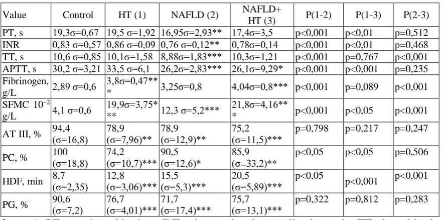
[image:5.595.74.520.296.515.2]SFMK level is higher by 9.6% (p<0.05) than in patients with HT (Table 1).
Table 1
Values of plasma hemostasis in different groups of patients
Value Control HT (1) NAFLD (2) NAFLD+
HT (3) P(1-2) P(1-3) P(2-3) PT, s 19,3σ=0,67 19,5 σ=1,92 16,95σ=2,93** 17,4σ=3,5 p<0,001 p<0,01 p=0,512 INR 0,83 σ=0,57 0,86 σ=0,09 0,76 σ=0,12** 0,78σ=0,14 p<0,001 p<0,01 p=0,468 TT, s 10,6 σ=0,85 10,1σ=1,58 8,88σ=1,83*** 10,3σ=1,21 p<0,001 p=0,767 p<0,001 APTT, s 30,2 σ=3,21 33,5 σ=6,1 26,2σ=2,83*** 26,1σ=9,29* p<0,001 p<0,001 p=0,235 Fibrinogen,
g/L 2,89 σ=0,6
3,8σ=0,47**
* 3,25σ=0,8 4,04σ=0,8*** p<0,001 p=0,089 p<0,001 SFMC 10–2
g/L 4,1 σ=0,6
19,9σ=3,75*
** 12,3 σ=5,2***
21,8σ=4,16**
* p<0,001 p<0,05 p<0,001
AT III, % 94,4 (σ=16,8) 78,9 (σ=7,96)** 78,9 (σ=12,9)** 75,2 (σ=11,5)*** p=0,798 p=0,217 p=0,247
PC, % 100 (σ=18,8) 74,2 (σ=10,7)*** 90,5 (σ=12,6)* 85,9 (σ=33,2)** p<0,05 p<0,05 p=0,506
HDF, min 8,7 (σ=2,35) 12,8 (σ=3,06)*** 15,5 (σ=5,3)*** 20,5 (σ=5,89)*** p<0,05 p<0,001 p<0,001
PG, % 90,6 (σ=7,2) 76,7 (σ=4,01)*** 71,7 (σ=17,4)*** 75,7 (σ=13,1)*** p=0,322 p=0,812 p=0,283
Notes: 1. PT - prothrombin time; INR - international normalization ratio; TT, thrombin time; APTT - activated partial thromboplastin time; SFMC - soluble fibrin-monomer complexes; AT III - antithrombin III; PC - protein C; HDF - Hageman-dependent fibrinolysis; PG - plasminogen; HT - hypertension; NAFLD - non-alcoholic fatty liver disease 2. * - significance of change according to the Mann-Whitney criterion (U Test) compared with the control group: * – p<0.05; ** – p<0.01; *** – p<0.001.
According to the results of our own studies, when compared with the control group,
there is a decrease in AT III by 16.4% in both the HT group (p<0.01) and the NAFLD group
(p<0.01), combined pathology leads to more significant inhibition AT III - by 20.3%
(p<0.001). Significant differences in PC levels when compared with the control cohort were
found in the HT group - the value decreases by 26% (p<0.001), in the NAFLD group - by
9.5% (p<0.05), in the NAFLD + HT group - by 14.1% (p<0.01). According to our studies, the
(p<0.001), NAFLD + HT - by 16.5% (p<0.001) control group, and has no significant
differences in intergroup comparison. The definition of HDF is the determination of the time
of dissolution of a retraged fibrin clot under the influence of the plasmin proteolytic enzyme.
We observe the lengthening of this time in all groups compared with the control: in the HT
group - by 47% (p<0.001), in the NAFLD group - by 78% (p<0.001), in the NAFLD + HT
group - 2.4 times (p<0.001). Unlike patients with HT, the duration of HDF is 21% longer with
NAFLD (p<0.05), and 60% with NAFLD + HT (p<0.001). Comparison of groups with liver
damage demonstrates a longer clot lysis time in the case of a combined pathology - by 32%
(p<0.001).
Discussion
In the group of patients with AH, there were no significant differences in PT, INR,
APTT and TT levels from control indicators, although a tendency for their levels to increase
is in accordance with the data described in the literature (Hassan & Merghani, 2016; JiskaniS,
Memon, & Naseem, 2017; Nnenna Adaeze, Uchenna Emeribe, Abdullahi Nasiru, Babayo, &
Uko, 2014). NAFLD is characterized by a decrease in PT, INR, TT, APTT, which indicates
an acceleration of coagulation in this group of patients, and corresponds to the literature data
(Fargion, Porzio, & Fracanzani, 2014; Stine, Intagliata, Northup, & Caldwell, 2017). In cases
of accession of hypertension to NAFLD, a significant shortening of the time of clot formation
is observed by the internal mechanism of activation of coagulant hemostasis. While the value
of PCMC significantly exceeds the benchmarks in all groups, with a high level in patients
with hypertension, connected with NAFLD. Since SFMC is a marker of thrombosis (Elazab
Elged, El-Gamal, Bastawy, & Saeed, 2016), an increase in its level indicates an increase in
the prothrombogenic activity of the blood when NAFLD is attached to hypertension.
Fibrinogen level rises in response to systemic inflammation, tissue damage and the presence
of cancer. It has been established that an increased level of fibrinogen in inflammation, cancer
and other diseases is the cause of thrombosis (Davalos & Akassoglou, 2012; Repetto & De
Re, 2017). According to the results of our own research, the value of fibrinogen increased in
HT patients, as well as the levels of this indicator significantly increase when HT is added to
NAFLD, by 19.6% (p <0.001) as compared with an isolated course of NAFLD. Since SFMC
and fibrinogen characterizes the final link of thrombosis in the blood coagulation cascade, it is
possible to consider the comorbid course of hypertension and NAFLD as a factor of
According to the results of our studies in patients with hypertension and patients with
NAFLD suppression of anticoagulant hemostasis observed by reducing levels of AT III and
sun, consistent with the data described in the literature [8, 14]. In the case of a combination of
HT and NAFLD, the level of AD BP decreases to the same extent as with an isolated course
of HT or NAFLD. Decrease in PC is more marked in patients with hypertension than in
patients with NAFLD and in patients with comorbid course of these diseases. There is a
depletion of anti-bursting potential in all groups of patients, due to a decrease in the activity
of protein anticoagulants. According to the results of our own research, a decrease in blood
fibrinolytic activity was also found in the HT and NAFLD groups, which corresponds to the
literature data. In addition, for the first time it was found that the combination of these
diseases leads to a more significant depression of fibrinolysis due to the prolongation of the
Hageman-dependent time, and a decrease in the level of plasminogen is the same in all groups
of patients.
Determination of coagulation state, anticoagulant and fibrinolytic links of hemostasis
do not take into account the entire volume of hemostatic changes occurring during the
comorbid course of hypertension and NAFLD. There is a need for a more extensive study of
the hemostatic system, including the platelet part of hemostasis in this cohort of patients.
Conclusions
1. In patients with hypertension and patients with NAFLD suppression of
anticoagulant hemostasis is observed by reducing the activity of AT III and PC. Reduction of
blood fibrinolytic activity in patients with hypertension, NAFLD and their combination
manifests itself in lengthening the time of dissolution of the fibrin clot and inhibition of
plasminogen. The increase in the time of Hageman-dependent fibrinolysis is more significant
in the HT group combined with NAFLD, whereas the level of plasminogen decreases equally
in all groups of patients.
2.Patients with NAFLD has accelerating of coagulation at all stages of thrombus
formation, while the combination of HT and NAFLD is accompanied by an increase in
thrombogenic potential, predominantly at the final stage of blood coagulation.
3. For patients with NAFLD and patients with hypertension associated with NAFLD,
activation of the internal coagulation pathway is characteristic, as evidenced by the shortening
of the APTT. These changes reflect the presence of thrombophilic changes, creating an
additional risk of thrombotic complications in this category of patients.
4. In hypertension connected with NAFLD, depletion of anticoagulant and fibrinolytic
of prothrombogenic changes, therefore comorbidity of hypertension and NAFLD can be
considered a factor of thrombophilic changes of hemostasis.
References
1. Byrne, C. D., & Targher, G. (2015). NAFLD: A multisystem disease. Journal
of Hepatology, 62(S1), S47–S64. https://doi.org/10.1016/j.jhep.2014.12.012
2. Davalos, D., & Akassoglou, K. (2012). Fibrinogen as a key regulator of
inflammation in disease. Seminars in Immunopathology, 34(1), 43–62.
https://doi.org/10.1007/s00281-011-0290-8
3. Elazab Elged, A. A., El-Gamal, R. A., Bastawy, S., & Saeed, M. (2016).
Soluble fibrin monomer complex assay enhances early and accurate diagnosis of acute
myocardial infarction. International Journal of Clinical and Experimental Pathology, 9(5),
5801–5809.
4. Fargion, S., Porzio, M., & Fracanzani, A. L. (2014). Nonalcoholic fatty liver
disease and vascular disease: state-of-the-art. World Journal of Gastroenterology, 20(37),
13306–13324. https://doi.org/10.3748/wjg.v20.i37.13306
5. Hall, J. E., do Carmo, J. M., da Silva, A. A., Wang, Z., & Hall, M. E. (2015).
Obesity-Induced Hypertension: Interaction of Neurohumoral and Renal Mechanisms.
Circulation Research, 116(6), 991–1006. https://doi.org/10.1161/CIRCRESAHA.116.305697
6. Hassan, F. M., & Merghani, M. M. (2016). Coagulation Disturbance among
Essential Hypertensive and Diabetes Mellitus Type II Patients-Khartoum State Merghani
MM. Bangladesh Journal of Medical Science (Vol. 15).
https://doi.org/http://dx.doi.org/10.3329/bjms.v15i3.30199
7. JiskaniS, A., Memon, S., & Naseem, L. (2017). Prothrombin time (PT),
activated partial thromboplastin time (APTT) and international normalized ratio (INR) as
predictive factors of coagulopathy in newly diagnosed hypertensive patients. Hematology &
Transfusion International Journal, 4(3), 1–0. https://doi.org/10.15406/HTIJ.2017.4.00086
8. Junker, R., Heinrich, J., Schulte, H., Erren, M., & Assmann, G. (1998).
Hemostasis in normotensive and hypertensive men: results of the PROCAM study. The
prospective cardiovascular Münster study. Journal of Hypertension, 16(7), 917–923.
Retrieved from http://www.ncbi.nlm.nih.gov/pubmed/9794731
9. Lallukka, S., Orho-Melander, M., Lundbom, N., Olkkonen, V. M., &
Yki-Järvinen, H. (2016). Activity of Coagulation Factors IX and XIII and Fibrinogen are
https://doi.org/10.1016/S0168-8278(16)00860-6
10. Lip, G. Y., Blann, A. D., Jones, A. F., Lip, P. L., & Beevers, D. G. (1997).
Relation of endothelium, thrombogenesis, and hemorheology in systemic hypertension to
ethnicity and left ventricular hypertrophy. The American Journal of Cardiology, 80(12),
1566–1571. Retrieved from http://www.ncbi.nlm.nih.gov/pubmed/9416937
11. Makris, T. K., Tsoukala, C., Krespi, P., Hatzizacharias, A., Gialeraki, A.,
Papargyriou, J., … Mandalaki, T. (1997). Haemostasis balance disorders in patients with
essential hypertension. Thrombosis Research, 88(2), 99–107. Retrieved from
http://www.ncbi.nlm.nih.gov/pubmed/9361364
12. Mancia, G., Fagard, R., Narkiewicz, K., Redon, J., Zanchetti, A., Böhm, M., …
Wood, D. A. (2013). 2013 ESH/ESC guidelines for the management of arterial hypertension:
The Task Force for the management of arterial hypertension of the European Society of
Hypertension (ESH) and of the European Society of Cardiology (ESC). European Heart
Journal, 34(28), 2159–2219. https://doi.org/10.1093/eurheartj/eht151
13. Nnenna Adaeze, N., Uchenna Emeribe, A., Abdullahi Nasiru, I., Babayo, A., &
Uko, E. K. (2014). Evaluation of Prothrombin Time and Activated Partial Thromboplastin
Time in Hypertensive Patients Attending a Tertiary Hospital in Calabar, Nigeria. Advances in
Hematology, 2014, 1–7. https://doi.org/10.1155/2014/932039
14. Poulter, N. R., Prabhakaran, D., & Caulfield, M. (2015). Hypertension. The
Lancet, 386(9995), 801–812. https://doi.org/10.1016/S0140-6736(14)61468-9
15. Repetto, O., & De Re, V. (2017). Coagulation and fibrinolysis in gastric
cancer. Annals of the New York Academy of Sciences, 1404(1), 27–48.
https://doi.org/10.1111/nyas.13454
16. Rinella, M. E. (2015). Nonalcoholic Fatty Liver Disease. JAMA, 313(22),
2263. https://doi.org/10.1001/jama.2015.5370
17. Stine, J. G., Intagliata, N., Northup, P. G., & Caldwell, S. H. (2017).
Nonalcoholic Fatty Liver Disease, Portal Vein Thrombosis and Coagulation. Transplantation,
101(8), e281–e282. https://doi.org/10.1097/TP.0000000000001807
18. Tripodi, A., Fracanzani, A. L., Primignani, M., Chantarangkul, V., Clerici, M.,
Mannucci, P. M., … Fargion, S. (2014). Procoagulant imbalance in patients with
non-alcoholic fatty liver disease. Journal of Hepatology, 61(1), 148–154.
https://doi.org/10.1016/j.jhep.2014.03.013
19. Woodward, M., Lowe, G. D., Rumley, A., Tunstall-Pedoe, H., Philippou, H.,
activation markers: The Third Glasgow MONICA Survey. II. Relationships to cardiovascular
risk factors and prevalent cardiovascular disease. British Journal of Haematology, 97(4), 785–