ISSN Online: 2160-5874 ISSN Print: 2160-5866
DOI: 10.4236/jbbs.2017.79031 Sep. 25, 2017 425 Journal of Behavioral and Brain Science
Effect of Dimethoate on Object Recognition
Memory in Wistar Rats and Essay of
Treatment with Nettle
Majda Samih
1*, Pacôme Kouadio N’Go
2, Sabah Belaaouja
1, Amina Ouazzani Touhami
3,
Ahmed Omar Touhami Ahami
11Unit of Clinic and Cognitive Neuroscience, Laboratory of Biology and Health, Department of Biology, Faculty of Sciences, Ibn
Tofail University, Kenitra, Morocco
2Training and Research Unit of Biology Sciences, Peleforo Gon Coulibaly University, Korhogo, Ivory Coast
3Botanical and Plant Protection Unit, Laboratory of Biology and Health, Department of Biology, Faculty of Sciences, Ibn Tofaïl
University, Kenitra, Morocco
Abstract
Pesticides were economically important chemicals in agriculture. Their use has permitted agricultural progress, through the eradication of harmful insect and the fight against vectors of disease. However, several studies question the beneficial effects of organophosphorus compounds, showing that their dere-gulated use causes various problems of environmental pollution and human health. The present study shows that chronic exposure to a subtoxic dose of dimethoate is likely to affect cognitive and behavioral functions of rats (both males and females). Our results show that exposure to dimethoate affects both short and long-term memory capacities. The short-term memory results are more pronounced. Treatment with nettle extract allowed a significant im-provement in cognitive and behavioral performance of the rats after their ex-posure to dimethoate.
Keywords
Dimethoate, Dioic Nettle, Acetylcholinesterase, Chelation, Memory
1. Introduction
It was in the 1940s that the first synthetic pesticides appeared on the market with very positive results in increasing agricultural yields. Twenty years later, the first accusations of harm to people’s health and the environment were heard [1].
The debate about the risks and benefits of chemical control has since been
ex-How to cite this paper: Samih, M., N’Go, P.K., Belaaouja, S., Touhami, A.O. and Ahami, A.O.T. (2017) Effect of Dimethoate on Object Recognition Memory in Wistar Rats and Essay of Treatment with Nettle. Journal of Behavioral and Brain Science, 7, 425-445.
https://doi.org/10.4236/jbbs.2017.79031
Received: July 29, 2017 Accepted: September 22, 2017 Published: September 25, 2017
Copyright © 2017 by authors and Scientific Research Publishing Inc. This work is licensed under the Creative Commons Attribution International License (CC BY 4.0).
DOI: 10.4236/jbbs.2017.79031 426 Journal of Behavioral and Brain Science
Inhibition of AChE causes an accumulation of acetylcholine released in the synaptic cleft upon nerve stimulation, leading to hyperstimulation of the nico-tinic and muscarinic receptors. Consequently, the passage of nerve information is disrupted [2]. In addition, oxidative stress caused by lipid peroxidation and promoted by dimethoate is considered to be a second mechanism of toxicity of this organophosphorus [3] [4].
In this study, we will highlight the effect of sub-chronic intoxication by dime-thoate on short-term and long-term memory. The poisoning took place on male and female Wistar rats. These intoxicated rats will undergo a trial of treatment by phytochelation. Extract of a medicinal plant, nettle, was obtained by decoc-tion.
Chelation therapy has started as a branch of medicine to remove heavy metals and other toxins from the body by introducing powerful chemical agents that associate with toxins and are then excreted in the urine.
From the point of view of a medical use, several clinical trials carried out in this context confirmed these pharmacological properties in humans.
Urtica dioica L., is a ubiquitous plant of the Urticaceae family, renowned for its unpleasant irritating effects, the nettle is actually rich in vitamins, minerals and numerous medicinal virtues [5]. Over the last few decades, several studies have focused on the pharmacological implications and on the analysis of the chemical composition of the plant. Although all its potentialities have yet to be realized, numerous studies have strengthened its claims in traditional medicine.
[6].
Indeed, put into practice in vitro and in vivo in animals, these studies have proved that this plant possesses numerous pharmacological properties in terms of antiproliferative, anti-inflammatory, antioxidant, analgesic, antiulcer, immu-nostimulating, anti-Infectious, anti-hypertensive and protective against cardi-ovascular disease. In addition, and in view of its richness in protein, minerals and vitamins, nettle provides a proven nutritional interest. In terms of toxicolo-gy, nettle remains innocuous and significant doses, administered orally to hu-mans, showed no side effects.
DOI: 10.4236/jbbs.2017.79031 427 Journal of Behavioral and Brain Science
hydroxyl radical OH˚ and the nitric oxide radical NO˚, was determined by spec-trophotometry. Numerous studies have shown that the methanolic and ethanolic extracts of the leaves have a remarkable antioxidant effect with respect to the 1,1-diphenyl-2-picrylhydrazyl radical (DPPH) [7] [8] [9]. Chelation of ferrous iron was evaluated using ferrozine which forms a red chromophore with residual iron (Fe (II)-Ferrozine) having an absorption maximum at 562 nm. The absor-bances obtained show that nettle has an important chelating activity with respect to the ferrous ion [10]. Another study, carried out on rats treated with tetrach-loromethane (CCl 4), showed that nettle decreased lipid peroxidation and in-creased the activity of the antioxidant defense system thus playing a protective role against hepatotoxicity. This antioxidant activity is essentially correlated with the content of phenolic compounds [7] [11].
Nettle is very rich in polyphenols and is located in different parts of plants according to the plant species and the polyphenolic group considered. These compounds group together a multitude of molecules and represent one of the most important groups present in the plant kingdom. Although they are very diversified, they all have in common the presence of one or more benzene rings carrying one or more hydroxyl functions. Polyphenols are divided into several categories, including flavonoids [SFA, 2005].
Flavonoids act mainly as primary antioxidants, stabilizing peroxide radicals, but they can also deactivate reactive oxygen species, inhibit lipoxygenase or che-late metals [12].
Flavonoids are in vitro enzyme inhibitors of: histidine decarboxylase (by que-retol and naringenin), elastase, hyaluronidase (by flavones and proanthocyani-dols) and catechol-O-methyltransferase.
2. Material and Methods
2.1. Animals Studied and Condition of Breeding
The animals used in this study are wistar albino rats (male and female), and are obtained from the Faculty of Science animal house. The animals are reared in transparent plastic cages (32 × 15 × 13 cm), covered by a stainless steel grill. The breeding takes place under standard conditions of ventilation, light (cycle of al-ternation 12H light/12H darkness), temperature (21 ± 2 degrees C) and relative humidity (65% ± 15%). Rats have ad libitum access to fresh water and seca-lin-based food (SNV, Temara, Morocco).
2.2. Treatment of Animals
2.2.1. Chronic Toxicity to Dimethoate
DOI: 10.4236/jbbs.2017.79031 428 Journal of Behavioral and Brain Science
for only a few hours. A dose of 1 ml/100mg was administered orally to each of the intoxicated animals and this for duration of one month. The results of the treatment are finally compared with those of the control and intoxicated rats.
2.3. Parameters Studied
2.3.1. Body Weight Measurements
Signs of systemic toxicity such as convulsion, weakness and trembling were mo-nitored. Weight is an important parameter in any experimental study because it is an indicator of physical health. The weight of each animal is monitored weekly with an electronic scale throughout the study.
2.3.2. Behavioral Testing: Novel Object Recognition Memory • Description of the device
The object recognition memory test involves spontaneous exploration beha-vior in rodents, so it does not require food or water restriction or negative reinforcement of the animals. It is based on the spontaneous tendency of rats to preferentially explore a new object in relation to a familiar object. This de-vice was used for the first time by [14].
This test takes place in a square open enclosure of 50 × 50 cm2 (L × l), sur-rounded by four walls of 50 cm2 height. It is made of wood painted with black color, which gives the image of an open cube. The device is illuminated by a light source positioned at a height of one meter.
• Experimental procedure
dup-DOI: 10.4236/jbbs.2017.79031 429 Journal of Behavioral and Brain Science
licate. The floor and objects are rinsed with 10% ethanol to remove any trace left by the animal in the device.
• Parameters taken into account
When the memory of the animal is not altered, it will explore the new object more than the old one, since it has memorized it. On the other hand, when the memory is defective, it will give more exploration time to the old object or it will explore both in an identical way. To evaluate the state of the mem-ory, we calculated an index of recognition according to the following equa-tion: Recognition index = time spent exploring the object B / Sigma time spent exploring object A and B [14].
2.4. Statistical Analysis
We performed the statistical analyzes using the SPSS 20.0 software. Parametric ANOVA repeat tests and 1-factor ANOVA were used to compare the data series. When there was a difference between more than two batches, a further analysis was carried out with Tukey’s post hoc test. The normality of distribution was first atested by the Kolmogorov-Smirnov test and homogeneity by the Levens test. The values of p < 0.05 are considered significant.
2.5. Limitations of Study
Although the research has reached its aims, there are some unavoidable limita-tions.
The main of them are expressed as follows:
• Lack of prior research studies on the topic.
• The environmental factor (temperature, light...) influences the behavior of
the rats and must be taken into consideration.
• The sample size is an important feature of any study. The higher the number,
the more reliable the results.
• If the mode of administration is by gavage, the rats should be handled with
care to avoid throat irritation.
3. Results
3.1. Macroscopic Observation
Signs of systemic toxicity such as weight loss, resting quake, decreased food in-take were observed in groups of rats poisoned compared to controls.
3.2. Effect on Body Weight (Drinking Water)
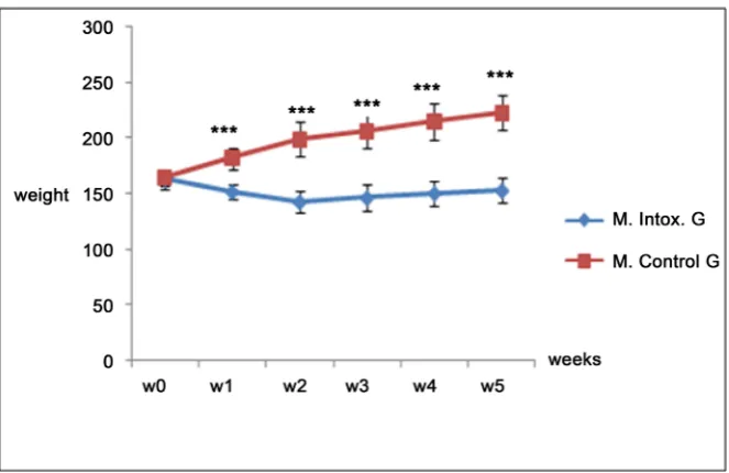
In males rats: sub-chronic intoxication with dimethoate for four weeks induces a progressive decrease in body weight in treated rats groups, unlike control rats
Figure 1. The Friedman test followed by the Bonferroni post-hoc test shows that the differences observed are highly significant between the two study groups (p < 0.001).
DOI: 10.4236/jbbs.2017.79031 430 Journal of Behavioral and Brain Science
Figure 1. Effect of dimethoate on body weight in male rats Number of average errors ± mean standard error (SEM). ***p < 0.001, comparison between intoxicated groups and controls.
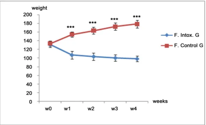
Figure 2. Effect of dimethoate on body weight in female rats Number of average errors ± mean standard error (SEM). ***p < 0.001, comparison between intoxicated groups and controls.
rats intoxicated during the study Figure 2. The Friedman test followed by the Bonferroni post-hoc test shows that this decrease is very highly significant (p < 0.001).
3.3. Effect on Body Weight (Gavage)
In males: Body weight remained steady throughout the study period in intox-icated rats, while it increased steadily in control rats Figure 3. The differences observed between the two study groups are highly significant (p < 0.001).
[image:6.595.209.541.327.529.2]DOI: 10.4236/jbbs.2017.79031 431 Journal of Behavioral and Brain Science
[image:7.595.209.541.213.428.2]compared with those poisoned by dimethoate gavage. The statistical analysis also showed a highly significant difference between the two study groups (p < 0.001)
Figure 4.
3.4. Evaluation of Memory Functions
3.4.1. Effect on Short Term Memory Recognition (STM): Case of Gavage
[image:7.595.211.537.481.679.2]In male rats: Figure 5 shows a decrease in the short-term recognition index. This index is below the recognition threshold in rats intoxicated with Dime-thoate (IR = 40%). This decrease is highly significant p < 0.001.
Figure 3. Effect of dimethoate on body weight in male rats Number of average errors ± mean standard error (SEM). ***p < 0.001, comparison between intoxicated groups and controls.
DOI: 10.4236/jbbs.2017.79031 432 Journal of Behavioral and Brain Science
Figure 5. Effect of Dimethoate Exposure on Object Recognition STM in Male Rats Rec-ognition index expressed on average ± mean standard error (SEM). ***p < 0.001, com-parison between intoxicated groups and controls.
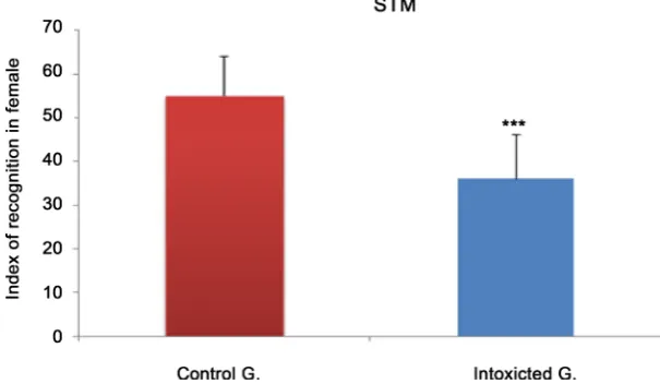
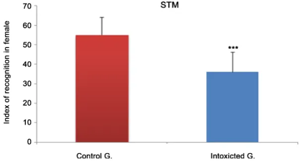
Figure 6. Effect of Dimethoate Exposure on Object Recognition STM in Female Rats Recognition index expressed on average ± mean standard error (SEM). ***p < 0.001, comparison between intoxicated groups and controls.
In female rats: Dimethoate poisoning alters the short-term recognition mem-ory in female rats. The expressed IR is highly significant between the two groups studied p < 0.001, either 43% to poisoned Figure 6.
3.4.2. Effects on STM Recognition: Drinking Water Case
In male rats: Dimethoate poisoning in drinking water induces an alteration of (STM) in rats. Indeed, the index of recognition is highly reduced in intoxicated rats. P < 0.001, compared to controls in Figure 7.
In female rats: There was also a highly significant reduction in the index of rec-ognition in Dimethoate addicts p < 0.001, compared with controls in Figure 8.
3.4.3. Effects on Long Term Memory Recognition (LTM): Case of Gavage
[image:8.595.220.523.311.487.2]DOI: 10.4236/jbbs.2017.79031 433 Journal of Behavioral and Brain Science
Figure 7. Effect of Dimethoate Exposure on Object Recognition STM In male rats Rec-ognition index expressed on average ± mean standard error (SEM). ***p < 0.001, com-parison between intoxicated groups and controls.
Figure 8. Effect of Dimethoate Exposure on Object Recognition STM in Female Rats Recognition index expressed on average ± mean standard error (SEM). ***p < 0.001, comparison between intoxicated groups and controls.
Dimethoate gavage compared to controls, even though this index was above the 50% recognition threshold. The difference revealed is very statistically significant p < 0.01, Figure 9.
In female rats: the long-term RI of 54% in intoxicated females did not differ significantly with controls p > 0.05 Figure 10.
3.4.4. Effects on Long Term Memory Recognition (LTM): Case of Gavage
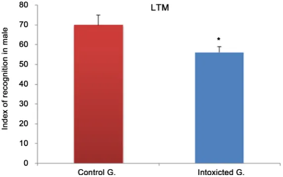
In male rats: As shown in Figure 11, RI is above the recognition threshold in rats treated with Dimethoate. However, this index differs significantly from that obtained by the control rats p < 0.05.
In female rats: It is also observed that the index of recognition is above the threshold of recognition of the objects in the rats intoxicated with Dimethoate, even if this index differs very significantly from that of the controls p < 0.01,
[image:9.595.221.523.301.464.2]DOI: 10.4236/jbbs.2017.79031 434 Journal of Behavioral and Brain Science
[image:10.595.230.518.300.467.2]Figure 9. Effect of Dimethoate Exposure on Object Recognition LTM in Male Rats Rec-ognition index expressed on average ± mean standard error (SEM). **p < 0.01, compari-son between intoxicated groups and controls.
Figure 10. Effect of Dimethoate Exposure on LTM Recognition Objects in female rats Recognition index expressed as mean ± mean standard error (SEM).
[image:10.595.230.517.506.685.2]DOI: 10.4236/jbbs.2017.79031 435 Journal of Behavioral and Brain Science
Figure 12. Effect of Dimethoate Exposure on LTM Recognition of Objects in female rats Discrimination index expressed as average ± mean standard error (SEM). **p < 0.01, comparison between intoxicated groups and controls.
Figure 13. Effect of Urtica Dioica Medicinal Plant Treatment on Short-term Recognition Memory (STM) in Male Rats Recognition% expressed as average ± mean standard error (SEM). ***p < 0.001, **p < 0.01, comparison between the 3 groups treated; ##p < 0.01, comparison between the dimethoate addicted group and plant-treated group (1-factor ANOVA/Tukey post-hoc analysis.
3.5. Effects of Chelation Treatment on Memory Functions
3.5.1. Effects of Nettle Plant on STM Recognition: Drinking Water Case
In male rats: Recognition STM is highly altered in rats treated with Dimethoate, the recognition index is 40% below the recognition threshold with a highly sig-nificant difference from that of the control rats p < 0.001. However, treatment with the medicinal plant Nettle Dioica has led to an improvement in this index, which has increased from 40% to 53%, with a very significant difference p < 0.01,
Figure 13.
In females: Observations similar to that of males were made. As shown in
[image:11.595.214.536.308.481.2]DOI: 10.4236/jbbs.2017.79031 436 Journal of Behavioral and Brain Science
[image:12.595.213.534.270.439.2]ter treatment with the plant Nettle dioica, this difference is not significant p < 0.05 (Figure 16).
Figure 14. Effect of treatment by the medicinal plant Urtica Dioica on the short-term recognition memory (STM) in female rats Recognition% expressed as average ± mean standard error (SEM). ***p < 0.001, *p < 0.05, comparison between the 3 treated groups; ##p < 0.01, comparison between the dimethoate addicted group and plant-treated group (1-factor ANOVA/Tukey post-hoc analysis.
[image:12.595.218.533.516.682.2]DOI: 10.4236/jbbs.2017.79031 437 Journal of Behavioral and Brain Science
Figure 16. Effect of Urtica Dioica medicinal plant on long-term recognition memory (LTM) in female rats Recognition% expressed as average ± mean standard error (SEM). **p < 0.01, comparison between the 3 groups treated (ANOVA with 1 factor/Post-hoc analysis of Tukey.
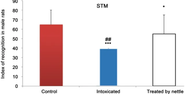
Figure 17. Effect of Urtica Dioica Medicinal Plant Treatment on Short-term Recognition Memory (STM) in Male Rats Recognition% expressed as average ± mean standard error (SEM). ***p < 0.001, *p < 0.05, comparison between the 3 treated groups; ##p < 0.01, comparison between the dimethoate addicted group and plant-treated group (1-factor ANOVA/Tukey post-hoc analysis.
3.5.3. Effects of the Nettle Plant on the STM of Recognition: Case of Gavage
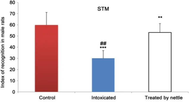
In male rats: In the case of gavage intoxication, it was demonstrated that the recognition STM was very much reduced (37%), compared to controls p < 0.001.
Treatment with the Nettle Dioica plant resulted in an improvement in this index, which rose from 37% to 60% in Figure 17.
In females: As shown in Figure 18, poisoning also induced a high alteration of STM recognition with 40% index versus 70% in p < 0.001 controls. On the other hand, the treatment with the plant caused an improvement of this index to 59% p < 0.001.
3.5.4. Effects of the Plant Nettle on the Recognition LTM: Case of Gavage
[image:13.595.213.534.306.469.2]DOI: 10.4236/jbbs.2017.79031 438 Journal of Behavioral and Brain Science
[image:14.595.228.520.333.480.2]Figure 18. Effect of Urtica Dioica Medicinal Plant Treatment on Short-term Recognition Memory (STM) in Female Rats Recognition% expressed as average ± mean standard er-ror (SEM). ***p < 0.001, *p < 0.05, comparison between the 3 treated groups; #P < 0.05, comparison between the dimethoate poisoning group and plant-treated group (1-factor ANOVA/Tukey post-hoc analysis.
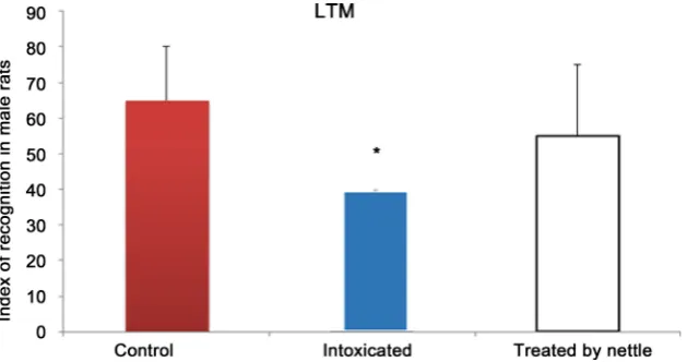
Figure 19. Effect of Urtica Dioica Medicinal Plant Treatment on Long-Term Recognition Memory (LTM) in Male Rats Recognition% expressed as average ± mean standard error (SEM). **p < 0.01, comparison between the 3 treatment groups (1-factor ANOVA/Tukey post-hoc analysis).
[image:14.595.227.516.543.693.2]DOI: 10.4236/jbbs.2017.79031 439 Journal of Behavioral and Brain Science
nificantly altered (54%) in intoxicated rats, compared to controls p < 0.05. However, treatment with the Nettle plant allowed a small increase in this index without significant effect p < 0.05.
4. Discussion
4.1. Effect of Dimethoate on Body Weight
The effect of the diméthoate on the physical weight gain for both modes of ad-ministration behaves appreciably in a different way. Indeed, in the case of the poisoning with diméthoate in drinking water, we observe a highly significant decrease in weight depending on the duration of the treatment for both rats’ sexes, as shown in Figure 1 and Figure 2. To the best of our knowledge, there have been no previous studies of this kind on the animal model. However these results indicate the significant impact of the diméthoate on the health of the agricultural populations living in the polluted middle. On the other hand, ob-servations made in groups of rats exposed to dimethoate gavage show a slight decrease in body weight for both sexes males and females, as shown in Figure 3
and Figure 4. Our results are in line with the works of Jallouli on mice poisoned by the diméthoate [15].
Another study of the developmental toxicity in Malathion also demonstrates a low physical weight gain for young rats to the (GD 6-PND 45) stage [16]. From the physio-pathological point of view, the low physical weight gain of the ex-posed groups could be attributed to the effect of overexpression of the ACh mo-lecules which increases the gastric motility and decreases the intestinal absorp-tion [17]. Indeed, the parasympathetic nervous system stimulates the processes of digestion, through the NT ACh. However, the excess of this NT due to the in-hibition of AChE by the OP could be the cause of the dysfunction of this process
[18].
4.2. Effect of Dimethoate on Memory Functions
The recognition memory capabilities of rats intoxicated with Dimethoate were highlighted by the NOR (Novel Object) recognition memory test. This metho-dology allowed to link chronic exposure to OPs insecticides and memory im-pairment which is a serious health problem for the general population. In this work, short and long-term types of memory have been studied, both of which are very much affected in Alzheimer’s disease [19].
The test is based on the spontaneous tendency of rodents to explore a new object in relation to another already seen. This cognitive test, evaluating the abil-ity to recognize a previous stimulus, is a fundamental study of human amnesia by animal model [20]. Indeed, the memory of recognition rests on the ability to judge whether we have already encountered any stimulus, in fact visual (face, object, place...).
DOI: 10.4236/jbbs.2017.79031 440 Journal of Behavioral and Brain Science
tum [21].
To evoke hypotheses concerning the mechanism of action of alteration of memory, it is useful to know the cerebral structures considered as support for this cognitive function. Indeed, the NOR test model is influenced by hippocam-pal and cortical lesions [22] [23]. It is widely demonstrated that in rats and large apes, parahippocampal regions including the perirhinal cortex play a crucial role in the process of object recognition memory [24], i.e. their ability to evaluate a stimulus already encountered as a factor of familiarity based on the integrity of the medial temporal lobe [25]. This cerebral structure plays an important role in the formation of recognition memory. Its lesion leads to impaired performance
[26]. A previous study has shown that exposure to subtoxic doses of Malathion induces a very significant inhibition of AChE in the hippocampus and associated regions [16]. The hippocampal system is rich in the cholinergic system, which has a central role in the encoding and consolidation of the new stimulus. This process therefore makes it possible to integrate and reorganize an already formed memory. By this means, in rodents, when a stimulus is previously expe-rienced and reactivated, the hippocampus permits discrimination between the familiar and the new with a spontaneous and natural tendency to interact more with novelty [27]. Dysfunction in the OP induced cholinergic system in the hip-pocampus and/or perirhinal cortex may be suggested as a cause of alteration of short-term recognition memory. Indeed, the perirhinal cortex represents the main cerebral site of transit of information of environmental origin such as visu-al, olfactory and somatosensory stimuli, through which the hippocampus rece-ives its “inputs” to form the memory [28].
DOI: 10.4236/jbbs.2017.79031 441 Journal of Behavioral and Brain Science
long-term recognition memory processes involve various components, such as subcortical and cortical structures. However, the sex effect that depends on the effect of dimethoate on recognition memory functions remains to be discussed. Indeed, the ability to recognize objects could be altered in both male rats and females exposed to Dimethoate. A developmental study showed that when the period of exposure to chlorpyrifos (CPF) occurs at the end of gestation (GD 17 - 20), only female mice with altered reference and working spatial memory. In this case, the muscarinic cholinergic system seems to be one of the systems whose functioning is modified or altered [29] [30]. On the other hand, if the period of exposure to CPF occurs postnatally, during the first days of life (PND 1 - 4), it is only the male mice that have spatial memory problems [31]. This implies that in order to better understand the sex-effect of dimethoate, exposure studies on dif-ferent stages of development should be undertaken.
4.3. Effect of Nettle on the Functions of Memory
Although we have studied the effects of dimethoate intoxication in rats, we have tested the therapeutic effects of a medicinal plant, Nettle, which has antioxidant properties. In order to better support its properties, it is desirable to evoke the pro-oxidizing and proinflammatory effects induced by OPs in general and Di-methoate in particular. Most authors agree that chronic or acute exposure in-duces oxidative and proinflammatory stress effects in animals [4] [31] [32] [33] [34] [35].
In addition, dimethoate intoxication has been shown to cause cellular altera-tions and oxidative stress, leading to lipid peroxidation and the generation of free radicals [4] [36] [37]. In this study it was demonstrated that Nettle treat-ment resulted in a remarkable improvetreat-ment in short-term memory recognition capacity, which was strongly altered by dimethoate. Indeed, antioxidants consti-tute the defense system that limits the toxicity associated with free radicals [38]. In addition, a dietary intake rich in antioxidant products has shown protective effects against oxidative stress [38] [39], this has been demonstrated for vitamin E [40] [41] and for Selenium [39].
These results suggest that nettle treatment has a positive effect by eliminating free radicals associated with exposure to Dimethoate. In addition, it would be appropriate to investigate the effects of dimethoate on the serotonergic system and on the catecholamines involved in the regulation of memory performance
[42]. These systems have been disturbed during intoxication with CPF [43] [44] [45] [46] and methyl parathion [47].
5. Conclusions
DOI: 10.4236/jbbs.2017.79031 442 Journal of Behavioral and Brain Science [3] Halliwell, B. (2006) Reactive Species and Antioxidants. Redox Biology Is a
Funda-mental Theme of Aerobic Life. Plant Physiology, 141, 312-322.
https://doi.org/10.1104/pp.106.077073
[4] Yukti, S., Bashir, S., Irshad, M., Gupta, S.D. and Dogra, T.D. (2005) Effects of Acute Dimethoate Administration on Antioxidant Status of Liver and Brain of Experi-mental Rats. Toxicology, 206, 49-57. https://doi.org/10.1016/j.tox.2004.06.062
[5] Wichtl, M. and Anton, R. (2003) Plantes thérapeutiques, tradition, pratique offici-nale, science et thérapeutique. [Therapeutic Plants, Tradition, Officinal Practice, Science and Therapeutics.] 2nd Edition, Inter/Tec & Doc Éditions, Paris, 587-589. [6] Henri, C. (1997) Traité pratique et raisonné des plantes médicinales indigènes.
[Practical and Rational Treaty of Native Medicinal Plants.] 3rd Edition, de l’Envol, Paris, 1251 p.
[7] Kataki, M.S., Murugamani, V., Rajkumari, A., Mehra, P.S., Awasthi, D. and Yadav, R.S. (2012) Antioxidant, Hepatoprotective, and Anthelmintic Activities of Methanol Extract of Urtica dioica L. Leaves. Pharmaceutical Crops, 3, 38-46.
https://doi.org/10.2174/2210290601203010038
[8] Khare, V., Kushwaha, P., Verma, S., Gupta, A., Srivastava, S. and Rawat, A.K.S. (2012) Pharmacognostic Evaluation and Antioxidant Activity of Urtica dioica L.
Chinese Medicine, 3, 128-135. https://doi.org/10.4236/cm.2012.33021
[9] Pourmorad, F., Hosseinimehr, S.J. and Shahabimajd, N. (2006) Antioxidant Activi-ty, Phenol and Flavonoid Contents of Some Selected Iranian Medicinal Plants.
African Journal of Biotechnology, 5, 1142 -1145.
[10] Gulcin, I., Kufrevioglu, O.I.., Oktay, M. and Buyukokuroglu, M.E. (2004) Antioxi-dant, Antimicrobial, Antiulcer and Analgesic Activities of Nettle (Urtica dioica L.).
Journal of Ethnopharmacology, 90, 205-215.
https://doi.org/10.1016/j.jep.2003.09.028
[11] Kanter, M., Coskun, O. and Budancamanak, M. (2005) Hepatoprotective Effects of
Nigella sativa L and Urtica dioica L on Lipid Peroxidation, Antioxidant Enzyme Systems and Liver Enzymes in Carbon Tetrachloride-Treated Rats. World Journal of Gastroenterology, 11, 6684-6688. https://doi.org/10.3748/wjg.v11.i42.6684
[12] Sarni-Manchado, P. and Cheynier, V., Eds. (2006) Les polyphénols en agroali- mentaire. [Polyphenols in Agroalimentary.] Techniques & Documentation. [13] Gallo, M.A. and Lawryk, N.J. (1991) Organic Phosphorus Pesticides. In: Hayes Jr.,
W.J. and Laws Jr., E.R., Eds., Handbook of Pesticide Toxicology, Vol. 2, Classes of Pesticides, Academic Press, San Diego, 917-1123.
DOI: 10.4236/jbbs.2017.79031 443 Journal of Behavioral and Brain Science Studies of Memory in Rats. 1: Behavioral Data. Behavioural Brain Research, 31, 47-59. https://doi.org/10.1016/0166-4328(88)90157-X
[15] Jallouli, M., El Bini Dhouib, I., Dhouib, H., Gharbi, N. and El Fazaa, S. (2015) Ef-fects of Dimethoate in Male Mice Reproductive Parameters. Regulatory Toxicology and Pharmacology, 73, 853-858. https://doi.org/10.1016/j.yrtph.2015.10.010
[16] N’Go, P.K., Azzaoui, F.-Z., Soro, P.R., Samih, M., Ahami, A.O.T., Najimi, M. and Chigr, F. (2013) Developmental Effects of Malathion Exposure on Recognition Memory and Spatial Learning in Males Wistar Rats. JBBS, 3, 331-340.
https://doi.org/10.4236/jbbs.2013.33033
[17] Jones, A.L. and Karalliedde, L. (2006) Davidson’s Principles and Practice of Medi-cine. In: Boon, N.A., Colledge, N.R., Davidson, S.S. and Walker, B.R., Eds., 2nd Edi-tion, Churchill Livingstone, Edinburgh, 203-226.
[18] Eskenazi, B., Bradman, A. and Castorina, R. (1999) Exposures of Children to Orga-nophosphate Pesticides and Their Potential Adverse Health Effects. Environmental Health Perspectives, 107, 409. https://doi.org/10.1289/ehp.99107s3409
[19] Dibo-Cohen, C.M. (2006) Mémoire spatiale contextuelle et schizophrénie. [Con- textual Spatial Memory and Schizophrenia.] Université Pierre et Marie Curie—Paris VI, Français.
[20] Baxter, M.G. (2009) Involvement of Medial Temporal Lobe Structures in Memory and Perception. Neuron, 61, 667-677. https://doi.org/10.1016/j.neuron.2009.02.007
[21] Mauricio, V.-H., Castillo, I., Díaz, C., Alés, I. and Rodríguez-Moreno, A. (2012) Dimethoate Accelerates the Extinction of Eyeblink Conditioning in Mice. Neuro-toxicology, 33, 105-110. https://doi.org/10.1016/j.neuro.2011.12.003
[22] Buckmaster, C.A., Eichenbaum, H., Amaral, D.G., Suzuki, W.A. and Rapp, P.R. (2004) Entorhinal Cortex Lesions Disrupt the Relational Organization of Memory in Monkeys. Journal of Neuroscience, 24, 9811-9825.
https://doi.org/10.1523/JNEUROSCI.1532-04.2004
[23] Clarke, B.O., Porter, N.A., Marriott, P.J. and Blackbeard, J.R. (2010) Investigating the Levels and Trends of Organochlorine Pesticides and Polychlorinated Biphenyl in Sewage Sludge. Environment International, 36, 323-329.
https://doi.org/10.1016/j.envint.2010.01.004
[24] Aggleton, J.P., O’Mara, S.M., Vann, S.D., Wright, N.F., Tsanov, M. and Erichsen, J.T. (2010) Hippocampal-Anterior Thalamic Pathways for Memory: Uncovering a Network of Direct and Indirect Actions. European Journal of Neuroscience, 31, 2292-2307. https://doi.org/10.1111/j.1460-9568.2010.07251.x
[25] Hammond, B., Dudek, R., Lemen, J. and Nemeth, M. (2004) Results of a 13 Week Safety Assurance Study with Rats Fed Grain from Glyphosate Tolerant Corn. Food and Chemical Toxicology, 42, 1003-1014. https://doi.org/10.1016/j.fct.2004.02.013
[26] Albasser, M.M., Amin, E., Lin, T.-C.E., Iordanova, M.D. and Aggleton, J.P. (2012) Evidence That the Rat Hippocampus Has Contrasting Roles in Object Recognition Memory and Object Recency Memory. Behavioral Neuroscience, 126, 659.
https://doi.org/10.1037/a0029754
[27] Antunes, M.S., Morey, K.J., Smith, J.J., Albrecht, K.D., Bowen, T.A., Zdunek, J.K., Troupe, J.F., Cuneo, M.J., Webb, C.T., Hellinga, H.W., et al. (2011) Programmable Ligand Detection System in Plants through a Synthetic Signal Transduction Path-way. PLoS One, 6, e16292. https://doi.org/10.1371/journal.pone.0016292
[28] Rosauro, C.J., Cammarota, M., Gruart, A., Izquierdo, I. and Delgado-García. J.M. (2010) Plastic Modifications Induced by Object Recognition Memory Processing.
DOI: 10.4236/jbbs.2017.79031 444 Journal of Behavioral and Brain Science
tives, 113, 1027-1031. https://doi.org/10.1289/ehp.7968
[32] Maryam, A., Abdollahi, M., Kebryaeezadeh, A., Hosseini, R. and Sabzevari, O. (2003) Biochemical Evidence for Free Radicalinduced Lipid Peroxidation as a Me-chanism for Subchronic Toxicity of Malathion in Blood and Liver of Rats. Human & Experimental Toxicology, 22, 205-211.
https://doi.org/10.1191/0960327103ht346oa
[33] Banks, C.N. and Lein, P.J. (2012) A Review of Experimental Evidence Linking Neu-rotoxic Organophosphorus Compounds and Inflammation. Neurotoxicology, 33, 575-584. https://doi.org/10.1016/j.neuro.2012.02.002
[34] Kathleen, R. and Xiong, S. (1997) Effect of Administration of Malathion for 14 Days on Macrophage Function and Mast Cell Degranulation. Toxicological Sciences, 37, 95-99. https://doi.org/10.1093/toxsci/37.1.95
[35] Singh, A.K. and Jiang, Y. (2003) Lipopolysaccharide (lps) Induced Activation of the Immune System in Control Rats and Rats Chronically Exposed to a Low Level of the Organothiophosphate Insecticide, Acephate. Toxicology and Industrial Health, 19, 93-108. https://doi.org/10.1191/0748233703th181oa
[36] Reuber, M.D. (1984) Carcinogenicity of Dimethoate. Environmental Research, 34, 193-211. https://doi.org/10.1016/0013-9351(84)90089-6
[37] Ferah, S. (2007) Histopathological Effects of Dimethoate on Testes of Rats. Bulletin of Environmental Contamination and Toxicology, 78, 479-484.
https://doi.org/10.1007/s00128-007-9196-5
[38] Joël, P., Bonjean, K., Cayeux, K. and Defraigne, J.-O. (2002) Mécanismes physiolo-giques de la défense antioxydante. [Physiological Mechanisms of Antioxidant De-fense.] Nutrition clinique and métabolisme, 16, 233-239.
https://doi.org/10.1016/S0985-0562(02)00166-8
[39] Ben Amara, I., Soudani, N., Troudi, A., Bouaziz, H., Boudawara, T. and Zeghal, N. (2011) Antioxidant Effect of Vitamin E and Selenium on Hepatotoxicity Induced by Dimethoate in Female Adult Rats. Ecotoxicology and Environmental Safety, 74, 811-819. https://doi.org/10.1016/j.ecoenv.2010.11.007
[40] Schwenke, D.C. and Behr, S.R. (1998) Vitamin E Combined with Selenium Inhibits Atherosclerosis in Hypercholesterolemic Rabbits Independently of Effects on Plas-ma Cholesterol Concentrations. Circulation Research, 83, 366-377.
https://doi.org/10.1161/01.RES.83.4.366
[41] Shireen, K.F., Pace, R.D., Mahboob, M. and Khan, A.T. (2008) Effects of Dietary Vitamin E, C and Soybean Oil Supplementation on Antioxidant Enzyme Activities in Liver and Muscles of Rats. Food and Chemical Toxicology, 46, 3290-3294.
DOI: 10.4236/jbbs.2017.79031 445 Journal of Behavioral and Brain Science [42] Ove, Ö.S., Eriksson, T.M., Elvander-Tottie, E., D’Addario, C., Ekström, J.C., Sven-ningsson, P., Meister, B., Kehr, J. and Stiedl, O. (2008) The Role of 5-ht 1a Recep-tors in Learning and Memory. Behavioural Brain Research, 195, 54-77.
https://doi.org/10.1016/j.bbr.2008.02.023
[43] Aldridge, J.E., Seidler, F.J., Meyer, A., Thillai, I. and Slotkin, T.A. (2003) Serotoner-gic Systems Targeted by Developmental Exposure to Chlorpyrifos: Effects during Different Critical Periods. Environmental Health Perspectives, 111, 1736.
https://doi.org/10.1289/ehp.6489
[44] Slotkin, T.A. (2002) Nicotine and the Adolescent Brain: Insights from an Animal Model. Neurotoxicology and Teratology, 24, 369-384.
https://doi.org/10.1016/S0892-0362(02)00199-X
[45] Slotkin, T.A., Lassiter, T.L., Ryde, I.T., Wrench, N., Levin, E.D. and Seidler. F.J. (2009) Consumption of a High-Fat Diet in Adulthood Ameliorates the Effects of Neonatal Parathion Exposure on Acetylcholine Systems in Rat Brain Regions. En-vironmental Health Perspectives, 117, 916. https://doi.org/10.1289/ehp.0800459
[46] van Dam, P.S., Bravenboer, B., van Asbeck, B.S., Marx, J.J.M. and Gispen, W.H. (1999) High Rat Food Vitamin E Content Improves Nerve Function in Streptozoto-cin-Diabetic Rats. European Journal of Pharmacology, 376, 217-222.
https://doi.org/10.1016/S0014-2999(99)00376-3
[47] Mohammed, M., Shireen, K.F., Atkinson, A. and Khan, A.T. (2001) Lipid Peroxida-tion and Antioxidant Enzyme Activity in Different Organs of Mice Exposed to Low Level of Mercury. Journal of Environmental Science and Health, Part B, 36, 687-697.
https://doi.org/10.1081/PFC-100106195
Submit or recommend next manuscript to SCIRP and we will provide best service for you:
Accepting pre-submission inquiries through Email, Facebook, LinkedIn, Twitter, etc. A wide selection of journals (inclusive of 9 subjects, more than 200 journals)
Providing 24-hour high-quality service User-friendly online submission system Fair and swift peer-review system
Efficient typesetting and proofreading procedure
Display of the result of downloads and visits, as well as the number of cited articles Maximum dissemination of your research work