ISSN Online: 2157-9415 ISSN Print: 2157-9407
DOI: 10.4236/ss.2017.89046 Sep. 29, 2017 422 Surgical Science
Neuro Endocrine Tumor of the Gall Bladder: A
Case Report
Eddy Oleko Ekuke
1*, Yassine Kdhissi
1, Fatoumata Djouldé Smith Diallo
2, Pierlesky Elion Ossibi
1,
Hicham El Bouhadoutti
1, El Bachir Benjelloun
1, Ouadii Mouaqit
1, Benajah Dafr-Allah
2,
Abdelmalek Ousadden
1, Khalid Mazaz
1, Khalid Ait Taleb
11Department of Visceral Surgery, Hassan II University Hospital, Fez, Morocco
2Department of Gastroenterology and Hepatology, Hassan II University Hospital, Fez, Morocco
Abstract
Neuroendocrine tumors (NET) of the gallbladder are a rare entity with only 0.2% of all NET located in the gall bladder. Well-differentiated NETs occur at a relatively lower age group unlike other gallbladder tumors, whereas neu-roendocrine carcinoma (NEC) occurs in an older category of patients. The aim of our study is to discuss the current level of evidence regarding this pa-thological entity by means of a rare case report on a neuroendocrine carcino-ma of the gall bladder in a 63-year-old patient with a history of diabetes. Pa-tient underwent cholecystectomy for acute cholecystitis. Pathology findings on surgical specimen came back for neuroendocrine tumour.
Keywords
Neuroendocrine Tumors, Endocrine Carcinoma, Gall Bladder
1. Introduction
Primary NET can occur throughout the entire gastro-intestinal tract (from the esophagus down to the anus), the pancreas and exceptionally in the liver or the gall bladder.
Gallbladder NET are uncommon, due to their extremely rare epidemiological character and their circumstances of discovery, mostly fortuitous. Only 0.2% of all neuroendocrine tumors are located in the gallbladder [1]. Clinical setting points to one of acute cholecystitis, but definite diagnosis is only made on the pathology examination of surgical specimen. We hereby report a case of gallbladder NET, discovered on pathology examination of cholecystectomy spe-cimen in a 63-year-old male with a history of diabetes.
How to cite this paper: Ekuke, E.O., Kdhissi, Y., Diallo, F.D.S., Ossibi. P.E., El Bouhadoutti, H., Benjelloun, E.B., Mouaqit, O., Dafr-Allah, B., Ousadden, A., Mazaz, K. and Ait Taleb, K. (2017) Neuro Endocrine Tumor of the Gall Bladder: A Case Report. Surgical Science, 8, 422-427.
https://doi.org/10.4236/ss.2017.89046
Received: July 3, 2017 Accepted: September 26, 2017 Published: September 29, 2017
Copyright © 2017 by authors and Scientific Research Publishing Inc. This work is licensed under the Creative Commons Attribution International License (CC BY 4.0).
DOI: 10.4236/ss.2017.89046 423 Surgical Science
2. Case
Patient, 63-year-old diabetic male on insulin, presented with a three-month his-tory of biliary colic with associating intermittent fever relieved by over the counter analgesics and antispasmodic drugs.
Symptoms worsened a week prior to his admission by the exacerbation of right upper quadrant pain and fever prompting his consultation at our department.
Physical examination found a conscious patient, stable vitals, HR 100 beats/minute, 39˚C febrile with right upper quadrant guarding on abdominal examination.
Lab test came back with leukocytosis 22000/mm3; CRP level at 212 mg/l and
236 mg/l blood sugar. The rest of the lab results notably urea and electrolytes as well calcitonin levels were unremarkable.
Abdominal ultrasound revealed a large gallbladder with a thickened wall, 12 mm thick, containing several gallstones and a perivesicular effusion.
After initial fluid resuscitation patient was admitted for surgery, with pero-perative discovery of a distended gall bladder with pseudomembranes (Figure 1) and a slightly purulent perivesicular abscess about 5 cc, which was aspirated. Macroscopically, the surrounding liver tissue was normal with no palpable mass. Retrograde cholecystectomy was performed.
Immediate postoperative recovery was marked by surgical wound infection, which responded favorably to adequate antibiotics and dressing for up to 10 days post operatively.
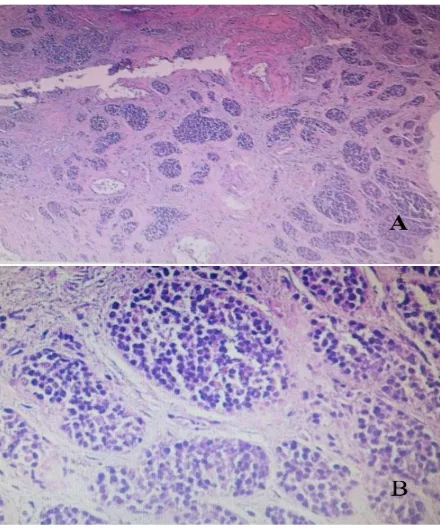
Pathology examination with immune histochemical marking of surgical spe-cimen came back for a stage 3 (WHO 2010, ENETS 2006) large cell neuroendo-crine carcinoma (Figure 2). There were no vascular emboli nor were perineural invasion and resection margins were clean. The tumor was staged pT2Nx.
The case was discussed at a multidisciplinary cancerology meeting where tho-racic-abdomino-pelvic CT was recommended.
[image:2.595.249.496.541.706.2]Thoracic-abdomino-pelvic CT revealed a metastatic lesion of segment V of the liver (Figure 3).
DOI: 10.4236/ss.2017.89046 424 Surgical Science
Figure 2. (A) Tumor proliferation infiltrating gall bladder wall, arranged in islets (HES ×
5); (B) Monomorphic tumor cells with irregular nuclei containing small nucleolus and abundant eosinophilic cytoplasm (HES × 40).
Figure 3. (A) Sagittal CT scan showing a metastatic lesion in segment V of the liver; (B)
Axial CT showing metastatic lesion located in segment V of the liver.
[image:3.595.208.538.389.601.2]DOI: 10.4236/ss.2017.89046 425 Surgical Science
3. Discussion
Primary NETs, all locations combined, are rare with 2 to 5 new cases per year per 100,000 inhabitants.
Primary NETs can occur throughout the GI tract (from the esophagus down to the anus), the pancreas, rarely the liver, and the gall bladder. Globally, its in-cidence is considerably low, about 0.5 - 5/100,000, with an estimated 70% of cas-es affecting the digcas-estive system, with only 0.2% located in the gall bladder.
Generally two broad spectrum can be identified: functional tumors (eliciting characteristic clinical symptoms pertaining to tumor secretion of peptides or amino acids) requiring specific anti-secretory treatment and non-functional tu-mors (not eliciting symptoms). The rarity and heterogeneity of NETs explain the low number of randomized studies and apparent lack of evidence. Their inci-dence and localizations varies with sex: men tend to have more NETs in the esophagus and stomach, whereas cases of hepatobiliary and colorectal PNETs involve women.
Neuroendocrine tumors in general are rare, accounting for only 0.5% of all gallbladder tumors and 0.2% of all neuroendocrine digestive neoplasms. Well-differentiated NET presents itself at a lower age compared to other gallbladder tumors [1], whereas NEC occurs mostly in an older category of pa-tients [1]. Neuroendocrine tumors of the gall bladder are common in women (68%) with ages ranging between 25 - 85 years [2].
Circumstances of discovery are extremely variably: as symptoms may relate to the local mass effect in the event of NF-NETs; right upper quadrant (RUQ) pain; jaundice, RUQ mass pointing to a large distended gall bladder. They could also be discovered for tuitouslyon cholecystectomy specimen in cases of non-complicated cholecystitis [3] [4]. Due to rapid growth of these tumors, me-tastases, mainly hepatic, may be revelatory in some cases (39.8%).
DOI: 10.4236/ss.2017.89046 426 Surgical Science be performed for pathology examination. It is also useful in identifying unre-sectable tumors thereby reducing the number of unnecessary laparotomies. 1 out of 3 of BDC patients are often considered operable after the radiological staging [6].
In principle, symptomatic gall bladder NETs are difficult to distinguish from other cancers of the gall bladder. Precise diagnoses are only made on pathology examination. Pathology findings not only confirm the diagnosis of NETs they also determine histo-prognostic factors. NETs are characterized by the presence of chromatin clumps (granulations with hyper dense material). The peculiar phenotype of these cells contribute to precise diagnosis immune histochemical marking. In fact, the following markers are expressed in varying degrees of spe-cificity in neuroendocrine tumors: synaptophysin, NSE, chromogranin A. The presence of at least two of these markers allows the precise diagnosis of neu-roendocrine carcinoma [7].
Treatment of NETs of the gallbladder should take into account the histologi-cal type and tumor staging. Surgery is the sole curative treatment, especially for carcinoid tumors with a poorer prognosis for poorly differentiated carcinomas that are aggressive and are rapidly metastatic at the time of diagnosis. Five-year survival rate varies from 0.0 to 8.3% [2]. The vast majority of gallbladder cancers require multi-visceral surgery or sometimes regional surgery. With the progress of anesthesia and intensive care coupled with a marked improvement in know-ledge of liver anatomy and surgery, multiple attempts at an aggressive surgical approach by certain teams have reported interesting survival at three and five years even in patients with advanced stage NEC [5]. Surgery for non-metastatic vesicular cancer remains rather less encouraging with an overall survival at five years not exceeding 5% even after complete resection. Surgery remains the sole curative treatment. Indications depend mainly on tumor staging. Consideration has to be given to patients’ age, general condition and associated co-morbidities. In general, about 20% of patients are inoperable at the time of diagnosis. The role of radiotherapy and chemotherapy in the treatment of non resectable NETs is not clear as recent studies generally suggest NEC are not sensitive to conven-tional radiotherapy.
4. Conclusion
Gallbladder NETs constitutes a rare pathological entity. These tumors are often discovered fortuitously postoperatively on pathology findings of surgical speci-men. Pathology examination should be carried out systematically on all chole-cystectomy specimens as this remains the only way to confirm the precise diag-nosis of neuroendocrine tumor of the gallbladder, determine histo-progdiag-nosis and guide management.
References
DOI: 10.4236/ss.2017.89046 427 Surgical Science Tumor-Like Lesions of the Hepatobiliary Tract, 27, 1-15.
[2] Buscemi, S., et al. (2016) “Pure” Large Cell Neuroendocrine Carcinoma of the Gallbladder: Report of a Case and Review of the Literature. International Journal of Surgery, 28, S128-S132.ttps://doi.org/10.1016/j.ijsu.2015.12.045
[3] Komatsuda, T., Ishida, H., Konno, K., et al. (2000) Gallbladder Carcinoma: Color Doppler Sonography. Abdominal Imaging, 25, 194-197.
https://doi.org/10.1007/s002619910044 https://doi.org/10.1007/s12157-011-0239-5
[4] Jacob, J., Chargari, C., Helissey, C., Ferrand, F.-R., Ceccaldi, B., Le Moulec, S., Bau-duceau, O., Fayolle, M. and Védrine, L. (2013) Neuroendocrine Carcinoma of the Digestive Tract: A Literature Review. La Revue de Médecine Interne, 34, 700-705.
https://doi.org/10.1016/j.revmed.2013.02.013
[5] El Fattach, H., Guerrache, Y., Eveno, C., Pocard, M., Kaci, R., Shaar-Chneker, C., Dautry, R., Boudiaf, M., Dohan, A. and Soyer, P. (2015) Primary Neuroendocrine Tumors of the Gallbladder: Ultrasonographic and MDCT Features with Pathologic Correlation. Diagnostic and Interventional Imaging, 96, 499-502.
https://doi.org/10.1016/j.diii.2014.11.027
[6] Gourgiotis, S., Kocher, H.M. et al. (2008) Gallbladder Cancer. The American Jour-nal of Surgery, 196, 252-264.https://doi.org/10.1016/j.amjsurg.2007.11.011
[7] El Mernissi, H., Benzoubeire, N., Salihoune, M., Errabih, I., Krami, H.E., Jahid, A., Ouazzani, L., Mahassini, N. and Ouazzani, H. (2011) Neuroendocrine Tumours of the Gallbladder with Pulmonary and Liver Metastasis: A Report on One Case.
Journal Africain d'Hépato Gastroentérologie, 5, 133-136.
https://doi.org/10.1007/s12157-011-0239-5
Submit or recommend next manuscript to SCIRP and we will provide best service for you:
Accepting pre-submission inquiries through Email, Facebook, LinkedIn, Twitter, etc. A wide selection of journals (inclusive of 9 subjects, more than 200 journals)
Providing 24-hour high-quality service User-friendly online submission system Fair and swift peer-review system
Efficient typesetting and proofreading procedure
Display of the result of downloads and visits, as well as the number of cited articles Maximum dissemination of your research work
Submit your manuscript at: http://papersubmission.scirp.org/

