http://wrap.warwick.ac.uk/
Original citation:Contino, Matteo, Lushi, Enkeleida, Tuval, Idan, Kantsler, Vasily and Polin, Marco. (2015) Microalgae scatter off solid surfaces by hydrodynamic and contact forces. Physical Review Letters, 115 (25). 258102.
Permanent WRAP url:
http://wrap.warwick.ac.uk/76012
Copyright and reuse:
The Warwick Research Archive Portal (WRAP) makes this work by researchers of the University of Warwick available open access under the following conditions. Copyright © and all moral rights to the version of the paper presented here belong to the individual author(s) and/or other copyright owners. To the extent reasonable and practicable the material made available in WRAP has been checked for eligibility before being made available.
Copies of full items can be used for personal research or study, educational, or not-for-profit purposes without prior permission or charge. Provided that the authors, title and full bibliographic details are credited, a hyperlink and/or URL is given for the original metadata page and the content is not changed in any way.
Publisher statement:
© 2015 American Physical Society
Published version: http://dx.doi.org/10.1103/PhysRevLett.115.258102
A note on versions:
Microalgae Scatter off Solid Surfaces by Hydrodynamic and Contact Forces
Matteo Contino,1 Enkeleida Lushi,2 Idan Tuval,3 Vasily Kantsler,1 and Marco Polin1,*
1
Physics Department, University of Warwick, Gibbet Hill Road, Coventry CV4 7AL, United Kingdom 2
School of Engineering, Brown University, Rhode Island 02912, USA 3
Mediterranean Institute for Advanced Studies (CSIC-UIB), E-07190 Esporles, Spain (Received 17 July 2015; published 17 December 2015)
Interactions between microorganisms and solid boundaries play an important role in biological processes, such as egg fertilization, biofilm formation, and soil colonization, where microswimmers move within a structured environment. Despite recent efforts to understand their origin, it is not clear whether these interactions can be understood as being fundamentally of hydrodynamic origin or hinging on the swimmer’s direct contact with the obstacle. Using a combination of experiments and simulations, here we study in detail the interaction of the biflagellate green algaChlamydomonas reinhardtii, widely used as a model puller microorganism, with convex obstacles, a geometry ideally suited to highlight the different roles of steric and hydrodynamic effects. Our results reveal that both kinds of forces are crucial for the correct description of the interaction of this class of flagellated microorganisms with boundaries.
DOI:10.1103/PhysRevLett.115.258102 PACS numbers: 47.63.Gd, 87.16.Qp, 87.17.Jj, 87.18.Tt
Microorganismal motility is often confined by solid objects. From biofilm formation within soil’s porous structure [1] to protistan parasites navigating through the densely packed blood of the host[2], and mammalian ova fertilization[3], solid boundaries alter both the motion and spatial distribution of microorganisms[4,5]in ways that are currently not well understood [6,7]. Explaining these interactions can pave the way for the use of extant microorganisms in technological applications ranging from bioremediation [8,9] to directed transport and delivery of pharmacological cargo at the microscale [10], as well as inform the design of artificial microswimmers[11]. One of the most basic types of interaction is the scattering off a solid plane. Bacteria and other microswimmers with rear-mounted flagella (“pusher” type) are well known to accumulate spontaneously on planar surfaces [12], a phenomenon that has been equally well explained by theories based on either purely steric[7]or hydrodynamic
[6,13] interactions. New experiments are finally prising these two effects apart, with results in clear support of the latter [14,15]. Our knowledge of cell-wall interaction for the other major class of microswimmers, those with front-mounted flagella (“puller”type) is distinctly less advanced. Recent experiments suggest that steric effects dominate the scattering of these flagellates off flat boundaries[5]. If true in general, this would place the two microswimmer types in clearly separated categories of interaction. However, sim-ilarly to the bacterial case [15], differentiating steric and hydrodynamic effects requires one to move beyond a plane wall. Here, we report the first detailed experimental study of the scattering of a model puller-type microswimmer, the biflagellate algaChlamydomonas reinhardtii(CR)[16], off a curved surface. Our results, supported also by numerical simulations, show that both hydrodynamic and steric forces
are needed to explain the microswimmer’s interaction with obstacles. At close contact, lubrication forces alone can lead to long-term entrapment, which is avoided through direct flagellar action following cell spinning.
CR strains CC125 and SHF1 (short flagella mutant) were grown axenically in a tris-acetate-phosphate medium[17]
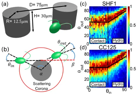
at 21°C under continuous fluorescent illumination (100μE=m2s, OSRAM Fluora). Cells from exponentially growing cultures at∼5×10⁶ cells=ml were harvested and loaded into30μm thick PDMS-based microfluidic chan-nels [Fig.1(a)] previously passivated with a bovine serum albumine solution. Individual channels contain a 4.5× 4.5mm2 region where 2R¼25μm diameter circular pillars are arranged in either hexagonal or slightly ran-domized square lattices of spacing75μm. Identical results
[image:2.612.320.553.494.653.2]were obtained with the two geometries. Cells were imaged under either brightfield or phase contrast illumination with a Nikon TE2000-U inverted microscope fitted with a long-pass filter (cutoff wavelength 765 nm, Knight Optical, UK) to prevent phototactic stimulation. High throughput measurements of CR scattering off individual pillars are based on low magnification (10×, Ph1, NA 0.25) low frame rate (50 fps) recordings (Pike F-100B, Allied Vision Technologies). Cells’trajectories during the>80k scatter-ing events recorded for CC125 (>45k for SHF1) were digitized with standard particle tracking routines [18,19]. Approximately 300 high magnification (40×, NA 1.3), high frame rate (1200 fps, Phantom V 5.2, Vision Research) movies complemented previous measurements. Experimentally, it is necessary to adopt a suitable criterion to define the beginning and end of the scattering. This needs to balance fully capturing the scattering process with reducing the impact of intrinsic swimming noise. We choose to define the scattering with reference to a circular impact area, concentric to the post and extending from its surface a distance ∼3μm larger than the flagellar length, giving a radius of27μm for CC125 (SHF1,25μm) [see Fig.1(b)]. Scattering coronae of different radii modify the angles describing the scattering process according to the simple geometric factor expected for straight trajectories, and hence the choice of radius is in principle largely arbitrary[20]. Our choice was based on the smallest radius that we were confident would capture the full interaction with the pillar surface. For each trajectory, the incoming and outgoing angles,θin andθout respectively, are defined as the signed angle between the local radial direction and the incoming or outgoing swimming directions. The latter are calculated by a linear fit to the five trajectory points immediately external to the impact area. The total deflection angle is indicated withβ (sign conventions, Fig.1(b)].
Figure 1(c) shows the experimentally determined conditional probabilities pðθoutjθinÞ. We will focus on the average hθoutiðθinÞ ¼
R
θoutpðθoutjθinÞdθout. For both strains, this function is well described by
hθoutiðθinÞ ¼
θ
out if θin<θin;
mθinþq if θin≥θin
ð1Þ
with the parameters summarized in Table I. Qualitatively similar results are borne out by the simulations of a three
bead puller swimmer, our reference minimal model for CR, which includes hydrodynamics (see Table I; details are given in Ref.[20]). We will refer toθin >θinandθin <θin as the hydrodynamic and contact regimes, respectively, although it will be seen that hydrodynamic forces play a role also forθin <θin. It should be kept in mind that Eq.(1) ignores the presence of a transition region between these regimes, which for CC125 happens over a∼20° wide range ofθin (SHF1, ∼15°). Within this region, for both strains,
hθoutiðθinÞdeviates from Eq.(1)by a small margin (≤3.4°, i.e., an error of ≲6%). These small errors justify our coarsened but conceptually convenient approach.
Within the hydrodynamic regime,hθoutidepends linearly on θin with a slope m≃0.6 (see Table I). Qualitatively,
[image:3.612.54.566.88.157.2]m≠1signals an interaction. Theoretical studies based on far-field hydrodynamics for puller microswimmers skim-ming off planar and spherical surfaces[13,21–23]predict consistently a repulsive reorientation of the microorgan-ism’s trajectory. Indeed, this can be directly observed from the angular deflectionβðθinÞmeasured in our experiments (Fig.2, inset). βranges fromð33.50.87Þ° forθin ¼57° (just past the transition) down toð5.11Þ° forθin ¼84°
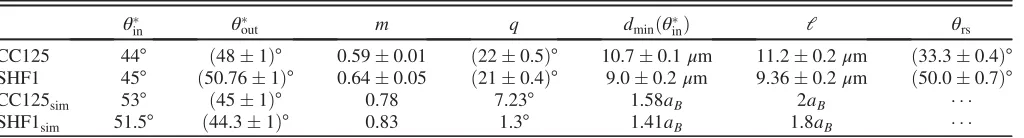
TABLE I. Synopsis of experimental and simulation parameters.ðθin;θout; m; qÞare defined in Eq.(1),dminðθinÞis the minimal distance ofChlamydomonasfrom the pillar surface forθin¼θin,lis the average flagellar length, andθrsis the characteristic decay angle for the probability of random scattering.
θ
in θout m q dminðθinÞ l θrs
CC125 44° ð481Þ° 0.590.01 ð220.5Þ° 10.70.1μm 11.20.2μm ð33.30.4Þ° SHF1 45° ð50.761Þ° 0.640.05 ð210.4Þ° 9.00.2μm 9.360.2μm ð50.00.7Þ°
CC125sim 53° ð451Þ° 0.78 7.23° 1.58aB 2aB
SHF1sim 51.5° ð44.31Þ° 0.83 1.3° 1.41aB 1.8aB
FIG. 2 (color online). Contact and hydrodynamic scattering (CC125).hθouti (blue circles) andðdmin−lÞ(green squares) vs θin.θin¼θin corresponds to l−dmin≃0.5μm. The solid red line corresponds to Eq.(1). Inset: deflection angleβvsθin. For SHF1, see Ref.[20].
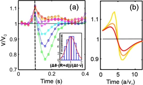
(CC125) [SHF1, fromð42.61Þ° toð6.91.3Þ°]. For the same range ofθin, the minimal swimmer separation from the pillar surface, dmin, increases from 11.530.04μm to 13.650.07μm (CC125, Fig. 2) (SHF1, from 9.420.04 to 10.730.03μm). Within most of the hydrodynamic regime, then,dminis larger than the average flagellar length l¼11.20.2μm (CC125) (SHF1, l¼9.360.22μm). Consequently, the observed inter-action can only be ascribed to hydrodynamic forces (from which comes the name “hydrodynamic” regime). These cause also an increase of the swimming speedv by up to ∼10%with respect to the average speed far from the pillars,
v0, as the cell enters the corona [Fig.3(a)]. This effect can be clearly seen in our simulations, but only when hydro-dynamics is present [Fig. 3(b)]. Conceptually similar evidence for the role of hydrodynamics has been reported for bacteria swimming at distances from a planar wall larger than the compound body and flagellar lengths[14]. We present it here for the first time for a much larger, puller-type eukaryotic microorganism. Notice that, contrary to experiments, the simulations also show the swimmer decelerating as it leaves the pillar. The difference suggests that flagella increase their power output under a moderate load increase, as observed already for CR in Ref.[24].
As θin decreases, so does dmin (Fig. 2), and direct flagellar contact becomes important. The transition from hydrodynamic to contact regimes, then, is at a critical angle θin¼θin where hydrodynamic and steric contributions to the deviation are equivalent. In our experiments, this happens when l−dmin≃0.4μm [ðl; dminÞ ¼
ð11.2;10.7Þμm for CC125, ð9.36;9Þ μm for SHF1]. This is strikingly similar to the length of the flagellar tip
(∼0.5μm), where outer microtubule doublets progressively disappear leaving only the central pair[25]. The tip is then likely significantly softer than the standard axoneme and therefore unable to provide a force sufficient to dominate the interaction with the wall.
Within the contact regime (θin <θin),dmin<land the alga makes close contact with the pillar. Following the average profile of the velocity vðtÞ during the scattering [Fig.3(a)], the process can be divided in three stages. The first corresponds to the initial collision of the microalga with the post, which appears as a sudden deceleration lasting between 40 ms (θin∈½20°;30°) and 60 ms (θin ∈½0°;10°). Steric arguments [26] would predict
v0sinðθinÞ (angle in radians) as the minimal speed. We observe a decrease of significantly smaller magnitude [Fig. 3(a)], reaching at most ∼25% for almost head-on events. The discrepancy is due to partial cell reorientation during slowing down, possibly due to a combination of hydrodynamics and direct flagellar-wall contact. The reor-ientation is completed during the second stage, ending with a fully recovered speed [vðtÞ ¼v0] and the cell aligned parallel to the pillar surface. Steric interactions imply that during this recovery the angleθthat the cell makes with the local surface normal should obey (angles in radians) _
θ¼vðθÞ=ðRþdÞ, where dis the swimmer-surface sepa-ration. Assuming vðθÞ ¼v0sinðθÞ, the angular variations within this stage, fΔθjg, can be obtained from the instantaneous speed vj. These should then satisfy
½arcsinðvjþ1=v0Þ−arcsinðvj=v0Þ¼Δtðvjþ1þvjÞ=2ðRþdÞ, whereΔt¼20ms is the inverse frame rate. The inset of Fig. 3(a) shows that this is the case, supporting the interpretation that cells reorient by simply sliding over the convex, curved surface. At the same time, the high speed movies[20] reveal that at the end of the recovery stage the cell’s flagellar plane is consistently parallel to the pillar surface. This subtle detail, probably resulting from direct contact interactions, is important in the final scatter-ing stage, as we will now discuss. Havscatter-ing completed its reorientation, the alga could be expected to simply swim away at speed v0. For θout∼50° and starting at dmin≃
9μm from the pillar surface, CC125 would take∼90ms to exit the corona. Experimentally, instead, this stage lasts substantially longer: 1708ms. Within this time, the alga—spinning at a frequency of 1.780.4 Hz—can complete slightly more than a 1=4 turn around its axis. Given the initial orientational bias, the flagellar plane should now be perpendicular to the wall. Indeed, the high speed movies show clearly that cells leave the pillar with their flagellar plane always perpendicular to the surface. This configuration maximizes direct flagellar interaction with the obstacle, leading to ahθoutiselected by the simple geometrical rule proposed in Ref.[5]. For a measured body radiusa¼5.70.1μm, this would predicthθouti≃90°− arctanðl=2aÞ ¼45.51° (CC125), which compares very well with the experimental value θout ¼48° (SHF1,
FIG. 3 (color online). Velocity during scatteringvðtÞ(CC125). (a)vðtÞ=v0forθin¼0°–10° (∘), 10°–20° (∘), 20°–30° (∘), 30°–40°
(∘), 30°–40° (∘), 50°–60° (∘), 60°–70° (∘), and 70°–80° (∘). Dashed line: scattering corona boundary. Inset: experimental distribution ofΔθðRþdÞ=ðΔtvÞduring the recovery stage (θin∈½0°;35°). The red line is a Gaussian fit (mean, 1.020.07; standard deviation, 0.42). (b) Hydrodynamic regime simulations:vðtÞ=v0
[image:4.612.53.296.48.193.2]50.61° vs 50.76°). The duration of this stage, remarkably constant throughout the contact regime (<5% variation), can then be understood as the total time required for the cell to spin by 90° (∼125ms) and then swim away at the observed angleθout(∼50ms). The simulations confirm the effect of the body rotation in helping an entrapped swimmer scatter off when captured at the pillar surface (see the supporting movies [20]).
This process hinges on a mechanism bending CR trajectories towards the pillar, an effective attraction even-tually opposed by direct flagellar contact with the obstacle. A quantitative measure of such an interaction is given by the radius of curvature of the experimental trajectories,ρexp. From the evolution of the swimmer’s distance to the pillar center during the last stage of the scattering, we obtain ρexp¼468μm [20]. At the same time, approximating CR’s body as a sphere of radiusa, near-field hydrodynamic torques (HT) can be estimated using lubrication theory[27]
to give a radius of curvature
ρHT¼a
10ð1þδÞ2 δð4þδÞ
1
lnð1=ϵÞ; ð2Þ
whereδandϵare the pillar radius and the gap between the swimmer and the pillar surface, nondimensionalized bya. For the experimentally determined valuesðδ¼2;ϵ¼0.5Þ Eq. (2) givesρHT¼65μm. This simple estimate reveals that lubrication forces alone already provide∼70%of the observed effective torque, supporting the interpretation of a fundamentally hydrodynamic origin for the observed effective attraction. Extra torques come possibly from a combination of further hydrodynamic contributions beyond lubrication, and unequal performance of the two flagella. Although this suggests that sufficiently large pillars should trap CRs hydrodynamically, in the present case the scatter-ing is terminated by contact forces as soon as the flagellar plane becomes perpendicular to the surface. As a result, we predict that scattering events in the contact regime should have the same duration even for larger obstacles. This is supported by experiments with 40-μm-radius pillars[20], and it is compatible with previous results from scattering off a plane [5]. Our observations, then, differ from theoretical estimates of microorganismal capture by curved obstacles, based either on hydrodynamic [23,28]or steric
[26] interactions, where escape results purely from noise (although Ref. [28] discusses using a constant torque to mimic flagellar activity).
The scattering discussed so far proceeds according to a largely predictable dynamics and could be described as deterministic. Together with this, however, we observe a qualitatively different type of interaction, which we call random scattering. Random scatterings are characterized by a prolonged (∼500ms) almost head-on collision of the alga with the obstacle, resulting in multiple events of direct flagellar interaction with the surrounding surfaces,
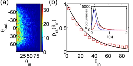
often including the upper and lower boundaries of the microfluidic chamber. A long duration is in fact the hallmark of random scatterings, and was used to distinguish these from deterministic events[20]. The outcome of such intrinsically complex dynamics is simple:θoutis uniformly distributed across the available range of values, independ-ently ofθin[Fig.4(a)], a behavior in sharp contrast to that of minimal models of puller microorganisms[23,28]. At the same time, the probabilityPrsof performing random rather than deterministic scattering depends strongly on θin. Empirically, we findPrsðθinÞ∝expð−θin=θrsÞ, whereθrs¼
33.3° for CC125 [Fig.4(b)and TableI]. This distribution can be recovered within a simple model where the initial dynamics of the orientation angleθfollows an advection-diffusion process in ½0°;90° with absorbing boundaries leading to either random (0°) or deterministic (90°) scatter-ing. Thenθrs¼Dr=ω, whereDrandωare the (rotational) diffusion and drift, respectively. From the deterministic scattering, we can estimate the effective drift towards 90° as ω¼ hθ_i ¼2v0=πR≃5rad=s. The experimental θrs then implies Dr≃3rad2=s (CC125), which agrees well with
Dr≃2rad2=s previously measured for the related species
Chlamydomonas nivalis[29]. Assuming the same Dr for CC125 and SHF1, the model predicts also that θCC125
rs =
θSHF1
rs ð≃0.67Þshould equalv0SHF1=vCC0 125ð≃0.615Þ, indeed verified experimentally within≲10%.
We have presented the first experimental study of the interaction of the model microalgaChlamydomonas rein-hardtii, commonly regarded as a prototypical puller micro-swimmer, with circular obstacles. The experiments reveal that the scattering is not simply steric, as previously suggested [5], but follows qualitatively different rules depending on the angle of incidence, with direct evidence of purely hydrodynamic interaction at large angles, and a multistage steric or hydrodynamic process at small angles. The latter is terminated by direct flagellar contact with the surface preventing the extended trapping around the convex structure predicted by minimal models [23,26,28], a behavior recapitulated by our simulations. The ability to
FIG. 4 (color online). Random scattering (CC125). (a) Distri-bution ofθoutvsθinfor random scatterings. (b)PrsðθinÞvsθin, experiments (□) and fit (−). Inset: distribution of all (−) and random (−) scattering events’duration. The difference represents deterministic events (−).
[image:5.612.322.545.49.160.2]avoid long-term trapping independently of obstacle size and shape might in fact represent a significant advantage for a soil alga such as Chlamydomonas, which in nature needs to navigate a heterogeneous porous material. Together with these deterministic interactions, we report the existence of random scatterings, and propose a mecha-nism to explain their likelihood. Although these events could be specific to our experimental configuration, their existence still suggests that front-mounted flagella pose a challenge to coarse grained descriptions of CR-like micro-organisms’interactions with surfaces.
ACKNOWLEDGMENTS
We acknowledge the support of a Ph.D. studentship from the Engineering and Physical Sciences Research Council (MC), the Spanish Ministry of Economy and Competitiveness Grant No. FIS2013-48444-C2-1-P, and the subprogram Ramón y Cajal (IT).
[1] W. M. Durham, O. Tranzer, A. Leombruni, and R. Stocker, Phys. Fluids24, 091107 (2012).
[2] N. Heddergott, T. Krüger, S. B. Babu, Ai Wei, E. Stellamanns, S. Uppaluri, T. Pfohl, H. Stark, M. Engstler, and S. M. Beverley,PLoS Pathogens8, e1003023 (2012). [3] P. Denissenko, V. Kantsler, D. J. Smith, and J. Kirkman-Brown,Proc. Natl. Acad. Sci. U.S.A. 109, 8007 (2012). [4] P. Galajda, J. E. Keymer, P. Chaikin, and R. H. Austin,
J. Bacteriol.189, 8704 (2007).
[5] V. Kantsler, J. Dunkel, M. Polin, and R. E. Goldstein, Proc. Natl. Acad. Sci. U.S.A.110, 1187 (2013).
[6] A. P. Berke, L. Turner, H. C. Berg, and E. Lauga,Phys. Rev. Lett.101, 038102 (2008).
[7] G. Li and J. X. Tang,Phys. Rev. Lett.103, 078101 (2009). [8] D. Valentine et al.,Science330, 208 (2010).
[9] J. D. Kessleret al.,Science331, 312 (2011).
[10] D. B. Weibel, P. Garstecki, D. Ryan, W. R. DiLuzio, M. Mayer, J. E. Seto, and G. M. Whitesides,Proc. Natl. Acad. Sci. U.S.A.102, 11963 (2005).
[11] A. T. Brown, I. D. Vladescu, A. Dawson, T. Vissers, J. Schwarz-Linek, J. S. Lintuvuori, and W. C. K. Poon, Soft Matter, doi: 10.1039/C5SM01831E (2016).
[12] P. D. Frymier, R. M. Ford, H. C. Berg, and P. T. Cummings, Proc. Natl. Acad. Sci. U.S.A.92, 6195 (1995).
[13] G. J. Li and A. M. Ardekani, Phys. Rev. E 90, 013010 (2014).
[14] M. Molaei, M. Barry, R. Stocker, and J. Sheng,Phys. Rev. Lett.113, 068103 (2014).
[15] O. Sipos, K. Nagy, R. Di Leonardo, and P. Galajda, Phys. Rev. Lett.114, 258104 (2015).
[16] R. E. Goldstein,Annu. Rev. Fluid Mech.47, 343 (2015). [17] J. D. Rochaix, S. Mayfield, M. Goldschmidt-Clermont, and
J. M. Erickson, in Plant Molecular Biology: A Practical Approach, edited by C. H. Schaw (IRL Press, Oxford, England, 1988), pp. 253–275.
[18] V. Pelletier, N. Gal, P. Fournier, and M. L. Kilfoil, Phys. Rev. Lett.102, 188303 (2009).
[19] The code can be downloaded athttp://people.umass.edu/ kilfoil/downloads.html.
[20] See Supplemental Material at http://link.aps.org/ supplemental/10.1103/PhysRevLett.115.258102for supple-mentary material and supporting movies showing typical deterministic (contact and hydrodynamic) and random scatterings for both strains; further plots characterizing the experimental scattering; a brief discussion of the simulations.
[21] E. Lauga and T. Powers, Rep. Prog. Phys. 72, 096601 (2009).
[22] S. E. Spagnolie and E. Lauga, J. Fluid Mech. 700, 105 (2012).
[23] S. E. Spagnolie, G. R. Moreno-Flores, D. Bartolo, and E. Lauga,Soft Matter11, 3396 (2015).
[24] I. Minoura and R. Kamiya,Cell Motil. Cytoskeleton31, 130 (1995).
[25] E. H. Harris,The Chlamydomonas Sourcebook(Academic Press, Oxford, England, 2009), Vol. 3, Chap. 8.
[26] J. Elgeti and G. Gompper, Europhys. Lett. 101, 48003 (2013).
[27] S. Kim and S. J. Karrilla,Microhydrodynamics: Principles and Selected Applications(Dover, Boston, 1991), Chap. 9. [28] K. Schaar, A. Zöttl, and H. Stark, Phys. Rev. Lett. 115,
038101 (2015).