research communications
Acta Cryst.(2017). E73, 1835–1839 https://doi.org/10.1107/S2056989017015985
1835
Received 28 October 2017 Accepted 2 November 2017
Edited by H. Stoeckli-Evans, University of Neuchaˆtel, Switzerland
Keywords:crystal structure; molecular confor-mation; hydrogen bonding; polymorphism.
CCDC references:1583686; 1583685
Supporting information:this article has supporting information at journals.iucr.org/e
Crystal structures of (
E
)-1-{3-[(5-fluoro-2-hydroxy-benzylidene)amino]phenyl}ethanone and of a
fourth polymorph of (
E
)-1-{3-[(2-hydroxy-3-meth-oxybenzylidene)amino]phenyl}ethanone
Marisiddaiah Girisha,aHemmige S. Yathirajan,a* Ravindranath S. Rathoreband Christopher Glidewellc*
aDepartment of Studies in Chemistry, University of Mysore, Manasagangotri, Mysuru 570 006, India,bCentre for
Biological Sciences (Bioinformatics), School of Earth, Biological and Environmental Sciences, Central University of South Bihar, Patna 800 014, India, andcSchool of Chemistry, University of St Andrews, St Andrews, Fife KY16 9ST, UK.
*Correspondence e-mail: [email protected], [email protected]
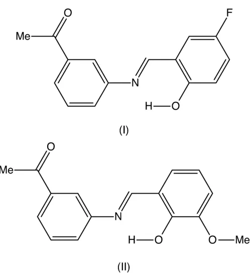
In the molecules of both (E )-1-{3-[(5-fluoro-2-hydroxybenzylidene)amino]-phenyl}ethanone, C15H12FNO2, (I), and (E
)-1-{3-[(2-hydroxy-3-methoxybenzyl-idene)amino]phenyl}ethanone, C16H15NO3, (II), which crystallizes withZ0= 2 in
space groupPca21, there are intramolecular O—H N hydrogen bonds, and the
non-H atoms in each molecule are essentially coplanar. In the crystal of (I), molecules are linked by a single C—H O hydrogen bond to form aC(8) chain, whereas in the crystal of (II), molecules are linked by three C—H O hydrogen bonds to form sheets within which orthogonalC2
2
(16) andC2 2
(17) chains can be identified. Comparisons are made with some related structures.
1. Chemical context
Schiff bases of general typeRR0C NR00can exhibit very wide
structural diversity and have found a wide range of applica-tions (Jia & Li, 2015), ranging from anti-bacterial, anti-fungal and anti-tumour activity (Rani et al., 2015), via catalysis (Kumar et al., 2009), to use as organic photovoltaic materials (Jeevadason et al., 2014). The extensive patent literature on their medicinal applications has recently been reviewed (Hameedet al., 2017). With this great diversity of use in mind, we report herein on the molecular and supramolecular structures of two closely related Schiff bases,(E )-1-{3-[(5-fluoro-2-hydroxybenzylidene)amino]phenyl}ethanone (I) and (E )-1-{3-[(2-hydroxy-3-methoxybenzylidene)amino]phenyl}-ethanone (II). Compounds (I) and (II) were prepared by straightforward condensation reactions between 3-acetyl-aniline (3-aminoacetophenone) and the appropriately substi-tuted salicylaldehydes. Their molecular constitutions differ only in the identity and location of a single substituent, 5-fluoro in (I)versus3-methoxy in (II), but their crystallization behaviour is different. Compound (I) crystallizes in the monoclinic space group P21/n with Z0 = 1 (Fig. 1), while
compound (II) crystallizes in the orthorhombic space group
Pca21withZ0= 2 (Figs. 2 and 3), and it will be convenient to
refer to the molecules of (II) which contain the atoms N11 and N21 as molecules of types 1 and 2, respectively. Compound (II), in fact, represents the fourth polymorphic form of this compound to be identified. Three other forms, one in Pna21
withZ0= 2, and two others inP2
12121, each withZ0= 1, have
recently been reported (Zbacˇniket al., 2015).
2. Structural commentary
In each of compounds (I) (Fig. 1) and (II) (Figs. 2 and 3), the non-H atoms are almost coplanar. Thus in (I), the r.m.s. deviation of the non-H atoms from their mean plane is only 0.085 A˚ , with a maximum individual deviation from the plane of 0.196 (2) A˚ for the acetyl atom C18. Similarly, in compound (II), the r.m.s. deviations of the non-H atoms from the mean planes of the two molecules are 0.086 and 0.071 A˚ for mol-ecules 1 and 2, respectively, with corresponding maximum deviations of 0.225 (5) and 0.211 (5) A˚ for atoms C118 and C218, respectively. In all of the molecules there is an intra-molecular O—H N hydrogen bond (Tables 1 and 2); although this probably influences the orientation of the hy-droxylated ring relative to the central spacer unit, it will not have any influence on the orientation of the acetylphenyl ring
relative to the rest of the molecule. In the two molecules of (II), the deviation of the methoxy C atoms C128 and C228 from the planes of their adjacent aryl rings are 0.107 (9) and 0.049 (11) A˚ , respectively. Consistent with this, the pair of exocyclic C—C—O angles at each of the atoms C123 and C223 differ byca10, as is generally observed in planar alkoxyarene derivatives (Seip & Seip, 1973; Ferguson et al., 1996). The dihedral angle between the mean planes of the two molecules in (II) is 80.74 (3).
1836
Girishaet al. C15H12FNO2and C16H15NO3 Acta Cryst.(2017). E73, 1835–1839
[image:2.610.47.296.78.353.2]research communications
Figure 3
The structure of molecule 2 in compound (II), showing the atom-labelling scheme. Displacement ellipsoids are drawn at the 30% probability level. Table 1
Hydrogen-bond geometry (A˚ ,) for (I).
D—H A D—H H A D A D—H A
O22—H22 N1 0.98 (3) 1.72 (3) 2.607 (2) 148 (3) C27—H27 O17i 0.93 2.58 3.475 (3) 163
Symmetry code: (i)xþ1 2;y
1 2;zþ
[image:2.610.312.567.190.261.2]3 2.
Table 2
Hydrogen-bond geometry (A˚ ,) for (II).
D—H A D—H H A D A D—H A
O122—H122 N11 1.06 (6) 1.68 (6) 2.604 (4) 142 (5) O222—H222 N21 0.92 (6) 1.79 (6) 2.603 (5) 147 (5) C116—H116 O223i 0.93 2.50 3.347 (6) 152
C127—H127 O217 0.93 2.59 3.496 (5) 164 C227—H227 O117ii 0.93 2.58 3.487 (5) 164
Symmetry codes: (i)xþ1;y;zþ1
2; (ii)x;y1;z.
Figure 1
[image:2.610.313.565.434.549.2] [image:2.610.45.293.584.715.2]The molecular structure of compound (I), showing the atom-labelling scheme. Displacement ellipsoids are drawn at the 30% probability level.
Figure 2
[image:2.610.311.567.595.714.2]3. Supramolecular features
The supramolecular assembly in compound (I) is very simple, as shown in Fig. 4. In addition to the intramolecular hydrogen bond noted above, there is a single C—H O hydrogen bond (Table 1), which links molecules related by a 21screw axis into C(8)chains running parallel to the [010] direction. Two chains of this type, related to one another by inversion, pass through each unit cell, but there are no direction-specific interactions between adjacent chains.
There are three C—H O hydrogen bonds in the structure of compound (II) (Table 2): one of these links the two mol-ecules within the selected asymmetric unit and the two others link these bimolecular aggregates into complex sheets, whose formation is readily analysed in terms of two one-dimensional sub-structures (Fergusonet al., 1998a,b; Gregsonet al., 2000). The hydrogen bond having atom C227 as the donor links bimolecular aggregates related by translation to form aC22(16)
chain running parallel to the [010] direction (Fig. 5), and that having atom C116 as the donor links aggregates related by a 21
screw axis into C2
2(17) chains running parallel to the [001]
direction (Fig. 6). The combination of the orthogonal chains along [010] and [001] generates a sheet lying parallel to (100). Two sheets of this type, related to one another by the glide planes, pass through each unit cell but there are no direction-specific interactions between adjacent sheets.
4. Database survey
The structures of Schiff bases derived from hydroxyaryl aldehydes have recently been the subject of a general survey, in which a number of structural errors, often involving misplaced H atoms, were pointed out (Blagus et al., 2010).
research communications
Acta Cryst.(2017). E73, 1835–1839 Girishaet al. C
[image:3.610.44.295.68.303.2] [image:3.610.384.494.367.702.2]15H12FNO2and C16H15NO3
1837
Figure 4Part of the crystal structure of compound (I), showing the formation of a hydrogen-bondedC(8) chain running parallel to the [010] direction. For the sake of clarity, the H atoms not involved in the motif shown have been omitted. Hydrogen bonds are drawn as dashed lines and the atoms marked with an asterisk (*) or a hash (#) are at the symmetry positions (1
2x, 1 2+y,
3
2z) and ( 1 2x,
1 2+y,
3
2z), respectively.
Figure 5
Part of the crystal structure of compound (II), showing the formation of a hydrogen-bondedC2
2(16) chain running parallel to the [010] direction.
[image:3.610.45.295.530.701.2]For the sake of clarity, the H atoms not involved in the motif shown have been omitted, and the hydrogen bonds are drawn as dashed lines.
Figure 6
Part of the crystal structure of compound (II), showing the formation of a hydrogen-bondedC2
2(17) chain running parallel to the [001] direction.
Closely related to the present structures are those of (E )-1-{3-[(2-hydroxy-3-methoxybenzylidene)amino]phenyl}ethanone (III) (De et al., 2009), and of the previously recorded poly-morphs of (II) (Zbacˇniket al., 2015).
Compound (III) is isomorphous with compound (I): as in (I), the structure of (III) contains an intramolecular O— H N hydrogen bond and the non-H atoms are effectively coplanar. The structure of (III) also contains an inter-molecular C—H O hydrogen bond, although this is nowhere mentioned in the original report (De et al., 2009); this inter-action formsC(8) chains along [010], exactly the same as those in the structure of (I), so that (I) and (III) are, in fact, isostructural despite their different patterns of substitution.
Three other polymorphic forms of compound (II) have recently been reported and are described as forms I, II and II,I respectively (Zbacˇniket al., 2015). Form I is orthorhombic in space group Pna21 with Z0 = 2, and forms II and III both
crystallize in space groupP212121withZ0= 1, so that thePca21
form reported here can be regarded as form IV. All three forms, I–III, can be crystallized from ethanol solutions under different conditions and a crucial factor in determining which polymorph is obtained appears to be the filtration process used prior to crystallization. By contrast, the form described here was crystallized from a solution in dichloromethane. In all of the molecules in forms I–III, there is an intramolecular
O—H N hydrogen bond and, in every case, the non-H atoms are effectively co-planar as found here for (I) and (II). The supramolecular assembly differs in all three polymorphs I–III: form II contains no intermolecular hydrogen bonds; in form III two C—H O hydrogen bonds generate a
C(8)C(10)[R1
2(6)] chain of rings; and in form I, three C—
H O hydrogen bonds generate sheets in which the compo-nent sub-structures both involve molecules related by an
n-glide plane, in contrast to the sheets found for form IV reported here.
5. Synthesis and crystallization
For the synthesis of compounds (I) and (II), 3-acetyl aniline (0.740 mmol) and a catalytic quantity of acetic acid were added to solution of the appropriate aldehyde, 5-fluoro-salicylaldehyde for (I) or 3-methoxy5-fluoro-salicylaldehyde for (II) (0.740 mmol) in ethanol (20 cm3), and these mixtures were then heated under reflux for 5 h. The mixtures were then cooled to ambient temperature and the solvent was removed under reduced pressure. The solid residues were then washed with cold ethanol and dried under reduced pressure. Crystals suitable for single crystal X-ray diffraction were grown by slow evaporation, at ambient temperature and in the presence of air, of solutions in dimethylsulfoxide for (I) and in
dichloro-1838
Girishaet al. C15H12FNO2and C16H15NO3 Acta Cryst.(2017). E73, 1835–1839
[image:4.610.46.563.91.413.2]research communications
Table 3
Experimental details.
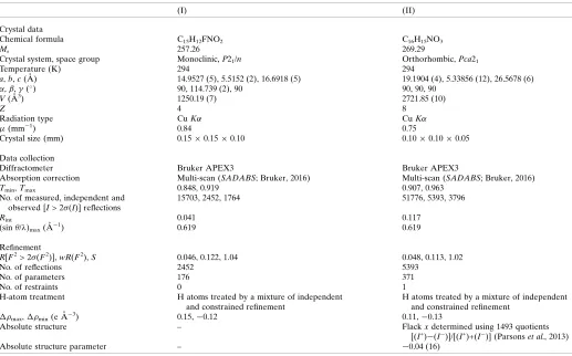
(I) (II)
Crystal data
Chemical formula C15H12FNO2 C16H15NO3
Mr 257.26 269.29
Crystal system, space group Monoclinic,P21/n Orthorhombic,Pca21
Temperature (K) 294 294
a,b,c(A˚ ) 14.9527 (5), 5.5152 (2), 16.6918 (5) 19.1904 (4), 5.33856 (12), 26.5678 (6)
,,(
) 90, 114.739 (2), 90 90, 90, 90
V(A˚3) 1250.19 (7) 2721.85 (10)
Z 4 8
Radiation type CuK CuK
(mm1) 0.84 0.75
Crystal size (mm) 0.150.150.10 0.100.100.05
Data collection
Diffractometer Bruker APEX3 Bruker APEX3
Absorption correction Multi-scan (SADABS; Bruker, 2016) Multi-scan (SADABS; Bruker, 2016)
Tmin,Tmax 0.848, 0.919 0.907, 0.963
No. of measured, independent and observed [I> 2(I)] reflections
15703, 2452, 1764 51776, 5393, 3796
Rint 0.041 0.117
(sin/)max(A˚
1) 0.619 0.619
Refinement
R[F2> 2(F2)],wR(F2),S 0.046, 0.122, 1.04 0.048, 0.113, 1.02
No. of reflections 2452 5393
No. of parameters 176 371
No. of restraints 0 1
H-atom treatment H atoms treated by a mixture of independent and constrained refinement
H atoms treated by a mixture of independent and constrained refinement
max, min(e A˚
3) 0.15,0.12 0.11,0.13
Absolute structure – Flackxdetermined using 1493 quotients
[(I+)(I
)]/[(I+)+(I
)] (Parsonset al., 2013)
Absolute structure parameter – 0.04 (16)
methane for (II): m.p. for (I) 362–364 K and m.p. for (II) 352– 354 K.
6. Refinement
Crystal data, data collection and structure refinement details are summarized in Table 3. For compound (II), one bad outlier reflection (8,1,3) was omitted from the data set before the final refinements. All H atoms were located in difference-Fourier maps. The C-bound H atoms were subsequently treated as riding atoms in geometrically idealized positions: C—H 0.93– 0.96 A˚ with Uiso(H) = 1.5Ueq(C-methyl) and 1.2Ueq(C) for
other C-bound H atoms. The methyl groups were permitted to rotate but not to tilt. For the H atoms bonded to O atoms, the atomic coordinates were refined with Uiso(H) = 1.5Ueq(O),
giving the O—H distances shown in Tables 1 and 2. The correct orientation of the structure of (II) relative to the polar axis direction was established by means of the Flackx para-meter (Flack, 1983),x=0.04 (16) calculated (Parsonset al., 2013) using 1493 quotients of the type [(I+)(I)]/[(I+)+(I)], and by means of the Hooft y parameter (Hooftet al., 2010),y=
0.03 (16). In the final analysis of variance for (I) there was a large value, 1.859, ofK= [mean(Fo2)/mean(Fc2)] for the group
of 4258 very weak reflections havingFc/Fc(max) in the range
0.000 < Fc/Fc(max) < 0.008; the corresponding value for (II)
was 2.1539 for 565 reflections havingFc/Fc(max) in the range
0.000 <Fc/Fc(max) < 0.009.
Acknowledgements
MG thanks the UGC (India) for the award of a Rajeev Gandhi fellowship and HSY thanks the University of Mysore for research facilities.
References
Blagus, A., Cincˇic´, D., Frisˇcˇic´, T., Kaitner, B. & Stilinovic´, V. (2010). Maced. J. Chem. Chem. Eng.29, 117–138.
Bruker (2016). APEX3, SAINT and SADABS. Bruker AXS Inc., Madison, Wisconsin, USA.
De, R. L., Mukherjee, J., Mandal, M., Roy, L., Bhowal, R. & Banerjee, I. (2009).Ind. J. Chem.48B, 595–598.
Ferguson, G., Glidewell, C., Gregson, R. M. & Meehan, P. R. (1998a). Acta Cryst.B54, 129–138.
Ferguson, G., Glidewell, C., Gregson, R. M. & Meehan, P. R. (1998b). Acta Cryst.B54, 139–150.
Ferguson, G., Glidewell, C. & Patterson, I. L. J. (1996).Acta Cryst. C52, 420–423.
Flack, H. D. (1983).Acta Cryst.A39, 876–881.
Gregson, R. M., Glidewell, C., Ferguson, G. & Lough, A. J. (2000). Acta Cryst.B56, 39–57.
Hameed, A., Al-Rashida, M., Uroos, M., Ali, S. A. & Khan, K. M. (2017).Expert Opin. Ther. Pat.27, 63–79.
Hooft, R. W. W., Straver, L. H. & Spek, A. L. (2010).J. Appl. Cryst. 43, 665–668.
Jeevadason, A. W., Murugavel, K. & Neelakantan, M. A. (2014). Renewable Sustainable Energy Rev.36, 220–227.
Jia, Y. & Li, J. (2015).Chem. Rev.115, 1597–1621.
Kumar, S., Dhar, D. N. & Saxena, P. N. (2009).J. Sci. Ind. Res.68, 181– 187.
Parsons, S., Flack, H. D. & Wagner, T. (2013).Acta Cryst.B69, 249– 259.
Rani, A., Kumar, M., Khare, R. & Tuli, H. S. (2015).J. Biol. Chem. Sci.2, 62–91.
Seip, H. M. & Seip, R. (1973).Acta Chem. Scand.27, 4024–4027. Sheldrick, G. M. (2015a).Acta Cryst.A71, 3–8.
Sheldrick, G. M. (2015b).Acta Cryst.C71, 3–8. Spek, A. L. (2009).Acta Cryst.D65, 148–155.
Zbacˇnik, M., Nogalo, I., Cincˇic´, D. & Kaitner, B. (2015). CrystEng-Comm,17, 7870–7877.
research communications
Acta Cryst.(2017). E73, 1835–1839 Girishaet al. C
supporting information
sup-1
Acta Cryst. (2017). E73, 1835-1839
supporting information
Acta Cryst. (2017). E73, 1835-1839 [https://doi.org/10.1107/S2056989017015985]
Crystal structures of (
E
)-1-{3-[(5-fluoro-2-hydroxybenzylidene)amino]phenyl}-ethanone and of a fourth polymorph of (
E
)-1-{3-[(2-hydroxy-3-methoxybenzyl-idene)amino]phenyl}ethanone
Marisiddaiah Girisha, Hemmige S. Yathirajan, Ravindranath S. Rathore and Christopher
Glidewell
Computing details
For both structures, data collection: APEX3 (Bruker, 2016); cell refinement: SAINT (Bruker, 2016); data reduction:
SAINT (Bruker, 2016); program(s) used to solve structure: SHELXT2014 (Sheldrick, 2015a); program(s) used to refine
structure: SHELXL2014 (Sheldrick, 2015b); molecular graphics: PLATON (Spek, 2009); software used to prepare
material for publication: SHELXL2014 (Sheldrick, 2015b) and PLATON (Spek, 2009).
(E)-1-{3-[(5-Fluoro-2-hydroxybenzylidene)amino]phenyl}ethanone (I)
Crystal data
C15H12FNO2 Mr = 257.26
Monoclinic, P21/n a = 14.9527 (5) Å
b = 5.5152 (2) Å
c = 16.6918 (5) Å
β = 114.739 (2)°
V = 1250.19 (7) Å3 Z = 4
F(000) = 536
Dx = 1.367 Mg m−3
Cu Kα radiation, λ = 1.54178 Å Cell parameters from 2452 reflections
θ = 3.3–72.5°
µ = 0.84 mm−1 T = 294 K Block, yellow
0.15 × 0.15 × 0.10 mm
Data collection
Bruker APEX3 diffractometer
Radiation source: microfocus sealed tube Multilayer mirror monochromator
φ and ω scans
Absorption correction: multi-scan (SADABS; Bruker, 2016)
Tmin = 0.848, Tmax = 0.919
15703 measured reflections 2452 independent reflections 1764 reflections with I > 2σ(I)
Rint = 0.041
θmax = 72.5°, θmin = 3.3° h = −18→18
k = −6→6
l = −20→20
Refinement
Refinement on F2
Least-squares matrix: full
R[F2 > 2σ(F2)] = 0.046 wR(F2) = 0.122 S = 1.03 2452 reflections
176 parameters 0 restraints
Primary atom site location: structure-invariant direct methods
supporting information
sup-2
Acta Cryst. (2017). E73, 1835-1839
Hydrogen site location: mixed
H atoms treated by a mixture of independent and constrained refinement
w = 1/[σ2(F
o2) + (0.0427P)2 + 0.3677P]
where P = (Fo2 + 2Fc2)/3
(Δ/σ)max < 0.001
Δρmax = 0.15 e Å−3
Δρmin = −0.12 e Å−3
Special details
Geometry. All esds (except the esd in the dihedral angle between two l.s. planes) are estimated using the full covariance matrix. The cell esds are taken into account individually in the estimation of esds in distances, angles and torsion angles; correlations between esds in cell parameters are only used when they are defined by crystal symmetry. An approximate (isotropic) treatment of cell esds is used for estimating esds involving l.s. planes.
Fractional atomic coordinates and isotropic or equivalent isotropic displacement parameters (Å2)
x y z Uiso*/Ueq
supporting information
sup-3
Acta Cryst. (2017). E73, 1835-1839
Atomic displacement parameters (Å2)
U11 U22 U33 U12 U13 U23
N1 0.0650 (9) 0.0630 (9) 0.0489 (8) 0.0097 (7) 0.0308 (7) 0.0060 (7) C11 0.0543 (9) 0.0585 (10) 0.0503 (9) 0.0096 (8) 0.0263 (7) 0.0124 (8) C12 0.0550 (9) 0.0571 (10) 0.0556 (9) −0.0021 (8) 0.0290 (8) 0.0028 (8) C13 0.0542 (9) 0.0569 (10) 0.0556 (10) 0.0015 (8) 0.0243 (8) 0.0026 (8) C14 0.0680 (12) 0.0636 (11) 0.0681 (11) −0.0095 (9) 0.0266 (9) 0.0000 (9) C15 0.0710 (13) 0.0798 (14) 0.0795 (13) −0.0147 (11) 0.0366 (11) 0.0115 (12) C16 0.0664 (11) 0.0795 (13) 0.0614 (11) 0.0038 (10) 0.0372 (9) 0.0150 (10) C17 0.0646 (11) 0.0673 (12) 0.0634 (11) 0.0009 (10) 0.0314 (9) −0.0068 (9) O17 0.1074 (11) 0.0916 (11) 0.0868 (10) −0.0230 (9) 0.0682 (9) −0.0232 (8) C18 0.0975 (16) 0.0946 (17) 0.0870 (15) −0.0151 (14) 0.0476 (13) −0.0330 (13) C27 0.0641 (10) 0.0641 (11) 0.0434 (8) 0.0101 (9) 0.0279 (8) 0.0076 (8) C21 0.0636 (10) 0.0573 (10) 0.0416 (8) 0.0157 (8) 0.0222 (7) 0.0076 (7) C22 0.0782 (12) 0.0663 (11) 0.0528 (10) 0.0190 (10) 0.0349 (9) 0.0058 (9) O22 0.1131 (12) 0.0961 (11) 0.0753 (9) −0.0030 (9) 0.0660 (9) −0.0058 (8) C23 0.1021 (16) 0.0819 (14) 0.0559 (11) 0.0201 (13) 0.0406 (11) −0.0040 (11) C24 0.0948 (15) 0.0731 (13) 0.0556 (11) 0.0175 (12) 0.0222 (10) −0.0091 (10) C25 0.0775 (13) 0.0646 (12) 0.0601 (11) 0.0036 (10) 0.0209 (10) 0.0051 (10) F25 0.1246 (11) 0.1000 (10) 0.0836 (9) −0.0353 (9) 0.0367 (8) −0.0144 (8) C26 0.0735 (12) 0.0698 (12) 0.0454 (9) 0.0064 (10) 0.0245 (8) 0.0051 (9)
Geometric parameters (Å, º)
N1—C27 1.279 (2) C18—H18B 0.9600 N1—C11 1.418 (2) C18—H18C 0.9600 C11—C12 1.389 (2) C27—C21 1.440 (2) C11—C16 1.390 (2) C27—H27 0.9300 C12—C13 1.385 (2) C21—C26 1.388 (3) C12—H12 0.9300 C21—C22 1.409 (2) C13—C14 1.387 (2) C22—O22 1.349 (2) C13—C17 1.496 (2) C22—C23 1.383 (3) C14—C15 1.377 (3) O22—H22 0.98 (3) C14—H14 0.9300 C23—C24 1.368 (3) C15—C16 1.371 (3) C23—H23 0.9300 C15—H15 0.9300 C24—C25 1.371 (3) C16—H16 0.9300 C24—H24 0.9300 C17—O17 1.213 (2) C25—F25 1.357 (2) C17—C18 1.487 (3) C25—C26 1.366 (3) C18—H18A 0.9600 C26—H26 0.9300
supporting information
sup-4
Acta Cryst. (2017). E73, 1835-1839
C11—C12—H12 119.6 C26—C21—C22 119.03 (17) C12—C13—C14 119.56 (16) C26—C21—C27 119.20 (15) C12—C13—C17 118.34 (16) C22—C21—C27 121.76 (17) C14—C13—C17 122.09 (17) O22—C22—C23 119.35 (17) C15—C14—C13 119.92 (19) O22—C22—C21 121.21 (17) C15—C14—H14 120.0 C23—C22—C21 119.4 (2) C13—C14—H14 120.0 C22—O22—H22 107.5 (17) C16—C15—C14 120.28 (18) C24—C23—C22 120.82 (18) C16—C15—H15 119.9 C24—C23—H23 119.6 C14—C15—H15 119.9 C22—C23—H23 119.6 C15—C16—C11 120.97 (17) C23—C24—C25 119.2 (2) C15—C16—H16 119.5 C23—C24—H24 120.4 C11—C16—H16 119.5 C25—C24—H24 120.4 O17—C17—C18 120.53 (17) F25—C25—C26 118.85 (18) O17—C17—C13 120.10 (17) F25—C25—C24 119.1 (2) C18—C17—C13 119.37 (18) C26—C25—C24 122.1 (2) C17—C18—H18A 109.5 C25—C26—C21 119.48 (17) C17—C18—H18B 109.5 C25—C26—H26 120.3 H18A—C18—H18B 109.5 C21—C26—H26 120.3
C27—N1—C11—C12 −5.5 (3) C11—N1—C27—C21 178.53 (15) C27—N1—C11—C16 176.20 (16) N1—C27—C21—C26 179.41 (16) C16—C11—C12—C13 1.8 (2) N1—C27—C21—C22 −0.1 (3) N1—C11—C12—C13 −176.54 (16) C26—C21—C22—O22 179.49 (17) C11—C12—C13—C14 −0.7 (3) C27—C21—C22—O22 −1.0 (3) C11—C12—C13—C17 −179.86 (16) C26—C21—C22—C23 −0.2 (3) C12—C13—C14—C15 −0.5 (3) C27—C21—C22—C23 179.31 (17) C17—C13—C14—C15 178.60 (18) O22—C22—C23—C24 −179.71 (18) C13—C14—C15—C16 0.6 (3) C21—C22—C23—C24 0.0 (3) C14—C15—C16—C11 0.4 (3) C22—C23—C24—C25 0.0 (3) C12—C11—C16—C15 −1.6 (3) C23—C24—C25—F25 179.98 (19) N1—C11—C16—C15 176.83 (17) C23—C24—C25—C26 0.3 (3) C12—C13—C17—O17 3.8 (3) F25—C25—C26—C21 179.80 (16) C14—C13—C17—O17 −175.26 (19) C24—C25—C26—C21 −0.5 (3) C12—C13—C17—C18 −175.50 (18) C22—C21—C26—C25 0.5 (3) C14—C13—C17—C18 5.4 (3) C27—C21—C26—C25 −179.09 (16)
Hydrogen-bond geometry (Å, º)
D—H···A D—H H···A D···A D—H···A
O22—H22···N1 0.98 (3) 1.72 (3) 2.607 (2) 148 (3) C27—H27···O17i 0.93 2.58 3.475 (3) 163
supporting information
sup-5
Acta Cryst. (2017). E73, 1835-1839
(E)-1-{3-[(2-Hydroxy-3-methoxybenzylidene)amino]phenyl}ethanone (II)
Crystal data
C16H15NO3 Mr = 269.29
Orthorhombic, Pca21 a = 19.1904 (4) Å
b = 5.33856 (12) Å
c = 26.5678 (6) Å
V = 2721.85 (10) Å3 Z = 8
F(000) = 1136
Dx = 1.314 Mg m−3
Cu Kα radiation, λ = 1.54184 Å Cell parameters from 5394 reflections
θ = 3.3–72.6°
µ = 0.75 mm−1 T = 294 K Block, yellow
0.10 × 0.10 × 0.05 mm
Data collection
Bruker APEX3 diffractometer
Radiation source: microfocus sealed tube Multilayer mirror monochromator
φ and ω scans
Absorption correction: multi-scan (SADABS; Bruker, 2016)
Tmin = 0.907, Tmax = 0.963
51776 measured reflections 5393 independent reflections 3796 reflections with I > 2σ(I)
Rint = 0.117
θmax = 72.6°, θmin = 3.3° h = −23→23
k = −6→6
l = −32→32
Refinement
Refinement on F2
Least-squares matrix: full
R[F2 > 2σ(F2)] = 0.048 wR(F2) = 0.113 S = 1.02 5393 reflections 371 parameters 1 restraint
Primary atom site location: structure-invariant direct methods
Secondary atom site location: difference Fourier map
Hydrogen site location: mixed
H atoms treated by a mixture of independent and constrained refinement
w = 1/[σ2(F
o2) + (0.0463P)2 + 0.4368P]
where P = (Fo2 + 2Fc2)/3
(Δ/σ)max < 0.001
Δρmax = 0.11 e Å−3
Δρmin = −0.13 e Å−3
Absolute structure: Flack x determined using 1493 quotients [(I+)-(I-)]/[(I+)+(I-)] (Parsons et al., 2013)
Absolute structure parameter: −0.04 (16)
Special details
Geometry. All esds (except the esd in the dihedral angle between two l.s. planes) are estimated using the full covariance matrix. The cell esds are taken into account individually in the estimation of esds in distances, angles and torsion angles; correlations between esds in cell parameters are only used when they are defined by crystal symmetry. An approximate (isotropic) treatment of cell esds is used for estimating esds involving l.s. planes.
Fractional atomic coordinates and isotropic or equivalent isotropic displacement parameters (Å2)
x y z Uiso*/Ueq
supporting information
sup-6
Acta Cryst. (2017). E73, 1835-1839
supporting information
sup-7
Acta Cryst. (2017). E73, 1835-1839
C222 0.3792 (2) −0.5367 (9) 0.09519 (15) 0.0544 (11) C223 0.3269 (2) −0.7193 (10) 0.09223 (16) 0.0629 (13) C224 0.3180 (3) −0.8857 (9) 0.13109 (18) 0.0626 (13) H224 0.2842 −1.0098 0.1286 0.075* C225 0.3588 (2) −0.8708 (9) 0.17392 (18) 0.0600 (12) H225 0.3517 −0.9833 0.2002 0.072* C226 0.4092 (2) −0.6926 (8) 0.17789 (17) 0.0553 (11) H226 0.4358 −0.6813 0.2071 0.066* O222 0.38787 (19) −0.3768 (8) 0.05600 (12) 0.0797 (12) H222 0.425 (3) −0.274 (12) 0.063 (2) 0.119* O223 0.28893 (19) −0.7128 (8) 0.04865 (12) 0.0957 (14) C228 0.2377 (3) −0.9052 (14) 0.0424 (2) 0.109 (2) H22A 0.2603 −1.0655 0.0410 0.163* H22B 0.2125 −0.8774 0.0117 0.163* H22C 0.2059 −0.9018 0.0703 0.163*
Atomic displacement parameters (Å2)
U11 U22 U33 U12 U13 U23
supporting information
sup-8
Acta Cryst. (2017). E73, 1835-1839
C218 0.082 (4) 0.078 (3) 0.067 (3) −0.031 (3) −0.010 (3) −0.011 (3) C227 0.038 (2) 0.059 (3) 0.051 (3) 0.006 (2) −0.0052 (18) −0.008 (2) C221 0.039 (2) 0.058 (3) 0.047 (2) 0.002 (2) 0.0019 (17) −0.010 (2) C222 0.047 (2) 0.073 (3) 0.043 (2) −0.015 (2) 0.0048 (17) −0.007 (2) C223 0.055 (3) 0.089 (4) 0.044 (2) −0.021 (3) 0.005 (2) −0.015 (2) C224 0.060 (3) 0.067 (3) 0.061 (3) −0.016 (2) 0.012 (2) −0.010 (2) C225 0.061 (3) 0.058 (3) 0.061 (3) −0.001 (2) 0.005 (2) 0.004 (2) C226 0.052 (2) 0.059 (3) 0.054 (2) 0.007 (2) −0.007 (2) −0.001 (2) O222 0.079 (2) 0.117 (3) 0.0437 (17) −0.045 (2) −0.0040 (16) 0.004 (2) O223 0.093 (3) 0.143 (4) 0.0508 (18) −0.067 (3) −0.0126 (18) 0.000 (2) C228 0.097 (4) 0.151 (6) 0.078 (4) −0.070 (4) −0.010 (3) −0.022 (4)
Geometric parameters (Å, º)
supporting information
sup-9
Acta Cryst. (2017). E73, 1835-1839
C128—H12B 0.9600 C228—H22B 0.9600 C128—H12C 0.9600 C228—H22C 0.9600
supporting information
sup-10
Acta Cryst. (2017). E73, 1835-1839
C124—C125—H125 120.0 C224—C225—H225 119.8 C125—C126—C121 120.8 (4) C225—C226—C221 120.1 (4) C125—C126—H126 119.6 C225—C226—H226 119.9 C121—C126—H126 119.6 C221—C226—H226 119.9 C122—O122—H122 109 (3) C222—O222—H222 108 (4) C123—O123—C128 116.8 (4) C223—O223—C228 116.5 (4) O123—C128—H12A 109.5 O223—C228—H22A 109.5 O123—C128—H12B 109.5 O223—C228—H22B 109.5 H12A—C128—H12B 109.5 H22A—C228—H22B 109.5 O123—C128—H12C 109.5 O223—C228—H22C 109.5 H12A—C128—H12C 109.5 H22A—C228—H22C 109.5 H12B—C128—H12C 109.5 H22B—C228—H22C 109.5
supporting information
sup-11
Acta Cryst. (2017). E73, 1835-1839
Hydrogen-bond geometry (Å, º)
D—H···A D—H H···A D···A D—H···A
O122—H122···N11 1.06 (6) 1.68 (6) 2.604 (4) 142 (5) O222—H222···N21 0.92 (6) 1.79 (6) 2.603 (5) 147 (5) C116—H116···O223i 0.93 2.50 3.347 (6) 152
C127—H127···O217 0.93 2.59 3.496 (5) 164 C227—H227···O117ii 0.93 2.58 3.487 (5) 164