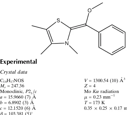
(
Z
)-2-[Methoxy(phenyl)methylidene]-3,4,5-trimethyl-2,3-dihydro-1,3-thiazole
Biplab Maji, Herbert Mayr and Peter Mayer*
Ludwig-Maximilians-Universita¨t, Department, Butenandtstrasse 5–13, 81377 Mu¨nchen, Germany
Correspondence e-mail: pemay@cup.uni-muenchen.de
Received 20 July 2012; accepted 31 July 2012
Key indicators: single-crystal X-ray study;T= 173 K; mean(C–C) = 0.002 A˚;
Rfactor = 0.036;wRfactor = 0.104; data-to-parameter ratio = 16.7.
In the title compound, C14H17NOS, the plane defined by the bridging methylene C atom and its three substituents makes dihedral angles of 14.37 (8) with the heterocycle and
26.17 (8) with the phenyl ring, while the dihedral angle
between the heterocycle and the phenyl ring is 36.29 (7). In
the crystal, molecules are linked by C—H contacts.
Related literature
For chemical background, see: Ukaiet al.(1943); Enderset al. (2007); Biju et al. (2011); Breslow (1958). For a related structure, see: Reisseret al.(2003).
Experimental
Crystal data
C14H17NOS Mr= 247.36
Monoclinic,P21=c a= 15.9660 (7) A˚ b= 6.8902 (3) A˚ c= 12.1520 (6) A˚
= 103.381 (5)
V= 1300.54 (10) A˚3 Z= 4
MoKradiation
= 0.23 mm1 T= 173 K
0.350.250.17 mm
Data collection
Oxford Diffraction Xcalibur diffractometer
Absorption correction: multi-scan (CrysAlis PRO; Oxford Diffraction, 2009) Tmin= 0.953,Tmax= 1.000
9041 measured reflections 2637 independent reflections 2023 reflections withI> 2(I) Rint= 0.029
Refinement
R[F2> 2(F2)] = 0.036 wR(F2) = 0.104 S= 1.08 2637 reflections
158 parameters
H-atom parameters constrained
max= 0.29 e A˚
3
min=0.19 e A˚
[image:1.610.45.248.456.641.2]3
Table 1
Hydrogen-bond geometry (A˚ ,).
Cg1 is the centroid of the C5–C10 ring.
D—H A D—H H A D A D—H A
C8—H8 Cg1i
0.95 2.83 3.6324 (16) 142 C13—H13B Cg1ii
0.98 2.74 3.5657 (15) 143
Symmetry codes: (i)x;y1 2;zþ
1
2; (ii)x;yþ1;z.
Data collection: CrysAlis PRO(Oxford Diffraction, 2009); cell refinement: CrysAlis PRO; data reduction: CrysAlis PRO; program(s) used to solve structure:SIR99 (Altomare et al., 1999); program(s) used to refine structure:SHELXL97(Sheldrick, 2008); molecular graphics:ORTEP-3(Farrugia, 1997) andPLATON(Spek, 2009); software used to prepare material for publication: SHELXL97.
The authors thank Prof. Thomas M. Klapo¨tke for generous allocation of diffractometer time.
Supplementary data and figures for this paper are available from the IUCr electronic archives (Reference: SU2484).
References
Altomare, A., Burla, M. C., Camalli, M., Cascarano, G. L., Giacovazzo, C., Guagliardi, A., Moliterni, A. G. G., Polidori, G. & Spagna, R. (1999).J. Appl. Cryst.32, 115–119.
Biju, A. T., Kuhl, N. & Glorius, F. (2011).Acc. Chem. Res.44, 1182–1195. Breslow, R. (1958).J. Am. Chem. Soc.80, 3719–3726.
Enders, D., Niemeier, O. & Henseler, A. (2007).Chem. Rev.107, 5606–5655. Farrugia, L. J. (1997).J. Appl. Cryst.30, 565.
Oxford Diffraction (2009).CrysAlis PRO. Oxford Diffraction Ltd, Yarnton, England.
Reisser, M., Maier, A. & Maas, G. (2003).Eur. J. Org. Chem.pp. 2071–2079. Sheldrick, G. M. (2008).Acta Cryst.A64, 112–122.
Spek, A. L. (2009).Acta Cryst.D65, 148–155.
Ukai, T., Tanaka, R. & Dokawa, T. (1943).J. Pharm. Soc. Jpn,63, 296–300.
Acta Crystallographica Section E Structure Reports
Online
supporting information
Acta Cryst. (2012). E68, o2644 [doi:10.1107/S1600536812034137]
(Z)-2-[Methoxy(phenyl)methylidene]-3,4,5-trimethyl-2,3-dihydro-1,3-thiazole
Biplab Maji, Herbert Mayr and Peter Mayer
S1. Comment
Thiazolium ions (Ukai et al., 1943) are known to catalyze benzoin condensations of aldehydes (Enders et al., 2007; Biju
et al., 2011) in presence of a base. An acyl anion equivalent, the so-called Breslow intermediate (Breslow, 1958) was
proposed to be the key intermediate of these transformations. To understand the structure of these intermediates we now
report the X-ray analysis of the O-methyl-protected Breslow intermediate derived from 3,4,5-trimethylthiazolium ion and
benzaldehyde.
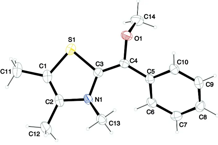
The molecular structure of the title compound is shown in Fig. 1. The exocyclic double bond has a length of 1.349 (2) Å
which is comparable to that observed for a related structure [1.353 Å; Reisser et al., 2003]. The endocyclic double bond
length is 1.330 (2) Å [1.332 Å; Reisser et al., 2003]. The angle sum around the methylene carbon atom, C4, amounts to
360° resulting in a trigonal planar environment of the methylene atom. However, this mean plane (C4/O1/C3/C5) is not
coplanar with either the plane of the heterocycle (S1/N1/C1-C3) or the plane of the phenyl ring (C5-C10). The
corresponding dihedral angles are 14.37 (8)° and 26.17 (8)°, respectively. The dihedral angle between the heterocycle and
the phenyl ring is 36.29 (7)°.
In the crystal, molecules are linked via C–H···π contacts (Table 1 and Fig. 2).
S2. Experimental
A solution of 2-(methoxy(phenyl)methyl)-3,4,5-trimethylthiazolium trifluoromethanesulfonate (397 mg, 1.00 mmol) in
THF (6 ml) was added dropwise to a stirred suspension of NaH (36 mg, 1.5 mmol) in dry THF (5 ml) at -20 °C under
nitrogen, and the reaction mixture was allowed to stir for 36 h in the dark. After warming to room temperature, the
solvent was removed under vacuum, and the residue was suspended in dry toluene (20 ml) and filtered through a celite
pad under nitrogen. Then the solvent was evaporated to give 205 mg (0.829 mmol, 83%) of the title compound as 2:1
mixture of Z:E isomers. Crystals of the title compound suitable for X-ray diffraction analysis were grown by slow
evaporation of a solution in n-pentane under nitrogen.
S3. Refinement
The C-bound H atoms were included in calculated positions and treated as riding atoms: C-H = 0.95 and 0.98 Å for CH
and CH3 -atoms, respectively, with Uiso(H) = k × Ueq(C), where k = 1.5 for CH3 H atoms and = 1.2 for other H atoms. The
Figure 1
The molecular structure of the title molecule, with atom labelling. Displacement ellipsoids are drawn at 50% probability
level.
Figure 2
The crystal packing of the title compound viewed along the b axis.
(Z)-2-[Methoxy(phenyl)methylidene]-3,4,5-trimethyl-2,3-dihydro- 1,3-thiazole
Crystal data
C14H17NOS
Mr = 247.36
Monoclinic, P21/c
Hall symbol: -P 2ybc
a = 15.9660 (7) Å
[image:3.610.128.479.365.584.2]c = 12.1520 (6) Å
β = 103.381 (5)°
V = 1300.54 (10) Å3
Z = 4
F(000) = 528
Dx = 1.263 (1) Mg m−3
Mo Kα radiation, λ = 0.71073 Å
Cell parameters from 4082 reflections
θ = 4.5–26.2°
µ = 0.23 mm−1
T = 173 K
Block, yellow
0.35 × 0.25 × 0.17 mm
Data collection
Oxford Diffraction Xcalibur diffractometer
Radiation source: fine-focus sealed tube Graphite monochromator
Detector resolution: 15.9809 pixels mm-1
ω scans
Absorption correction: multi-scan
(CrysAlis PRO; Oxford Diffraction, 2009)
Tmin = 0.953, Tmax = 1.000
9041 measured reflections 2637 independent reflections 2023 reflections with I > 2σ(I)
Rint = 0.029
θmax = 26.3°, θmin = 4.5°
h = −19→18
k = −7→8
l = −13→15
Refinement
Refinement on F2
Least-squares matrix: full
R[F2 > 2σ(F2)] = 0.036
wR(F2) = 0.104
S = 1.08
2637 reflections 158 parameters 0 restraints
Primary atom site location: structure-invariant direct methods
Secondary atom site location: difference Fourier map
Hydrogen site location: inferred from neighbouring sites
H-atom parameters constrained
w = 1/[σ2(F
o2) + (0.0601P)2] where P = (Fo2 + 2Fc2)/3 (Δ/σ)max < 0.001
Δρmax = 0.29 e Å−3
Δρmin = −0.19 e Å−3
Special details
Geometry. All e.s.d.'s (except the e.s.d. in the dihedral angle between two l.s. planes) are estimated using the full covariance matrix. The cell e.s.d.'s are taken into account individually in the estimation of e.s.d.'s in distances, angles and torsion angles; correlations between e.s.d.'s in cell parameters are only used when they are defined by crystal symmetry. An approximate (isotropic) treatment of cell e.s.d.'s is used for estimating e.s.d.'s involving l.s. planes.
Refinement. Refinement of F2 against ALL reflections. The weighted R-factor wR and goodness of fit S are based on F2,
conventional R-factors R are based on F, with F set to zero for negative F2. The threshold expression of F2 > 2σ(F2) is
used only for calculating R-factors(gt) etc. and is not relevant to the choice of reflections for refinement. R-factors based
on F2 are statistically about twice as large as those based on F, and R-factors based on all data will be even larger.
Fractional atomic coordinates and isotropic or equivalent isotropic displacement parameters (Å2)
x y z Uiso*/Ueq
S1 0.39851 (3) 0.51962 (7) 0.17439 (4) 0.03514 (16)
O1 0.33687 (7) 0.25112 (16) 0.31574 (8) 0.0298 (3)
N1 0.23682 (8) 0.61091 (19) 0.10461 (10) 0.0248 (3)
C1 0.36863 (11) 0.7059 (3) 0.07369 (13) 0.0314 (4)
C2 0.28401 (11) 0.7353 (2) 0.04813 (12) 0.0282 (4)
C3 0.28957 (10) 0.4751 (2) 0.17383 (12) 0.0229 (3)
C4 0.26767 (9) 0.3291 (2) 0.23561 (12) 0.0227 (3)
C5 0.18289 (9) 0.2433 (2) 0.22982 (12) 0.0213 (3)
H6 0.1286 0.3031 0.0642 0.030*
C7 0.04027 (10) 0.1508 (2) 0.12457 (13) 0.0271 (4)
H7 −0.0027 0.1525 0.0559 0.033*
C8 0.02412 (10) 0.0577 (2) 0.21848 (14) 0.0297 (4)
H8 −0.0297 −0.0040 0.2149 0.036*
C9 0.08769 (11) 0.0561 (2) 0.31769 (13) 0.0294 (4)
H9 0.0775 −0.0084 0.3824 0.035*
C10 0.16563 (10) 0.1466 (2) 0.32384 (12) 0.0252 (3)
H10 0.2083 0.1435 0.3928 0.030*
C11 0.43748 (12) 0.8120 (3) 0.03234 (16) 0.0437 (5)
H11A 0.4110 0.9126 −0.0216 0.065*
H11B 0.4689 0.7204 −0.0049 0.065*
H11C 0.4775 0.8725 0.0966 0.065*
C12 0.23515 (12) 0.8848 (3) −0.03065 (15) 0.0425 (5)
H12A 0.2160 0.9881 0.0133 0.064*
H12B 0.1849 0.8242 −0.0806 0.064*
H12C 0.2725 0.9401 −0.0762 0.064*
C13 0.15990 (10) 0.6830 (2) 0.13668 (13) 0.0288 (4)
H13A 0.1086 0.6296 0.0853 0.043*
H13B 0.1585 0.8250 0.1319 0.043*
H13C 0.1610 0.6429 0.2144 0.043*
C14 0.35969 (12) 0.0583 (3) 0.28898 (16) 0.0424 (5)
H14A 0.3103 −0.0282 0.2843 0.064*
H14B 0.4082 0.0121 0.3482 0.064*
H14C 0.3762 0.0594 0.2162 0.064*
Atomic displacement parameters (Å2)
U11 U22 U33 U12 U13 U23
S1 0.0248 (2) 0.0356 (3) 0.0462 (3) −0.00279 (18) 0.01062 (19) 0.00815 (19)
O1 0.0287 (6) 0.0251 (7) 0.0319 (6) 0.0017 (5) −0.0005 (5) 0.0051 (5)
N1 0.0293 (7) 0.0198 (7) 0.0265 (7) 0.0006 (6) 0.0094 (5) 0.0047 (5)
C1 0.0376 (10) 0.0279 (9) 0.0311 (9) −0.0085 (8) 0.0129 (7) −0.0003 (7)
C2 0.0385 (10) 0.0235 (9) 0.0241 (8) −0.0066 (7) 0.0105 (7) 0.0006 (6)
C3 0.0223 (8) 0.0220 (8) 0.0247 (8) 0.0002 (6) 0.0061 (6) −0.0015 (6)
C4 0.0235 (8) 0.0203 (8) 0.0237 (8) 0.0030 (6) 0.0040 (6) 0.0014 (6)
C5 0.0249 (8) 0.0150 (8) 0.0247 (8) 0.0017 (6) 0.0074 (6) −0.0015 (6)
C6 0.0311 (9) 0.0179 (8) 0.0261 (8) 0.0042 (7) 0.0091 (6) 0.0005 (6)
C7 0.0268 (8) 0.0195 (8) 0.0326 (9) 0.0032 (7) 0.0019 (7) −0.0051 (6)
C8 0.0269 (9) 0.0205 (9) 0.0437 (10) −0.0023 (7) 0.0119 (7) −0.0036 (7)
C9 0.0379 (10) 0.0217 (9) 0.0324 (9) −0.0027 (7) 0.0161 (7) 0.0013 (7)
C10 0.0311 (9) 0.0211 (8) 0.0237 (8) −0.0003 (7) 0.0070 (6) 0.0000 (6)
C11 0.0455 (11) 0.0442 (12) 0.0455 (10) −0.0170 (9) 0.0192 (9) 0.0017 (9)
C12 0.0496 (11) 0.0405 (12) 0.0375 (10) −0.0028 (9) 0.0103 (8) 0.0157 (8)
C13 0.0321 (9) 0.0225 (9) 0.0330 (9) 0.0036 (7) 0.0101 (7) 0.0026 (7)
Geometric parameters (Å, º)
S1—C1 1.7614 (17) C8—C9 1.385 (2)
S1—C3 1.7646 (16) C8—H8 0.9500
O1—C4 1.3999 (17) C9—C10 1.378 (2)
O1—C14 1.435 (2) C9—H9 0.9500
N1—C3 1.402 (2) C10—H10 0.9500
N1—C2 1.4186 (19) C11—H11A 0.9800
N1—C13 1.4589 (19) C11—H11B 0.9800
C1—C2 1.330 (2) C11—H11C 0.9800
C1—C11 1.500 (2) C12—H12A 0.9800
C2—C12 1.497 (2) C12—H12B 0.9800
C3—C4 1.349 (2) C12—H12C 0.9800
C4—C5 1.464 (2) C13—H13A 0.9800
C5—C6 1.399 (2) C13—H13B 0.9800
C5—C10 1.404 (2) C13—H13C 0.9800
C6—C7 1.383 (2) C14—H14A 0.9800
C6—H6 0.9500 C14—H14B 0.9800
C7—C8 1.384 (2) C14—H14C 0.9800
C7—H7 0.9500
C1—S1—C3 90.90 (8) C10—C9—H9 119.5
C4—O1—C14 113.50 (12) C8—C9—H9 119.5
C3—N1—C2 112.39 (13) C9—C10—C5 121.03 (14)
C3—N1—C13 119.55 (12) C9—C10—H10 119.5
C2—N1—C13 119.85 (13) C5—C10—H10 119.5
C2—C1—C11 129.13 (16) C1—C11—H11A 109.5
C2—C1—S1 111.73 (12) C1—C11—H11B 109.5
C11—C1—S1 119.09 (13) H11A—C11—H11B 109.5
C1—C2—N1 114.82 (15) C1—C11—H11C 109.5
C1—C2—C12 127.22 (15) H11A—C11—H11C 109.5
N1—C2—C12 117.94 (15) H11B—C11—H11C 109.5
C4—C3—N1 129.40 (14) C2—C12—H12A 109.5
C4—C3—S1 120.65 (12) C2—C12—H12B 109.5
N1—C3—S1 109.95 (11) H12A—C12—H12B 109.5
C3—C4—O1 114.14 (13) C2—C12—H12C 109.5
C3—C4—C5 129.08 (13) H12A—C12—H12C 109.5
O1—C4—C5 116.78 (12) H12B—C12—H12C 109.5
C6—C5—C10 117.31 (14) N1—C13—H13A 109.5
C6—C5—C4 122.12 (13) N1—C13—H13B 109.5
C10—C5—C4 120.40 (13) H13A—C13—H13B 109.5
C7—C6—C5 121.24 (14) N1—C13—H13C 109.5
C7—C6—H6 119.4 H13A—C13—H13C 109.5
C5—C6—H6 119.4 H13B—C13—H13C 109.5
C6—C7—C8 120.62 (14) O1—C14—H14A 109.5
C6—C7—H7 119.7 O1—C14—H14B 109.5
C8—C7—H7 119.7 H14A—C14—H14B 109.5
C7—C8—H8 120.6 H14A—C14—H14C 109.5
C9—C8—H8 120.6 H14B—C14—H14C 109.5
C10—C9—C8 120.94 (14)
C3—S1—C1—C2 3.05 (13) S1—C3—C4—O1 13.63 (19)
C3—S1—C1—C11 −179.13 (14) N1—C3—C4—C5 14.8 (3)
C11—C1—C2—N1 −178.57 (16) S1—C3—C4—C5 −166.26 (12)
S1—C1—C2—N1 −1.02 (18) C14—O1—C4—C3 −110.21 (16)
C11—C1—C2—C12 0.3 (3) C14—O1—C4—C5 69.70 (17)
S1—C1—C2—C12 177.82 (15) C3—C4—C5—C6 28.4 (2)
C3—N1—C2—C1 −2.3 (2) O1—C4—C5—C6 −151.44 (14)
C13—N1—C2—C1 146.04 (15) C3—C4—C5—C10 −156.27 (16)
C3—N1—C2—C12 178.72 (14) O1—C4—C5—C10 23.8 (2)
C13—N1—C2—C12 −32.9 (2) C10—C5—C6—C7 1.3 (2)
C2—N1—C3—C4 −176.49 (15) C4—C5—C6—C7 176.69 (14)
C13—N1—C3—C4 35.0 (2) C5—C6—C7—C8 −0.7 (2)
C2—N1—C3—S1 4.49 (15) C6—C7—C8—C9 −0.2 (2)
C13—N1—C3—S1 −143.98 (12) C7—C8—C9—C10 0.6 (2)
C1—S1—C3—C4 176.64 (13) C8—C9—C10—C5 0.0 (2)
C1—S1—C3—N1 −4.25 (11) C6—C5—C10—C9 −0.9 (2)
N1—C3—C4—O1 −165.29 (14) C4—C5—C10—C9 −176.45 (14)
Hydrogen-bond geometry (Å, º)
Cg1 is the centroid of the C5–C10 ring.
D—H···A D—H H···A D···A D—H···A
C8—H8···Cg1i 0.95 2.83 3.6324 (16) 142
C13—H13B···Cg1ii 0.98 2.74 3.5657 (15) 143