research communications
Acta Cryst.(2018). E74, 1609–1612 https://doi.org/10.1107/S2056989018014238
1609
Received 1 October 2018Accepted 8 October 2018
Edited by M. Weil, Vienna University of Technology, Austria
Keywords:crystal structure; picket fence porphyrin; zinc(II) porphyrin; atropisomer; iso-thiocyanate.
CCDC reference:1872076
Supporting information:this article has supporting information at journals.iucr.org/e
Crystal structure of (diethyl ether-
j
O
)[5,10,15,20-
tetrakis(2-isothiocyanatophenyl)porphyrinato-j
4N
]zinc diethyl ether solvate
Lisa Leben,aEike Schaub,aChristian Na¨therb and Rainer Hergesa*
a
Institut fu¨r Organische Chemie, Universita¨t Kiel, Otto-Hahn-Platz 4, 24118, Kiel, Germany, andbInstitut fu¨r Anorganische Chemie, Universita¨t Kiel, Otto-Hahn-Platz 6/7, 24118, Kiel, Germany. *Correspondence e-mail: rherges@oc.uni-kiel.de
The crystal structure of the title compound, [Zn(C48H24N8S4)(C4H10O)]C4H10O,
consists of discrete porphyrin complexes that are located on a twofold rotation axis. The ZnIIcation is fivefold coordinated by four N atoms of the porphyrin moiety and one O atom of a diethyl ether molecule in a slightly distorted square-pyramidal environment with the diethyl ether molecule in the apical position. The porphyrin backbone is nearly planar with the metal cation slightly shifted out of the plane towards the coordinating diethyl ether molecule. All four isothiocyanato groups of the phenyl substituents at themeso-positions face the same side of the porphyrin, as is characteristic for picket fence porphyrins. In the crystal structure, the discrete porphyrin complexes are arranged in such a way that cavities are formed in which additional diethyl ether solvate molecules are located around a twofold rotation axis. The O atom of the solvent molecule is not positioned exactly on the twofold rotation axis, thus making the whole molecule equally disordered over two symmetry-related positions.
1. Chemical context
Isothiocyanates serve as versatile starting materials for a variety of functional groups (Batey & Powell, 2000; Dinget al., 2011; Serra et al., 2014; Guo et al., 2010; Shin et al., 2000; Kosurkaret al., 2014; Alizadehet al., 2016; Raoet al., 2015). Included in porphyrin scaffolds, isothiocyanates may serve as precursors for the synthesis of tetratopic ligands with fourfold symmetry. In the case where all fourortho-substituents of the
meso-phenyl groups face the same side of the porphyrin plane, these porphyrins are denominated picket fence porphyrins. These compounds are widely used as model compounds for hemoproteins (Collman et al., 1975; Tabushi et al., 1985; Schappacheret al., 1989). With a bulkyortho-substituent and ZnII as the central metal cation, the rotational barriers are
sufficiently high to isolate the different atropisomers (Freitag & Whitten, 1983). A variety of picket fence porphyrins has been reported (Collman et al., 1975; Mansour et al., 2017; Cormodeet al., 2006; Le Mauxet al., 1993; Wuenschellet al., 1992). In most cases, amides are used as functional groups in theortho-positions of themeso-phenyl groups, which hampers further functionalization. The title compound now opens new avenues for the synthesis of functionalized picket fence porphyrins and is a promising starting material for the design of anion binding ligands. The title compound can be obtained in one step using a method reported by Jha et al. (Fig. 1), starting from the all-isomer of the amino derivative we have
published previously (Jhaet al., 2007; Lebenet al., 2018). It is important to note that the reaction has to be carried out at 273 K, because at room temperature a mixture of the atrop-isomers is obtained. After dissolving the tetrakis(isothio-cyanatophenyl) porphyrin in acetone and precipitating with diethyl ether, single crystals were obtained, which were char-acterized by single crystal X-ray diffraction.
2. Structural commentary
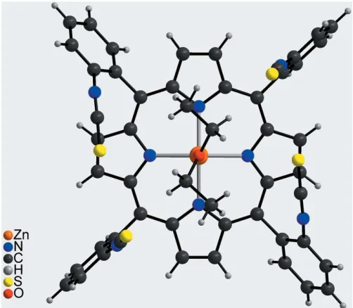
The asymmetric unit of the title compound, Zn(C48H24N8S4)(C4H10O)C4H10O, comprises one Zn
II
cation, one half of the porphyrin molecule and one half of a coordi-nating diethyl ether molecule as well as one half of a diethyl ether solvate molecule. The complex porphyrin molecule and the coordinating diethyl ether molecule are located on a twofold rotation axis whereas the solvent diethyl ether mol-ecule is in a general position and is equally disordered around a twofold rotation axis (Fig. 2). The four isothiocyanate substituents of the phenyl groups at themeso-positions point to the same side of the porphyrin moiety, which proves that the tetra-isomer has formed. The porphyrin plane is close to planar with a maximum deviation from the mean plane of 0.276 (3) A˚ . The phenyl rings are rotated out of the porphyrin plane by 63.16 (5) and 82.06 (6). The ZnII
cation is fivefold coordinated by the four N atoms of the porphyrin molecule in the basal positions and by one O atom of a diethyl ether
molecule in the apical position, leading to a distorted square-pyramidal coordination environment (Table 1, Fig. 3). The Zn—N distances of 2.0622 (13) and 2.0684 (14) A˚ and the Zn—O distance of 2.1352 (19) A˚ are in characteristic ranges. The angles around the ZnII cation range from 88.54 (6) to 99.69 (4) for the basal N
4 plane and from 160.61 (8) to
164.44 (8) involving the apical O atom, demonstrating that
the square pyramid is slightly distorted (Table 1). The ZnII cation is located 0.4052 (9) A˚ out of the mean porphyrin plane and is shifted towards the coordinating diethyl ether molecule (Fig. 4).
3. Supramolecular features
In the crystal structure of the title compound, each two discrete complexes form centrosymmetric pairs with the coordinating diethyl ether molecules pointing in opposite directions (Fig. 5). The complexes are arranged into columns along [001]. This arrangement leads to the formation of cavities between two neighbouring coordinating diethyl ether
1610
Lebenet al. [Zn(C [image:2.610.313.566.80.180.2]48H24N8S4)(C4H10O)]C4H10O Acta Cryst.(2018). E74, 1609–1612
research communications
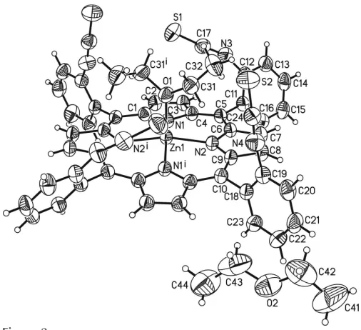
Figure 2
The molecular entities of the title compound with the atom labelling and displacement ellipsoids drawn at the 50% probability level. Only one orientation of the disordered diethyl ether solvent is given. [Symmetry code: (i)x+ 2,y,z+3
[image:2.610.42.298.161.369.2] [image:2.610.313.566.475.706.2]2.] Table 1
Selected geometric parameters (A˚ ,).
Zn1—N2 2.0622 (13) Zn1—N1 2.0685 (14) Zn1—N2i 2.0622 (13) Zn1—O1 2.1352 (19)
Zn1—N1i 2.0684 (14)
N2—Zn1—N2i 164.44 (8) N1i—Zn1—N1 160.61 (8)
N2—Zn1—N1i 88.85 (6) N2i—Zn1—O1 97.78 (4)
N2i—Zn1—N1i 88.54 (6) N1i—Zn1—O1 99.69 (4)
N2—Zn1—N1 88.54 (6) N1—Zn1—O1 99.69 (4) N2i—Zn1—N1 88.85 (6)
Symmetry code: (i)xþ1;y;zþ3 2.
Figure 1
[image:2.610.44.299.640.731.2]molecules, in which the disordered diethyl ether solvate mol-ecules are embedded (Fig. 5). There are no notable inter-molecular interactions between the inter-molecular moieties in the crystal structure.
4. Database survey
The synthesis of the metal-free oxygen derivative 5,10,15,20-tetrakis ,,, 2-isocyanatophenyl porphyrin has been known for several years (Collmanet al., 1998). However, the crystal structure of this compound has not yet been reported. A CSD database search (Version 5.39; Groom et al., 2016) revealed the crystal structures of several metal porphyrins with isothiocyanate entities as axial ligands (Dhifetet al., 2010; Scheidtet al., 1982; Ezzayaniet al., 2014; Dendenet al., 2015). In addition, the crystal structure of a para
-isothiocyanato-phenyl porphyrin has been reported (Sibrian-Vazquezet al., 2005).
5. Synthesis and crystallization
The metal-free all-isomer of 2-aminophenyl porphyrin was synthesized according to reported procedures (Collmanet al., 1975; Lindsey, 1980). Metallation followed standard metalla-tion condimetalla-tions as reported previously (Strohmeieret al., 1997; Lebenet al., 2018). For the introduction of the isothiocyanato groups, a modified synthesis was used (Jha et al., 2007). 5,10,15,20-Tetrakis(,,, 2-aminophenyl)zinc(II) porphyrin (150 mg, 203mmol) was dissolved in 30 ml of dichloromethane and cooled to 273 K. 1,10-Thiocarbonyldi-2,20-pyridone (TDP,
377 mg, 1.62 mmol) was added and the mixture stirred for 50 minutes at 273 K. Removing the solvent and filtration over silica gel (cyclohexane/ethyl acetate,v:v= 1:1) gave the title compound in quantitative yield. For crystallization, a small amount was dissolved in acetone and crystallized by adding diethyl ether.
1
H NMR (500 MHz, CDCl3, 300 K):= 8.80 (s, 8H, H-),
8.21 (dd,3J= 7.5 Hz,4J= 1.2 Hz, 4H, H-6), 7.78 (dt,3J= 7.9 Hz,
4
J= 1.5 Hz, 4H, H-4), 7.68 (dt,3J= 7.6 Hz,4J= 1.3 Hz, 4H, H-5), 7.61 (dd,3J= 8.2 Hz,4J= 1.0 Hz, 4H, H-3) ppm.13C NMR (125 MHz, CDCl3, 300 K):= 149.9 (C-), 141.0 (C1), 134.8
(C6), 134.5 (C2), 131.6 (C-), 129.3 (C4), 125.7 (C5), 124.4 (C3), 115.7 (C-meso) ppm. EI–MS (70 eV): m/z (%) = 904.1 (100) [M]+.
6. Refinement
Crystal data, data collection and structure refinement details are summarized in Table 2. The C—H hydrogen atoms were positioned with idealized geometries (C—H = 0.95–0.99 A˚ ; methyl H atoms of the coordinating diethyl ether molecule were allowed to rotate but not to tip) and were refined with
research communications
Acta Cryst.(2018). E74, 1609–1612 Lebenet al. [Zn(C
[image:3.610.45.296.71.290.2]48H24N8S4)(C4H10O)]C4H10O
1611
Figure 4
[image:3.610.315.566.523.723.2]Molecular structure of the discrete complex in a view parallel to the porphyrin plane.
Figure 5
Crystal structure of the title compound viewed along [001].
Figure 3
[image:3.610.46.294.563.716.2]Uiso(H) = 1.2Ueq(C) (1.5 for methyl H atoms) using a riding model. The O atom of the diethyl ether solvate molecule is not located exactly on the twofold rotation axis and thus the complete molecule is equally disordered over two sets of sites because of symmetry. Therefore for each atom the occupancy was set to 0.5, and atoms were treated with SADI and SIMU commands (Sheldrick, 2015b) to achieve similar displacement ellipsoids.
Acknowledgements
We thank Professor Dr Wolfgang Bensch for access to his experimental facility.
Funding information
The authors gratefully acknowledge financial support by the Deutsche Forschungsgemeinschaft within the Sonder-forschungsbereich 677.
References
Alizadeh, A., Bagherinejad, A., Bayat, F. & Zhu, L.-G. (2016). Tetrahedron,72, 7070–7075.
Batey, R. A. & Powell, D. A. (2000).J. Am. Chem. Soc.2, 3237–3240. Brandenburg, K. (2014).DIAMOND. Crystal Impact GbR, Bonn,
Germany.
Collman, J. P., Gagne, R. R., Reed, C., Halbert, T. R., Lang, G. & Robinson, W. T. (1975).J. Am. Chem. Soc.97, 1427–1439. Collman, J. P., Wang, Z. & Straumanis, A. (1998).J. Org. Chem.63,
2424–2425.
Cormode, D. P., Murray, S. S., Cowley, A. R. & Beer, P. D. (2006). Dalton Trans.pp. 5135–5140.
Denden, Z., Ezzayani, K., Saint-Aman, E., Loiseau, F., Najmudin, S., Bonifa´cio, C., Daran, J.-C. & Nasri, H. (2015).Eur. J. Inorg. Chem.
2015, 2596–2610.
Dhifet, M., Belkhiria, M. S., Daran, J.-C., Schulz, C. E. & Nasri, H. (2010).Inorg. Chim. Acta,363, 3208–3213.
Ding, Q., Liu, X., Cao, B., Zong, Z. & Peng, Y. (2011).Tetrahedron Lett.52, 1964–1967.
Ezzayani, K., Denden, Z., Najmudin, S., Bonifa´cio, C., Saint-Aman, E., Loiseau, F. & Nasri, H. (2014).Eur. J. Inorg. Chem.2014, 5348– 5361.
Freitag, R. A. & Whitten, D. G. (1983).J. Phys. Chem.87, 3918–3925. Groom, C. R., Bruno, I. J., Lightfoot, M. P. & Ward, S. C. (2016).Acta
Cryst.B72, 171–179.
Guo, Y.-J., Tang, R.-Y., Zhong, P. & Li, J.-H. (2010).Tetrahedron Lett.
51, 649–652.
Jha, S. C., Lorch, M., Lewis, R. A., Archibald, S. J. & Boyle, R. W. (2007).Org. Biomol. Chem.5, 1970–1974.
Kosurkar, U. B., Dadmal, T. L., Appalanaidu, K., Khageswara Rao, Y., Nanubolu, J. B. & Kumbhare, R. M. (2014).Tetrahedron Lett.
55, 1296–1298.
Leben, L., Na¨ther, C. & Herges, R. (2018). Acta Cryst.E74, 1285– 1289.
Le Maux, P., Bahri, H. & Simonneaux, G. (1993). Tetrahedron,49, 1401–1408.
Lindsey, J. (1980).J. Org. Chem.45, 5215.
Mansour, A., Zaied, M., Ali, I., Soliman, S. & Othmani, M. (2017). Polyhedron,127, 496–504.
Rao, D. S., Madhava, G., Rasheed, S., Thahir Basha, S., Lakshmi Devamma, M. N. & Naga Raju, C. (2015). Phosphorus Sulfur Silicon,190, 574–584.
Schappacher, M., Ricard, L., Fischer, J., Weiss, R., Montiel-Montoya, R., Bill, E. & Trautwein, A. X. (1989).Inorg. Chem.28, 4639–4645. Scheidt, W. R., Lee, Y. J., Geiger, D. K., Taylor, K. & Hatano, K.
(1982).J. Am. Chem. Soc.104, 3367–3374.
Serra, S., Moineaux, L., Vancraeynest, C., Masereel, B., Wouters, J., Pochet, L. & Fre´de´rick, R. (2014).Eur. J. Med. Chem.82, 96–105. Sheldrick, G. M. (2008).Acta Cryst.A64, 112–122.
Sheldrick, G. M. (2015a).Acta Cryst.A71, 3–8. Sheldrick, G. M. (2015b).Acta Cryst.C71, 3–8.
Shin, K. J., Koo, K. D., Yoo, K. H., Kim, D. C., Kim, D. J. & Park, S. W. (2000).Bioorg. Med. Chem. Lett.10, 1421–1425.
Sibrian-Vazquez, M., Jensen, T. J., Fronczek, F. R., Hammer, R. P. & Vicente, M. G. H. (2005).Bioconjugate Chem.16, 852–863. Stoe (2008). X-AREA, X-RED and X-SHAPE. Stoe & Cie,
Darmstadt, Germany.
Strohmeier, M., Orendt, A. M., Facelli, J. C., Solum, M. S., Pugmire, R. J., Parry, R. W. & Grant, D. M. (1997).J. Am. Chem. Soc.119, 7114–7120.
Tabushi, I., Kodera, M. & Yokoyama, M. (1985).J. Am. Chem. Soc.
107, 4466–4473.
Westrip, S. P. (2010).J. Appl. Cryst.43, 920–925.
Wuenschell, G. E., Tetreau, C., Lavalette, D. & Reed, C. A. (1992).J. Am. Chem. Soc.114, 3346–3355.
1612
Lebenet al. [Zn(C [image:4.610.313.554.88.385.2]48H24N8S4)(C4H10O)]C4H10O Acta Cryst.(2018). E74, 1609–1612
research communications
Table 2
Experimental details.
Crystal data
Chemical formula [Zn(C48H24N8S4)(C4H10O)]
-C4H10O
Mr 1054.60
Crystal system, space group Monoclinic,C2/c
Temperature (K) 200
a,b,c(A˚ ) 19.8830 (4), 17.1781 (3), 14.8684 (3)
() 91.667 (1)
V(A˚3) 5076.18 (17)
Z 4
Radiation type MoK (mm1) 0.70
Crystal size (mm) 0.140.110.07 Data collection
Diffractometer Stoe IPDS2
Absorption correction Numerical (X-REDand X-SHAPE; Stoe, 2008)
Tmin,Tmax 0.807, 0.951
No. of measured, independent and observed [I> 2(I)] reflections
39705, 5530, 5042
Rint 0.039
(sin/)max(A˚
1) 0.639
Refinement
R[F2> 2(F2)],wR(F2),S 0.036, 0.103, 1.05
No. of reflections 5530 No. of parameters 346 No. of restraints 26
H-atom treatment H-atom parameters constrained
max, min(e A˚
3
) 0.39,0.35
Computer programs:X-AREA(Stoe, 2008),SHELXT(Sheldrick, 2015a),SHELXL2014
(Sheldrick, 2015b),XP(Sheldrick, 2008),DIAMOND(Brandenburg, 2014) andpublCIF
supporting information
sup-1 Acta Cryst. (2018). E74, 1609-1612
supporting information
Acta Cryst. (2018). E74, 1609-1612 [https://doi.org/10.1107/S2056989018014238]
Crystal structure of (diethyl ether-
κ
O
)[5,10,15,20-tetrakis(2-isothiocyanato-phenyl)porphyrinato-
κ
4N
]zinc diethyl ether solvate
Lisa Leben, Eike Schaub, Christian N
ä
ther and Rainer Herges
Computing details
Data collection: X-AREA (Stoe, 2008); cell refinement: X-AREA (Stoe, 2008); data reduction: X-AREA (Stoe, 2008); program(s) used to solve structure: SHELXT (Sheldrick, 2015a); program(s) used to refine structure: SHELXL2014
(Sheldrick, 2015b); molecular graphics: XP (Sheldrick, 2008) and DIAMOND (Brandenburg, 2014); software used to prepare material for publication: publCIF (Westrip, 2010).
(Diethyl ether-κO)[5,10,15,20-tetrakis(2-isothiocyanatophenyl)porphyrinato-κ4N]zinc diethyl ether solvate
Crystal data
[Zn(C48H24N8S4)(C4H10O)]·C4H10O Mr = 1054.60
Monoclinic, C2/c a = 19.8830 (4) Å
b = 17.1781 (3) Å
c = 14.8684 (3) Å
β = 91.667 (1)°
V = 5076.18 (17) Å3
Z = 4
F(000) = 2184
Dx = 1.380 Mg m−3
Mo Kα radiation, λ = 0.71073 Å Cell parameters from 39705 reflections
θ = 1.6–27.0°
µ = 0.70 mm−1 T = 200 K Block, red
0.14 × 0.11 × 0.07 mm
Data collection
Stoe IPDS-2 diffractometer
ω scans
Absorption correction: numerical (X-Red and X-Shape; Stoe, 2008)
Tmin = 0.807, Tmax = 0.951
39705 measured reflections
5530 independent reflections 5042 reflections with I > 2σ(I)
Rint = 0.039
θmax = 27.0°, θmin = 1.6° h = −25→25
k = −21→21
l = −18→18
Refinement
Refinement on F2
Least-squares matrix: full
R[F2 > 2σ(F2)] = 0.036 wR(F2) = 0.103 S = 1.05 5530 reflections 346 parameters 26 restraints
Hydrogen site location: mixed
H-atom parameters constrained
w = 1/[σ2(F
o2) + (0.0603P)2 + 2.7141P]
where P = (Fo2 + 2Fc2)/3
(Δ/σ)max = 0.001
Δρmax = 0.39 e Å−3
Δρmin = −0.35 e Å−3
Extinction correction: SHELXL, Fc*=kFc[1+0.001xFc2λ3/sin(2θ)]-1/4
supporting information
sup-2 Acta Cryst. (2018). E74, 1609-1612
Special details
Geometry. All esds (except the esd in the dihedral angle between two l.s. planes) are estimated using the full covariance matrix. The cell esds are taken into account individually in the estimation of esds in distances, angles and torsion angles; correlations between esds in cell parameters are only used when they are defined by crystal symmetry. An approximate (isotropic) treatment of cell esds is used for estimating esds involving l.s. planes.
Fractional atomic coordinates and isotropic or equivalent isotropic displacement parameters (Å2)
x y z Uiso*/Ueq Occ. (<1)
supporting information
sup-3 Acta Cryst. (2018). E74, 1609-1612
H23 0.7436 0.4946 0.6569 0.063* N4 0.73517 (9) 0.75053 (11) 0.62762 (13) 0.0587 (4) C24 0.72587 (9) 0.81716 (12) 0.63222 (14) 0.0510 (4) S2 0.71060 (3) 0.90666 (3) 0.63860 (5) 0.07015 (18) O1 0.5000 0.76596 (11) 0.7500 0.0511 (4) C31 0.54792 (10) 0.81107 (12) 0.70173 (15) 0.0555 (5) H31A 0.5892 0.7798 0.6939 0.067* H31B 0.5604 0.8578 0.7374 0.067* C32 0.52032 (14) 0.83590 (17) 0.61075 (17) 0.0761 (7) H32A 0.5068 0.7898 0.5759 0.114* H32B 0.5550 0.8644 0.5788 0.114* H32C 0.4812 0.8697 0.6184 0.114*
O2 0.9968 (9) 0.5749 (3) 0.7693 (6) 0.092 (3) 0.5 C41 1.0756 (8) 0.5710 (9) 0.6491 (11) 0.155 (6) 0.5 H41A 1.0997 0.6007 0.6059 0.233* 0.5 H41B 1.1070 0.5431 0.6873 0.233* 0.5 H41C 1.0464 0.5346 0.6182 0.233* 0.5 C42 1.0345 (7) 0.6237 (9) 0.7061 (9) 0.114 (4) 0.5 H42A 1.0636 0.6605 0.7364 0.136* 0.5 H42B 1.0035 0.6521 0.6679 0.136* 0.5 C43 0.9492 (8) 0.6115 (8) 0.8328 (10) 0.124 (5) 0.5 H43A 0.9091 0.6246 0.7988 0.148* 0.5 H43B 0.9660 0.6586 0.8600 0.148* 0.5 C44 0.9249 (6) 0.5580 (6) 0.8994 (8) 0.117 (3) 0.5 H44A 0.8920 0.5822 0.9363 0.176* 0.5 H44B 0.9064 0.5111 0.8736 0.176* 0.5 H44C 0.9642 0.5455 0.9356 0.176* 0.5
Atomic displacement parameters (Å2)
U11 U22 U33 U12 U13 U23
supporting information
sup-4 Acta Cryst. (2018). E74, 1609-1612
C15 0.0495 (10) 0.0705 (13) 0.0418 (9) −0.0087 (9) 0.0040 (7) −0.0132 (9) C16 0.0465 (9) 0.0516 (10) 0.0443 (9) −0.0033 (7) 0.0032 (7) −0.0037 (7) N3 0.0655 (10) 0.0604 (10) 0.0485 (9) 0.0124 (8) 0.0023 (7) −0.0006 (8) C17 0.0506 (10) 0.0545 (11) 0.0540 (11) 0.0066 (8) −0.0021 (8) 0.0003 (9) S1 0.0758 (4) 0.0729 (4) 0.0663 (4) 0.0059 (3) 0.0115 (3) −0.0197 (3) C18 0.0377 (8) 0.0522 (9) 0.0368 (8) −0.0008 (7) 0.0036 (6) −0.0005 (7) C19 0.0423 (9) 0.0526 (10) 0.0417 (8) −0.0045 (7) −0.0004 (7) −0.0002 (7) C20 0.0430 (9) 0.0734 (13) 0.0529 (10) −0.0128 (9) 0.0049 (8) 0.0062 (9) C21 0.0389 (9) 0.0876 (16) 0.0592 (12) 0.0013 (10) 0.0104 (8) 0.0032 (11) C22 0.0483 (10) 0.0702 (14) 0.0707 (13) 0.0137 (10) 0.0129 (9) 0.0031 (11) C23 0.0463 (10) 0.0548 (11) 0.0579 (11) 0.0035 (8) 0.0105 (8) 0.0038 (8) N4 0.0569 (10) 0.0526 (10) 0.0665 (11) −0.0068 (8) 0.0004 (8) 0.0012 (8) C24 0.0418 (9) 0.0569 (12) 0.0543 (10) −0.0058 (8) 0.0004 (7) 0.0018 (8) S2 0.0700 (4) 0.0529 (3) 0.0870 (4) 0.0033 (2) −0.0071 (3) −0.0033 (3) O1 0.0499 (10) 0.0455 (10) 0.0586 (11) 0.000 0.0160 (8) 0.000 C31 0.0510 (10) 0.0541 (11) 0.0620 (12) −0.0087 (8) 0.0112 (9) 0.0022 (9) C32 0.0862 (18) 0.0802 (16) 0.0623 (14) −0.0162 (14) 0.0082 (12) 0.0123 (12) O2 0.073 (3) 0.087 (2) 0.116 (8) 0.005 (3) −0.004 (7) −0.006 (3) C41 0.152 (10) 0.135 (10) 0.179 (13) 0.050 (8) −0.008 (10) −0.063 (9) C42 0.091 (7) 0.125 (8) 0.122 (9) −0.016 (5) −0.039 (6) 0.025 (7) C43 0.125 (11) 0.099 (8) 0.145 (13) 0.046 (7) −0.036 (9) −0.035 (8) C44 0.108 (6) 0.071 (5) 0.175 (11) −0.013 (4) 0.030 (7) −0.002 (6)
Geometric parameters (Å, º)
Zn1—N2 2.0622 (13) C20—C21 1.374 (3) Zn1—N2i 2.0622 (13) C20—H20 0.9500
Zn1—N1i 2.0684 (14) C21—C22 1.376 (3)
Zn1—N1 2.0685 (14) C21—H21 0.9500 Zn1—O1 2.1352 (19) C22—C23 1.387 (3) N1—C4 1.368 (2) C22—H22 0.9500 N1—C1 1.370 (2) C23—H23 0.9500 N2—C9 1.368 (2) N4—C24 1.162 (3) N2—C6 1.370 (2) C24—S2 1.571 (2) C1—C10i 1.397 (2) O1—C31 1.436 (2)
C1—C2 1.443 (2) O1—C31i 1.436 (2)
C2—C3 1.348 (2) C31—C32 1.507 (3) C2—H2 0.9500 C31—H31A 0.9900 C3—C4 1.440 (2) C31—H31B 0.9900 C3—H3 0.9500 C32—H32A 0.9800 C4—C5 1.401 (2) C32—H32B 0.9800 C5—C6 1.404 (2) C32—H32C 0.9800 C5—C11 1.494 (2) O2—C42ii 1.11 (2)
C6—C7 1.440 (2) O2—C42 1.479 (12) C7—C8 1.355 (3) O2—C43 1.495 (11) C7—H7 0.9500 O2—C41ii 1.912 (18)
C8—C9 1.440 (2) C41—C44ii 0.755 (17)
supporting information
sup-5 Acta Cryst. (2018). E74, 1609-1612
C9—C10 1.401 (2) C41—C42 1.499 (14) C10—C1i 1.397 (2) C41—O2ii 1.912 (18)
C10—C18 1.494 (2) C41—H41A 0.9600 C11—C16 1.391 (3) C41—H41B 0.9599 C11—C12 1.398 (3) C41—H41C 0.9600 C12—C13 1.390 (3) C42—C43ii 0.703 (16)
C12—N3 1.393 (2) C42—O2ii 1.11 (2)
C13—C14 1.370 (3) C42—C42ii 1.92 (3)
C13—H13 0.9500 C42—H42A 0.9601 C14—C15 1.384 (3) C42—H42B 0.9599 C14—H14 0.9500 C43—C42ii 0.703 (16)
C15—C16 1.385 (3) C43—C41ii 0.900 (18)
C15—H15 0.9500 C43—C44 1.446 (14) C16—H16 0.9500 C43—H43A 0.9599 N3—C17 1.170 (3) C43—H43B 0.9600 C17—S1 1.576 (2) C44—C41ii 0.755 (17)
C18—C23 1.389 (3) C44—H44A 0.9600 C18—C19 1.394 (3) C44—H44B 0.9600 C19—N4 1.392 (3) C44—H44C 0.9599 C19—C20 1.394 (3)
N2—Zn1—N2i 164.44 (8) C31—O1—Zn1 122.65 (11)
N2—Zn1—N1i 88.85 (6) C31i—O1—Zn1 122.65 (11)
N2i—Zn1—N1i 88.54 (6) O1—C31—C32 111.82 (17)
N2—Zn1—N1 88.54 (6) O1—C31—H31A 109.3 N2i—Zn1—N1 88.85 (6) C32—C31—H31A 109.3
N1i—Zn1—N1 160.61 (8) O1—C31—H31B 109.3
N2—Zn1—O1 97.78 (4) C32—C31—H31B 109.3 N2i—Zn1—O1 97.78 (4) H31A—C31—H31B 107.9
N1i—Zn1—O1 99.69 (4) C31—C32—H32A 109.5
N1—Zn1—O1 99.69 (4) C31—C32—H32B 109.5 C4—N1—C1 106.50 (14) H32A—C32—H32B 109.5 C4—N1—Zn1 126.59 (11) C31—C32—H32C 109.5 C1—N1—Zn1 126.82 (11) H32A—C32—H32C 109.5 C9—N2—C6 106.88 (13) H32B—C32—H32C 109.5 C9—N2—Zn1 126.26 (11) C42ii—O2—C42 94.7 (13)
C6—N2—Zn1 126.16 (11) C42ii—O2—C43 26.4 (9)
N1—C1—C10i 125.29 (15) C42—O2—C43 120.3 (9)
N1—C1—C2 109.66 (14) C42ii—O2—C41ii 51.6 (8)
C10i—C1—C2 125.05 (16) C42—O2—C41ii 146.0 (10)
C3—C2—C1 106.91 (15) C43—O2—C41ii 27.3 (8)
C3—C2—H2 126.5 C44ii—C41—C43ii 122 (3)
C1—C2—H2 126.5 C44ii—C41—C42 137 (3)
C2—C3—C4 107.20 (15) C43ii—C41—C42 18.1 (13)
C2—C3—H3 126.4 C44ii—C41—O2ii 129 (2)
C4—C3—H3 126.4 C43ii—C41—O2ii 49.6 (12)
N1—C4—C5 125.53 (15) C42—C41—O2ii 35.6 (7)
supporting information
sup-6 Acta Cryst. (2018). E74, 1609-1612
C5—C4—C3 124.76 (16) C43ii—C41—H41A 94.4
C4—C5—C6 125.25 (16) C42—C41—H41A 110.3 C4—C5—C11 117.74 (15) O2ii—C41—H41A 143.7
C6—C5—C11 116.99 (15) C44ii—C41—H41B 114.2
N2—C6—C5 125.30 (15) C43ii—C41—H41B 124.1
N2—C6—C7 109.32 (15) C42—C41—H41B 108.7 C5—C6—C7 125.28 (16) O2ii—C41—H41B 97.5
C8—C7—C6 107.28 (16) H41A—C41—H41B 109.5 C8—C7—H7 126.4 C44ii—C41—H41C 50.2
C6—C7—H7 126.4 C43ii—C41—H41C 108.5
C7—C8—C9 106.85 (15) C42—C41—H41C 109.4 C7—C8—H8 126.6 O2ii—C41—H41C 82.6
C9—C8—H8 126.6 H41A—C41—H41C 109.5 N2—C9—C10 125.61 (15) H41B—C41—H41C 109.5 N2—C9—C8 109.64 (15) C43ii—C42—O2ii 109 (2)
C10—C9—C8 124.73 (16) C43ii—C42—O2 127 (2)
C1i—C10—C9 125.76 (16) O2ii—C42—O2 20.8 (8)
C1i—C10—C18 116.90 (15) C43ii—C42—C41 23 (2)
C9—C10—C18 117.33 (15) O2ii—C42—C41 92.9 (12)
C16—C11—C12 117.55 (16) O2—C42—C41 108.1 (13) C16—C11—C5 120.98 (17) C43ii—C42—C42ii 156 (3)
C12—C11—C5 121.45 (16) O2ii—C42—C42ii 50.1 (8)
C13—C12—N3 117.92 (18) O2—C42—C42ii 35.3 (7)
C13—C12—C11 121.16 (18) C41—C42—C42ii 142.7 (10)
N3—C12—C11 120.92 (16) C43ii—C42—H42A 107.4
C14—C13—C12 119.9 (2) O2ii—C42—H42A 132.6
C14—C13—H13 120.0 O2—C42—H42A 112.6 C12—C13—H13 120.0 C41—C42—H42A 109.5 C13—C14—C15 120.15 (18) C42ii—C42—H42A 96.5
C13—C14—H14 119.9 C43ii—C42—H42B 88.1
C15—C14—H14 119.9 O2ii—C42—H42B 102.7
C14—C15—C16 119.83 (18) O2—C42—H42B 109.6 C14—C15—H15 120.1 C41—C42—H42B 108.8 C16—C15—H15 120.1 C42ii—C42—H42B 86.7
C15—C16—C11 121.34 (19) H42A—C42—H42B 108.1 C15—C16—H16 119.3 C42ii—C43—C41ii 138 (3)
C11—C16—H16 119.3 C42ii—C43—C44 158 (2)
C17—N3—C12 157.6 (2) C41ii—C43—C44 26.4 (16)
N3—C17—S1 176.6 (2) C42ii—C43—O2 44.9 (18)
C23—C18—C19 117.82 (16) C41ii—C43—O2 103.2 (17)
C23—C18—C10 121.48 (16) C44—C43—O2 113.4 (10) C19—C18—C10 120.68 (17) C42ii—C43—H43A 83.7
N4—C19—C20 119.79 (18) C41ii—C43—H43A 83.3
N4—C19—C18 118.78 (16) C44—C43—H43A 102.7 C20—C19—C18 121.44 (19) O2—C43—H43A 107.3 C21—C20—C19 119.2 (2) C42ii—C43—H43B 86.1
C21—C20—H20 120.4 C41ii—C43—H43B 135.5
supporting information
sup-7 Acta Cryst. (2018). E74, 1609-1612
C20—C21—C22 120.49 (18) O2—C43—H43B 113.6 C20—C21—H21 119.8 H43A—C43—H43B 107.3 C22—C21—H21 119.8 C41ii—C44—C43 32.0 (18)
C21—C22—C23 120.1 (2) C41ii—C44—H44A 115.3
C21—C22—H22 119.9 C43—C44—H44A 111.3 C23—C22—H22 119.9 C41ii—C44—H44B 82.6
C22—C23—C18 120.90 (19) C43—C44—H44B 113.0 C22—C23—H23 119.5 H44A—C44—H44B 109.5 C18—C23—H23 119.5 C41ii—C44—H44C 126.1
C24—N4—C19 161.5 (2) C43—C44—H44C 104.0 N4—C24—S2 178.01 (19) H44A—C44—H44C 109.5 C31—O1—C31i 114.7 (2) H44B—C44—H44C 109.5