A second monoclinic polymorph of
2-(3,5-dimethyl-1
H
-pyrazol-1-yl)-2-hydroxyimino-
N
000-[1-(pyridin-2-yl)ethyl-idene]acetohydrazide
Maxym O. Plutenko,a* Rostislav D. Lampeka,aMatti Haukkaband Ebbe Nordlanderc
aDepartment of Chemistry, National Taras Shevchenko University, Volodymyrska Street 64, 01601 Kyiv, Ukraine,bDepartment of Chemistry, University of Jyvaskyla, PO Box 35, FI-40014 Jyvaskyla, Finland, andcInorganic Chemistry, Center for Chemistry and Chemical Engineering, Lund University, Box 124, SE-221 00 Lund, Sweden
Correspondence e-mail: [email protected]
Received 2 April 2013; accepted 8 April 2013
Key indicators: single-crystal X-ray study;T= 100 K; mean(C–C) = 0.001 A˚; Rfactor = 0.038;wRfactor = 0.106; data-to-parameter ratio = 23.8.
The title compound, C14H16N6O2, is a second monoclinic
polymorph of
2-[1-(3,5-dimethyl)pyrazolyl]-2-hydroxyimino-N0-[1-(2-pyridyl)ethylidene] acetohydrazide, with two
crystal-lographically independent molecules per asymmetric unit. The non-planar molecules are chemically equal having similar geometric parameters. The previously reported polymorph [Plutenkoet al.(2012).Acta Cryst.E68, o3281] was described in space groupCc(Z= 4). The oxime group and the O atom of the amide group areantiwith respect to the C—C bond. In the crystal, molecules are connected by N—H N hydrogen bonds into zigzag chains extending along thebaxis.
Related literature
For uses of oxime ligands, see: Penkovaet al.(2009); Kanderal
et al. (2005). For uses of oximes having additional donor functions as versatile ligands, see: Fritsky et al. (1998, 2004, 2006), Kanderalet al.(2005), Onindoet al.(1995); Slivaet al.
(1997). For related structures, see: Duda et al.(1997); Kand-eralet al.(2005); Kra¨mer & Fritsky (2000); Morozet al.(2010, 2012); Sliva et al. (1997); S´wia˛tek-Kozłowska et al. (2000); Mokhiret al.(2002); Penkova et al.(2010); Strotmeyeret al.
(2003); Fritsky et al. (2000). For structure of the first poly-morph, see Plutenko et al. (2012). For the synthesis, see: Kozikowski & Adamczyk (1983).
Experimental
Crystal data
C14H16N6O2
Mr= 300.33
Monoclinic,P21=n
a= 19.4734 (4) A˚
b= 7.7679 (2) A˚
c= 19.8042 (4) A˚ = 97.552 (1)
V= 2969.74 (11) A˚3
Z= 8
MoKradiation = 0.10 mm1
T= 100 K
0.390.330.28 mm
Data collection
Bruker Kappa APEXII DUO CCD diffractometer
Absorption correction: multi-scan (SADABS; Sheldrick, 2008)
Tmin= 0.964,Tmax= 0.974
64574 measured reflections 9973 independent reflections 8213 reflections withI> 2(I)
Rint= 0.027
Refinement
R[F2> 2(F2)] = 0.038
wR(F2) = 0.106
S= 1.03 9973 reflections 419 parameters
H atoms treated by a mixture of independent and constrained refinement
max= 0.49 e A˚ 3
[image:1.610.312.555.85.517.2]min=0.25 e A˚ 3
Table 1
Hydrogen-bond geometry (A˚ ,).
D—H A D—H H A D A D—H A
O2—H2O N5i
0.964 (17) 1.664 (17) 2.6193 (10) 170.1 (16) O4—H4O N10ii 0.978 (18) 1.670 (18) 2.6341 (10) 167.8 (17)
Symmetry codes: (i)xþ1 2;y
1 2;zþ
3 2; (ii)xþ
1 2;y
1 2;zþ
1 2.
Data collection:APEX2(Bruker, 2010); cell refinement:SAINT
(Bruker, 2009); data reduction:SAINT; program(s) used to solve structure:SHELXS97(Sheldrick, 2008); program(s) used to refine structure: SHELXL97 (Sheldrick, 2008); molecular graphics:
DIAMOND(Brandenburg, 2008); software used to prepare material for publication:SHELXL97.
Financial support from the State Fund for Fundamental Research of Ukraine (grant No. F40.3/041) and the Swedish Institute (Visby Program) is gratefully acknowledged.
Supplementary data and figures for this paper are available from the IUCr electronic archives (Reference: FK2070).
Acta Crystallographica Section E
Structure Reports Online
References
Brandenburg, K. (2008).DIAMOND. Crystal Impact GbR, Bonn, Germany. Bruker (2009).SAINT. Bruker AXS Inc., Madison, Wisconsin, USA. Bruker (2010).APEX2. Bruker AXS Inc., Madison, Wisconsin, USA. Duda, A. M., Karaczyn, A., Kozłowski, H., Fritsky, I. O., Głowiak, T.,
Prisyazhnaya, E. V., Sliva, T. Yu. & S´wia˛tek-Kozłowska, J. (1997).J. Chem. Soc. Dalton Trans.pp. 3853–3859.
Fritsky, I. O., Kozłowski, H., Kanderal, O. M. M., S´wia˛tek-Kozłowska, J., Gumienna-Kontecka, E. & Meyer, F. (2006).Chem. Commun. pp. 4125– 4127.
Fritsky, I. O., Kozłowski, H., Sadler, P. J., Yefetova, O. P., S´wia˛tek-Kozłowska, J., Kalibabchuk, V. A. & Głowiak, T. (1998).J. Chem. Soc. Dalton Trans.pp. 3269–3274.
Fritsky, I. O., Ott, R. & Kra¨mer, R. (2000).Angew. Chem. Int. Ed.39, 3255– 3258.
Fritsky, I. O., S´wia˛tek-Kozłowska, J., Dobosz, A., Sliva, T. Y. & Dudarenko, N. M. (2004).Inorg. Chim. Acta,357, 3746–3752.
Kanderal, O. M., Kozłowski, H., Dobosz, A., S´wia˛tek-Kozłowska, J., Meyer, F. & Fritsky, I. O. (2005).Dalton Trans.pp. 1428–1437.
Kozikowski, A. P. & Adamczyk, M. (1983).J. Org. Chem.48, 366–372. Kra¨mer, R. & Fritsky, I. O. (2000).Eur. J. Org. Chem.pp. 3505–3510. Mokhir, A. A., Gumienna-Kontecka, E., S´wia˛tek-Kozłowska, J., Petkova,
E. G., Fritsky, I. O., Jerzykiewicz, L., Kapshuk, A. A. & Sliva, T. Y. (2002).
Inorg. Chim. Acta,329, 113–121.
Moroz, Y. S., Demeshko, S., Haukka, M., Mokhir, A., Mitra, U., Stocker, M., Mu¨ller, P., Meyer, F. & Fritsky, I. O. (2012).Inorg. Chem.51, 7445–7447. Moroz, Y. S., Szyrweil, L., Demeshko, S., Kozłowski, H., Meyer, F. & Fritsky,
I. O. (2010).Inorg. Chem.49, 4750–4752.
Onindo, C. O., Sliva, T. Yu., Kowalik-Jankowska, T., Fritsky, I. O., Buglyo, P., Pettit, L. D., Kozłowski, H. & Kiss, T. (1995).J. Chem. Soc. Dalton Trans. pp. 3911–3915.
Penkova, L. V., Demeshko, S., Pavlenko, V. A., Dechert, S., Meyer, F. & Fritsky, I. O. (2010).Inorg. Chim. Acta,363, 3036–3040.
Penkova, L. V., Maciag, A., Rybak-Akimova, E. V., Haukka, M., Pavlenko, V. A., Iskenderov, T. S., Kozłowski, H., Meyer, F. & Fritsky, I. O. (2009).
Inorg. Chem.48, 6960–6971.
Plutenko, M. O., Lampeka, R. D., Haukka, M. & Nordlander, E. (2012).Acta Cryst.E68, o3381.
Sheldrick, G. M. (2008).Acta Cryst.A64, 112–122.
Sliva, T. Yu., Kowalik-Jankowska, T., Amirkhanov, V. M., Głowiak, T., Onindo, C. O., Fritsky, I. O. & Kozłowski, H. (1997).J. Inorg. Biochem.65, 287–294. Strotmeyer, K. P., Fritsky, I. O., Ott, R., Pritzkow, H. & Kra¨mer, R. (2003).
Supramol. Chem.15, 529–547.
supporting information
Acta Cryst. (2013). E69, o765–o766 [https://doi.org/10.1107/S1600536813009628]
A second monoclinic polymorph of 2-(3,5-dimethyl-1
H
-pyrazol-1-yl)-2-hy-droxyimino-
N
′
-[1-(pyridin-2-yl)ethylidene]acetohydrazide
Maxym O. Plutenko, Rostislav D. Lampeka, Matti Haukka and Ebbe Nordlander
S1. Comment
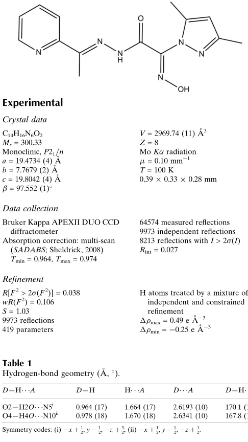
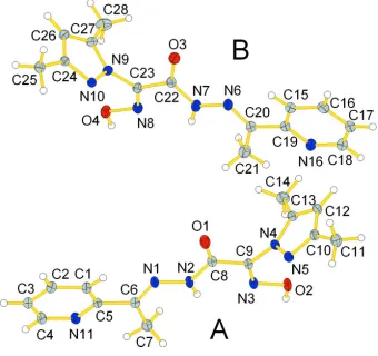
Oximes are one of the most efficient bridging ligands class. Polydentate ligands containing both oxime and other donor functions are of special interest due to their potential for the bridging coordination modes and mediation of strong magnetic superexchange between metal ions (Penkova et al., 2009; Kanderal et al., 2005; Moroz et al., 2010). Oxime ligands having the pyridyl groups in the molecule have been used in the preparation of complexes with a variety of transition metals, binding to metals in different modes most commonly as chelates or serving as bridge to metals, and the resulting species have been employed in molecular magnetism and supramolecular chemistry (Moroz et al., 2010, 2012). Herein we report a second polymorph of 2-[1-(3,5-dimethyl)pyrazolyl]-2-hydroxyimino-N′-[1-(2-pyridyl)ethylidene] acetohydrazide (II) (Fig. 1). In comparison, the first polymorph I described previously (Plutenko et al., 2012),
crystallized in monoclinic space group Cc, Z = 4, while the title compound II crystallized in space group P21/n with Z = 8 (Fig. 1).
Bond lengths N-N′, N-C and C-O of the amide group are 1.3693 (10), 1.3589 (11) and 1.2162 (11) respectively for molecule A and 1.3705 (11), 1.3603 (11) and 1.2156 (11) respectively for molecule B. Such bond lengths are typical for the protonated amide groups (Kanderal et al., 2005). The oxime group is situated in anti- position to the amide group which was shown earlier in the structures of the amide derivatives of 2-hydroxyiminopropanoic acid (Onindo et al., 1995; Sliva et al., 1997; Fritsky et al., 2006). The NC(=NOH)C(O)NH fragment deviates from planarity because of a twist between the oxime and the amide groups about the C(8)-C9 and C(22)-C(23) bonds; the O(1)-C(8)-C(9)-N(3) and O(22)-C(23)-C(9)-N(8) torsion angles are -175.953 (2)° and 164.073 (2)°. The bond lengths N-O and C-N of the oxime group are 1.3615 (9) and 1.2836 (11) Å respectively for A and 1.3645 (10) and 1.2819 (11) Å respectively for B. The bond lengths and angles within the oxime groups are normal and comparable to those in the related structures (Świątek-Kozłowska et al., 2000; Mokhir et al., 2002; Fritsky et al., 1998). The C=N and N—O bond lengths in the oxime moiety of the molecule clearly indicates that the oxime group exists in the nitroso rather than in the isonitroso form (Duda et al., 1997; Kanderal et al., 2005; Fritsky et al., 2004).
The C-C, C-N and N-N′ (1,331 (0) – 1,409 (0) Å) bond lengths in the pyrazole ring exhibit normal values (Penkova et al., 2010). The angles C-C′-C′′, C-N-C′, N-C-C′ and N-N′-C are near 108°. The pyrazole ring deviates from the plane formed by other atoms of ligand molecule. The N(4)-C(9)-N(5)-N(6) and torsion angles are -103.589 (2) and 105.359 (2)° respectively. The C-N and C-C bond lengths in the pyridine rings are normal for 2-substituted pyridine derivatives (Fritsky et al., 2000; Krämer et al., 2000; Strotmeyer et al., 2003).
S2. Experimental
The present compound was synthesized according to Plutenko et al. (2012):
Synthesis of ethyl 2-[1-(3,5-dimethyl)pyrazolyl]-2-hydroxyiminoacetate: A mixture of ethyl 2-chloro-2-hydroxyimino-acetate synthesized according to Kozikowski et al. (1983) (0.906g, 6 mmol) and 3,5-dimethylpyrazol (1.152g, 12 mmol) in 10 ml of chloroform was left for evaporation in the air overnight. The resulting precipitate was crystallized from water. Yield: 1.12g (88 %).
Synthesis of 2-[1-(3,5-dimethyl)pyrazolyl]-2-hydroxyiminoacetohydrazide: A solution of hydrazine hydrate (0.57 ml, 60%, 10.6 mmol) in water was added to a solution of ethyl 2-[1-(3,5-dimethyl)pyrazolyl]-2-hydroxyiminoacetate (1.12g, 5.3 mmol) in methanol (30 ml). The resulting mixture was heating under reflux for 1.5 hours. After that solvent was evaporated and product was crystallized from methanol. Yield 0.5g (48 %).
Synthesis of 2-[1-(3,5-dimethyl)pyrazolyl]-2-hydroxyimino-N′-[1-(2-pyridyl)ethylidene]acetohydrazide: A solution of [1-(3,5-dimethyl)pyrazolyl]-hydroxyiminoacetohydrazide (0.5g, 2.54 mmol) in methanol (30 ml) was treated with 2-acetylpyridine (0.307g, 2.54 mmol) and the mixture was heated under reflux for 3 hours. After that the solvent was evaporated in vacuum and the product was crystallized from methanol. Yield 0.65g (85 %).
S3. Refinement
OH, NH and CH3 hydrogen atoms were located from difference Fourier maps, other hydrogen atoms were positioned
geometrically and all but H(N) and H(O) were refined at idealized positions riding on the parent atoms, with C—H = 0.95–0.98 Å, and Uiso = 1.2–1.5 Ueq(parent atom). H(N) and H(O) atoms were refined freely with Uiso(H) = 1.2Ueq(N) or
1.5Ueq(O). All CH3 hydrogen atoms were allowed to rotate but not to tip. The highest peak is located 0.74 Å from atom
Figure 1
Figure 2
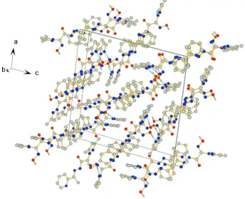
Crystal packing viewed along b-axis. Hydrogen bonds are indicated by dashed lines. H atoms not involved in H-bonds are omitted for clarity.
2-(3,5-Dimethyl-1H-pyrazol-1-yl)-2-hydroxyimino-N′-[1-(pyridin-2-yl)ethylidene]acetohydrazide
Crystal data C14H16N6O2
Mr = 300.33
Monoclinic, P21/n
Hall symbol: -P 2yn a = 19.4734 (4) Å b = 7.7679 (2) Å c = 19.8042 (4) Å β = 97.552 (1)° V = 2969.74 (11) Å3
Z = 8
F(000) = 1264 Dx = 1.343 Mg m−3
Mo Kα radiation, λ = 0.71073 Å Cell parameters from 9866 reflections θ = 2.8–31.6°
µ = 0.10 mm−1
T = 100 K Block, colourless 0.39 × 0.33 × 0.28 mm
Data collection
Bruker Kappa APEXII DUO CCD diffractometer
Radiation source: fine-focus sealed tube Curved graphite crystal monochromator Detector resolution: 16 pixels mm-1
φ scans and ω scans with κ offset Absorption correction: multi-scan
(SADABS; Sheldrick, 2008) Tmin = 0.964, Tmax = 0.974
64574 measured reflections 9973 independent reflections 8213 reflections with I > 2σ(I) Rint = 0.027
θmax = 31.7°, θmin = 1.6°
Refinement Refinement on F2
Least-squares matrix: full R[F2 > 2σ(F2)] = 0.038
wR(F2) = 0.106
S = 1.03 9973 reflections 419 parameters 0 restraints
Primary atom site location: structure-invariant direct methods
Secondary atom site location: difference Fourier map
Hydrogen site location: difference Fourier map H atoms treated by a mixture of independent
and constrained refinement w = 1/[σ2(F
o2) + (0.0555P)2 + 0.8835P]
where P = (Fo2 + 2Fc2)/3
(Δ/σ)max = 0.001
Δρmax = 0.49 e Å−3
Δρmin = −0.25 e Å−3
Special details
Geometry. All esds (except the esd in the dihedral angle between two l.s. planes) are estimated using the full covariance matrix. The cell esds are taken into account individually in the estimation of esds in distances, angles and torsion angles; correlations between esds in cell parameters are only used when they are defined by crystal symmetry. An approximate (isotropic) treatment of cell esds is used for estimating esds involving l.s. planes.
Refinement. Refinement of F2 against ALL reflections. The weighted R-factor wR and goodness of fit S are based on F2,
conventional R-factors R are based on F, with F set to zero for negative F2. The threshold expression of F2 > 2sigma(F2) is
used only for calculating R-factors(gt) etc. and is not relevant to the choice of reflections for refinement. R-factors based on F2 are statistically about twice as large as those based on F, and R- factors based on ALL data will be even larger.
Fractional atomic coordinates and isotropic or equivalent isotropic displacement parameters (Å2)
x y z Uiso*/Ueq
H28A 0.3918 −0.1995 0.1815 0.039* H28B 0.3509 −0.2533 0.1092 0.039* H28C 0.3104 −0.2383 0.1742 0.039*
Atomic displacement parameters (Å2)
U11 U22 U33 U12 U13 U23
C26 0.0159 (4) 0.0212 (4) 0.0149 (4) −0.0013 (3) 0.0027 (3) −0.0037 (3) C27 0.0158 (4) 0.0162 (4) 0.0183 (4) −0.0019 (3) 0.0048 (3) −0.0031 (3) C28 0.0349 (5) 0.0151 (4) 0.0311 (5) −0.0007 (4) 0.0126 (4) −0.0015 (4)
Geometric parameters (Å, º)
O1—C8 1.2162 (11) C7—H7B 0.9800 O2—N3 1.3615 (9) C7—H7C 0.9800 O2—H2O 0.964 (17) C8—C9 1.5035 (12) O3—C22 1.2156 (11) C10—C12 1.4061 (13) O4—N8 1.3645 (10) C10—C11 1.4931 (13) O4—H4O 0.978 (18) C11—H11A 0.9800 N1—C6 1.2915 (11) C11—H11B 0.9800 N1—N2 1.3693 (10) C11—H11C 0.9800 N2—C8 1.3589 (11) C12—C13 1.3721 (12) N2—H2N 0.859 (15) C12—H12 0.9500 N3—C9 1.2836 (11) C13—C14 1.4868 (13) N4—C13 1.3619 (11) C14—H14A 0.9800 N4—N5 1.3703 (10) C14—H14B 0.9800 N4—C9 1.4115 (11) C14—H14C 0.9800 N5—C10 1.3313 (11) C15—C16 1.3846 (13) N6—C20 1.2921 (12) C15—C19 1.3974 (13) N6—N7 1.3705 (11) C15—H15 0.9500 N7—C22 1.3603 (11) C16—C17 1.3916 (14) N7—H7N 0.882 (15) C16—H16 0.9500 N8—C23 1.2819 (11) C17—C18 1.3840 (15) N9—C27 1.3607 (11) C17—H17 0.9500 N9—N10 1.3683 (10) C18—H18 0.9500 N9—C23 1.4090 (11) C19—C20 1.4862 (13) N10—C24 1.3327 (11) C20—C21 1.5010 (13) N11—C5 1.3396 (11) C21—H21A 0.9800 N11—C4 1.3408 (12) C21—H21B 0.9800 N16—C18 1.3391 (13) C21—H21C 0.9800 N16—C19 1.3425 (12) C22—C23 1.5010 (12) C1—C2 1.3841 (12) C24—C26 1.4105 (13) C1—C5 1.3959 (12) C24—C25 1.4927 (13) C1—H1 0.9500 C25—H25A 0.9800 C2—C3 1.3909 (14) C25—H25B 0.9800 C2—H2 0.9500 C25—H25C 0.9800 C3—C4 1.3834 (14) C26—C27 1.3747 (13) C3—H3 0.9500 C26—H26 0.9500 C4—H4 0.9500 C27—C28 1.4884 (13) C5—C6 1.4872 (12) C28—H28A 0.9800 C6—C7 1.5019 (13) C28—H28B 0.9800 C7—H7A 0.9800 C28—H28C 0.9800
N3—C9—C8 118.31 (8) C27—C26—C24 106.14 (8) N4—C9—C8 117.99 (7) C27—C26—H26 126.9 N5—C10—C12 110.61 (8) C24—C26—H26 126.9 N5—C10—C11 120.42 (8) N9—C27—C26 106.07 (8) C12—C10—C11 128.96 (8) N9—C27—C28 121.87 (8) C10—C11—H11A 109.5 C26—C27—C28 132.05 (9) C10—C11—H11B 109.5 C27—C28—H28A 109.5 H11A—C11—H11B 109.5 C27—C28—H28B 109.5 C10—C11—H11C 109.5 H28A—C28—H28B 109.5 H11A—C11—H11C 109.5 C27—C28—H28C 109.5 H11B—C11—H11C 109.5 H28A—C28—H28C 109.5 C13—C12—C10 106.42 (8) H28B—C28—H28C 109.5
Hydrogen-bond geometry (Å, º)
D—H···A D—H H···A D···A D—H···A O2—H2O···N5i 0.964 (17) 1.664 (17) 2.6193 (10) 170.1 (16)
O4—H4O···N10ii 0.978 (18) 1.670 (18) 2.6341 (10) 167.8 (17)