Bystander responses impact accurate detection
of murine and human antigen-specific CD8
+
T
cells
Matthew D. Martin, … , Robert A. Seder, Vladimir P.
Badovinac
J Clin Invest.
2019;
129(9)
:3894-3908.
https://doi.org/10.1172/JCI124443
.
Induction of memory CD8
+T cells is important for controlling infections such as malaria and
HIV/AIDS and for cancer immunotherapy. Accurate assessment of antigen-specific
(Ag-specific) CD8
+T cells is critical for vaccine optimization and for defining correlates of
protection. However, conditions for determining Ag-specific CD8
+T cell responses ex vivo
using intracellular cytokine staining (ICS) may be variable, especially in humans with
complex antigens. Here, we used an attenuated whole parasite malaria vaccine model in
humans and various experimental infections in mice to show that the duration of antigenic
stimulation and timing of brefeldin A (BFA) addition influence the magnitude of Ag-specific
and bystander T cell responses. Indeed, after immunization with an attenuated whole
sporozoite malaria vaccine in humans, significantly higher numbers of IFN-g–producing
memory CD8
+T cells comprising Ag-specific and bystander responses were detected when
the duration of Ag stimulation prior to addition of BFA was increased. Mechanistic analyses
of virus-specific CD8
+T cells in mice revealed that the increase in IFN-g–producing CD8
+T
cells was due to bystander activation of Ag-experienced memory CD8
+T cells, and
correlated with the proportion of Ag-experienced CD8
+T cells in the stimulated populations.
Incubation with anti-cytokine antibodies (e.g., IL-12) improved accuracy in detecting bona
fide memory CD8
+T cell responses, suggesting this as the mechanism for the bystander
activation. These data have […]
Research Article
Immunology
Find the latest version:
Introduction
CD8+ T cells have a role in mediating protection in humans against
diverse pathogens impacting public health, such as HIV, influenza, and Plasmodium, the causative agent of malaria, and tumors (1–5). The critical factors for mediating protective immunity by T cells include the magnitude, quality, breadth, and location. Indeed, subjects with greater numbers of memory CD8+ T cells are better
protected against infection (6, 7). Therefore, the development of preventive and therapeutic vaccines against infections and tumors requires a precise understanding of how to generate and accurate-ly assess CD8+ T cell responses.
There are now a variety of techniques to measure CD8+ T cell
responses, each with strengths and weaknesses. Peptide-MHC tetramer staining allows for an accurate enumeration of epi-tope-specific cells within the host and the evaluation of their phe-notypic traits, but provides limited information on the function-ality of the T cell response. Additionally, peptide-MHC tetramer analyses require knowledge of both the exact T cell epitope and MHC allele, which vary considerably between individuals, making assessment of the antigenic breadth of response resource-inten-sive. Antigen-stimulated detection of T cells by ICS, in contrast, allows for assessment of T cell responses in an MHC-agnostic
manner, since stimulation of T cells occurs through autologous antigen (Ag) presentation. However, a limitation of the intracel-lular cytokine staining (ICS) assay, in which overlapping peptides are often used to stimulate T cells, is that the exact T cell epitope is not determined, and thus, the breadth of the host responses is not as precisely defined. Additionally, while ICS assays can provide data on the functional and phenotypic traits of responding cells, cells typically require stimulation for greater than 6 hours, during which time the cell phenotype of activated cells may change.
While alternative methods for detecting Ag-specific cells are used by some groups, such as activation-induced expression of CD137 (8), IFN-γ is the canonical gold-standard cytokine for ICS-based enu-meration of Ag-specific CD8+ T cells. In a review of the literature, we
determined that methods of conducting ICS, including length of incu-bation and timing of addition of brefeldin A (BFA; a Golgi-disrupting compound used to prevent secretion of cytokines), vary widely. Most often, ICS stimulation occurs for between 5 and 24 hours, and BFA is added several hours after the beginning of stimulation (4, 6, 9–39). While delaying addition of BFA may be necessary with certain types of Ag stimulation, such as use of whole pathogens rather than pep-tides to allow for Ag processing and presentation on MHC needed to drive cytokine production by CD8+ T cells, delayed BFA addition may
also allow for the release of cytokines into the culture media when stimuli that have innate stimulatory activity are used. Inflammatory cytokines such as IL-12 could enhance IFN-γ production by Ag-spe-cific CD8+ T cells that respond to antigens used for stimulation, but
also by memory T cells that are specific for other antigens (bystander activation). This is supported by evidence that Ag-experienced effec-Induction of memory CD8+ T cells is important for controlling infections such as malaria and HIV/AIDS and for cancer
immunotherapy. Accurate assessment of antigen-specific (Ag-specific) CD8+ T cells is critical for vaccine optimization and for defining correlates of protection. However, conditions for determining Ag-specific CD8+ T cell responses ex vivo using intracellular cytokine staining (ICS) may be variable, especially in humans with complex antigens. Here, we used an attenuated whole parasite malaria vaccine model in humans and various experimental infections in mice to show that the duration of antigenic stimulation and timing of brefeldin A (BFA) addition influence the magnitude of Ag-specific and bystander T cell responses. Indeed, after immunization with an attenuated whole sporozoite malaria vaccine in humans, significantly higher numbers of IFN-γ–producing memory CD8+ T cells comprising Ag-specific and bystander responses were detected when the duration of Ag stimulation prior to addition of BFA was increased. Mechanistic analyses of virus-specific CD8+ T cells in mice revealed that the increase in IFN-γ–producing CD8+ T cells was due to bystander activation of Ag-experienced memory CD8+ T cells, and correlated with the proportion of Ag-experienced CD8+ T cells in the stimulated populations. Incubation with anti-cytokine antibodies (e.g., IL-12) improved accuracy in detecting bona fide memory CD8+ T cell responses, suggesting this as the mechanism for the bystander activation. These data have important implications for accurate assessment of immune responses generated by vaccines intended to elicit protective memory CD8+ T cells.
Bystander responses impact accurate detection of
murine and human antigen-specific CD8
+
T cells
Matthew D. Martin,1 Isaac J. Jensen,2 Andrew S. Ishizuka,3 Mitchell Lefebvre,2 Qiang Shan,4 Hai-Hui Xue,2,4,5 John T. Harty,1,2,4
Robert A. Seder,3 and Vladimir P. Badovinac1,2,4
1Department of Pathology and 2Interdisciplinary Graduate Program in Immunology, University of Iowa, Iowa City, Iowa, USA. 3Vaccine Research Center, National Institute of Allergy and Infectious Diseases,
NIH, Bethesda, Maryland, USA. 4Department of Microbiology and Immunology, University of Iowa, Iowa City, Iowa, USA. 5Iowa City Veterans Affairs Health Care System, Iowa City, Iowa, USA.
Conflict of interest: The authors have declared that no conflict of interest exists.
Copyright: © 2019, American Society for Clinical Investigation.
Submitted: August 23, 2018; Accepted: June 18, 2019; Published: August 19, 2019.
ed these data and incubated PBMCs from a vaccinated or nonim-munized subject with PfSPZs for 12, 16, 20, and 24 hours, adding BFA for the final 4 hours. While the nonimmunized subject had no IFN-γ production above background for the duration of stimu-lation, the percentage of CD8+ T cells that produced IFN-γ in the
PfSPZ-vaccinated subject increased substantially over time (Fig-ure 1A), suggesting that duration of ICS increased the detection of total IFN-γ–producing CD8+ T cells.
Addition of BFA is used in ICS assays to block release of IFN-γ within the T cell, thereby improving the sensitivity of the response. However, BFA also could inhibit efficient Ag processing and presentation of PfSPZs (44). To determine whether timing of BFA addition impacted the detection of IFN-γ–producing CD8+ T
cells, cells were stimulated with PfSPZs for a total of 24 hours, with BFA being added after the first 8, 16, or 20 hours of incubation. A substantially higher percentage of CD8+ T cells producing IFN-γ
was detected when BFA was added 16 or 20 hours after the begin-ning of stimulation (Figure 1B), indicating that the timing of BFA addition strongly influences the frequency of IFN-γ–producing CD8+ T cells detected.
Delayed addition of BFA would allow for soluble factors includ-ing inflammatory cytokines to be released into the culture, and potential bystander activation of Ag-experienced effector and mem-ory CD8+ T cells. Thus, it is unclear whether enhanced detection of
IFN-γ–producing CD8+ T cells following an increase in incubation
time or a delay in addition of BFA was due to increased sensitivity of detection of Plasmodium-specific CD8+ T cells, and/or to triggering of
IFN-γ production by nonmalaria Ag-experienced CD8+ T cells. Delayed addition of BFA leads to bystander activation of Ag-ex-perienced CD8+ T cells. To further determine how ICS conditions
altered detection of Ag-specific CD8+ T cells, we used a
well-de-fined mouse model that allows precise detection of CD8+ T cells
of known Ag specificity. Mice (C57BL/6, Thy1.2) received adop-tive transfer of naive T cell receptor–transgenic (TCR-transgenic) GP33-specific P14 cells (Thy1.1) before LCMV-Armstrong infec-tion (Figure 2A), and memory P14 cells were detected by Thy1.1 expression. Additionally, infection of C57BL/6 mice with LCMV elicits large Ag-specific CD8+ T cell responses that recognize
LCMV-derived GP33 and NP396 epitopes (45), and we used pep-tide-stimulated ICS to detect cytokine-producing memory CD8+
T cells responding to GP33 and NP396 peptides (Figure 2, B–D). Mice generated in this manner contain endogenous (Thy1.1–,
blue gates) CD8+ T cells comprising naive cells and Ag-specific
cells that recognize GP33 and NP396 epitopes as well as additional LCMV-derived epitopes, and Ag-experienced P14 cells (Thy1.1+,
red gates) that recognize GP33 but not NP396 peptides. Addition of P14 cells allows for detection of true Ag-specific responses (in response to GP33 peptide) and bystander responses (in response to NP396 peptide), while IFN-γ production by endogenous CD8+
T cells, owing to mixed epitope specificities of this population, could represent either true Ag-specific responses or responses that include both Ag-specific and bystander responses. To high-light this, as can be seen in Figure 2, B–D, we present detection of IFN-γ–producing cells following ICS gated on total lympho-cytes (lymphocyte gate), total CD8+ T cells with identification of
endogenous (blue gates) or P14 (red gates) cells based on Thy1.1 expression (CD8 gate), or total P14 cells [Thy1.1+CD8+ (P14) gate].
tor or memory CD8+ T cells can be induced to produce IFN-γ not
only in response to cognate Ag, but also in a bystander manner that is solely driven by inflammatory cytokines (40–42). Thus, we sought to understand how variations in the conditions of ICS impact the accu-rate accounting of infection- or vaccine-induced Ag-specific CD8+ T
cells. These data have implications for improving vaccine design and accurately assessing correlates of CD8+ T cell–mediated protection.
Results
The frequency of IFN-γ–producing CD8+ T cells detected in humans
following vaccination is increased with increased time of ex vivo Ag stimulation. The potency and protective capacity of a vaccine
can be evaluated partly based on the magnitude of the induced memory CD8+ T cell population. To begin to address the impact
of varying the assay conditions on enumeration of Ag-specific CD8+ T cell memory, we assessed the frequency of Ag-specific
memory CD8+ T cells following vaccination of human subjects
with an attenuated whole sporozoite malaria vaccine (4, 6, 43). Using a peptide stimulation assay for this vaccine would be tech-nically challenging because of MHC polymorphism and the large number (>2000) of potential antigens presented to the immune system. Therefore, we used a previously developed assay in which stimulation of CD8+ T cells was achieved by incubation of PBMCs
from vaccinated subjects with the vaccine itself, which consists of aseptic, purified, irradiated, metabolically active Plasmodium
fal-ciparum sporozoites (PfSPZs), and in which 12-hour stimulation
was more sensitive than 6-hour stimulation (4). Here, we extend-Figure 1. Increased length of incubation and delayed addition of BFA lead to elevated detection of IFN-γ–producing human CD8+ T cells following
PfSPZ stimulation. (A) Percentage of CD8+ T cells producing IFN-γ when
PBMCs from a PfSPZ-vaccinated or unvaccinated (naive) subject were stimulated with PfSPZs for 12, 16, 20, or 24 hours. (B) Percentage of CD8+ T
[image:3.585.78.252.56.296.2]cells producing IFN-γ in response to GP33 or NP396 peptide was observed when BFA was added for the last hour (7+1) compared with when it was present during the entire incubation (0+8) (Fig-ure 2, B–D, CD8 gate, blue boxes; and Fig(Fig-ure 2E). Similar results were observed for LCMV-immune mice that did not receive P14 cells (Supplemental Figure 1, C and D).
A similar percentage of P14 cells produced IFN-γ following GP33 peptide stimulation regardless of timing of BFA addition (Fig-ure 2C, P14 gate), suggesting that delayed addition of BFA does not impact IFN-γ production of bona fide Ag-specific CD8+ T cells. To
determine whether increases in IFN-γ–producing CD8+ T cells that
occurred with delayed BFA addition were due to bystander activa-tion, we examined responses of “sensor” GP33-specific Thy1.1 P14 Notably, since detection of IFN-γ–producing CD8+ T cells is
impacted by peptide concentration and number of cells plated, we first identified conditions that allow for maximal detection of bona fide Ag-specific CD8+ T cells (2 × 10⁶ cells per well and
200 nm peptide concentration) (Supplemental Figure 1, A and B; supplemental material available online with this article; https:// doi.org/10.1172/JCI124443DS1). Inflammatory cytokines can potentially cause bystander activation of Ag-experienced CD8+ T
cells when BFA is not present during the entire incubation (40–42, 46), so we first sought to determine whether timing of BFA addi-tion impacted detecaddi-tion of IFN-γ–producing CD8+ T cells
[image:4.585.92.486.55.449.2]follow-ing specific peptide stimulation. Interestfollow-ingly, an approximately 1.5- to 2-fold increase in the percentage of endogenous CD8+ T
Figure 2. Delayed addition of BFA leads to bystander activation of CD8+ T cells. (A) Experimental design. Mice received adoptive transfer (AT) of naive P14
cells and were infected with LCMV-Armstrong. Approximately 3 weeks after infection, splenocytes were harvested and ICS was conducted. (B) Representative dot plots of IFN-γ production following 8-hour incubation without peptide and with BFA present for the entire incubation (0+8) or the final hour (7+1). Plots on the left are gated lymphocytes, plots in the middle are gated CD8+ T cells (Thy1.1– = endogenous CD8+ T cells, Thy1.1+ = P14 cells), and plots on the right are
gated P14 cells. Numbers inside plots indicate the percentage of cells producing IFN-γ out of all gated cells. (C) Representative dot plots of IFN-γ production following 8-hour incubation with GP33 peptide. (D) Representative dot plots of IFN-γ production following 8-hour incubation with NP396 peptide. (E) Left: Sum-mary graphs of the percentage of endogenous CD8+ T cells producing IFN-γ following stimulation with GP
33 peptide out of all CD8+ T cells with BFA present
for the entire incubation (0+8) or the final hour (7+1). Right: Percentage of endogenous CD8+ T cells producing IFN-γ when BFA was added for the final hour of
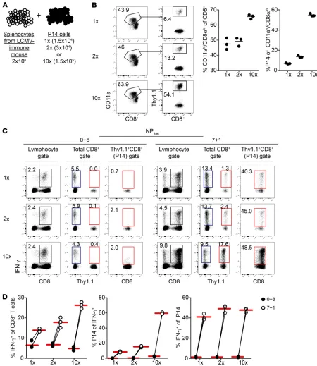
Figure 3. Contribution of bystander responses to IFN-γ–producing cells detected when addition of BFA is delayed is influenced by CD8+ T cell pool
composition. (A) Experimental design. Before 8-hour incubation with NP396 peptide, splenocytes from an LCMV-Armstrong–immune mouse were mixed with different numbers of sorted memory P14 cells. (B) Left: Representative dot plots of Ag-experienced (CD11ahiCD8lo) CD8+ T cells among all
CD8+ T cells (left plot) and percentage of P14 cells (Thy1.1+) among Ag-experienced CD8+ T cells (right plot) after mixing. Middle: Summary graph of the
percentage of Ag-experienced CD8+ T cells among all CD8+ T cells after mixing. Right: Summary graph of the percentage of memory P14 cells among
Ag-experienced CD8+ T cells after mixing. (C) Representative dot plots of IFN-γ production following 8-hour incubation with NP
396 peptide and with
BFA present for the entire incubation (0+8) or the final hour (7+1). Plots on the left are gated lymphocytes, plots in the middle are gated CD8+ T cells
(Thy1.1– = endogenous CD8+ T cells, Thy1.1+ = P14 cells), and plots on the right are gated P14 cells. Numbers inside plots indicate the percentage of cells
producing IFN-γ out of all gated cells. (D) Left: Summary graphs of the percentage of CD8+ T cells producing IFN-γ out of all CD8+ T cells with BFA
present for the entire incubation (0+8) or the final hour (7+1). Middle: Summary graphs of the percentage of P14 cells producing IFN-γ out of all IFN-γ+
CD8+ T cells with BFA present for the entire incubation (0+8) or the final hour (7+1). Right: Summary graphs of the percentage of gated P14 cells
cells could be detected when BFA was added before the last hour and when cells were stimulated for 8 or more hours, and bystand-er responses increased with greatbystand-er length of stimulation in the absence of BFA (Supplemental Figure 2). Thus, delayed addition of BFA in both the mouse and human models of viral and malaria- specific CD8+ T cells can result in bystander activation of
Ag-expe-rienced CD8+ T cells leading to inflation in frequencies and
num-bers of Ag-specific CD8+ T cells detected.
Contribution of bystander IFN-γ activation is dependent on CD8+ T cell pool composition and length of stimulation.
Ag-experi-enced CD8+ T cells can undergo bystander activation and produce
cytokines such as IFN-γ, while naive CD8+ T cells cannot (40,
46). Notably, the representation of Ag-experienced cells within the CD8+ T cell pool in human subjects can vary widely based on
[image:6.585.52.517.57.418.2]cells in response to NP396 peptide stimulation. While few IFN-γ– producing P14 cells were detected following stimulation with NP396 peptide in the presence of BFA for the entire incubation, 25%–30% of memory P14 cells produced IFN-γ in response to NP396 peptide when BFA was not present during the whole incubation time (Fig-ure 2D, red boxes). Analyses of T cell responses by ICS in human samples often rely on stimulation for greater than 8 hours, with addition of BFA before the last hour of incubation. To determine whether bystander responses contribute to IFN-γ–producing cells detected for incubation times of greater length and when BFA is added earlier in the culture, we stimulated splenocytes from LCMV-immune mice that contained memory P14 cells with NP396 peptide for 8 hours and added BFA after 4 or 7 hours, or for 12 hours and added BFA after 4, 8, or 11 hours. Bystander responses by P14
Figure 4. Contribution of bystander responses to IFN-γ–producing cells detected when BFA addition is delayed increases with extended length of stim-ulation but is seen following incubation times of 5 or more hours. Mice received adoptive transfer of naive P14 cells and were infected with LCMV-Arm-strong. ICS was conducted with GP33 (A and B) or NP396 (C and D) peptide approximately 3 weeks after infection. Total incubation times were 5, 8, 16, or 24 hours with BFA present for the whole incubation or the final hour. (A) Summary graphs of the percentage of endogenous (Thy1.1–) CD8+ T cells producing
IFN-γ out of all endogenous CD8+ T cells with BFA present for the entire incubation or the final hour. (B) Ratio of the percentage of endogenous CD8+ T
cells producing IFN-γ when BFA was present for the final hour of incubation over the percentage of endogenous CD8+ T cells producing IFN-γ when BFA
was present for the entire incubation. (C) Top: Summary graphs of the percentage of endogenous (Thy1.1–) CD8+ T cells producing IFN-γ out of all
endoge-nous CD8+ T cells with BFA present for the entire incubation or the final hour. Bottom: Summary graphs of the percentage of P14 cells (Thy1.1+) producing
IFN-γ out of all P14 cells with BFA present for the entire incubation or the final hour. (D) Ratio of the percentage of endogenous CD8+ T cells producing
IFN-γ when BFA was present for the final hour of incubation over the percentage of endogenous CD8+ T cells producing IFN-γ when BFA was present for
Figure 5. Delayed BFA addition leads to bystander activation of CD8+ T cells following stimulation
with pathogen-infected spleno-cytes. (A) Experimental design. Splenocytes from a naive mouse (stimulator cells) were CFSE-la-beled and either pulsed with GP33 peptide or infected with VacV-GP33 or VacV-OVA. Stimulator cells were mixed with splenocytes from an LCMV-Armstrong–immune mouse (sensor cells) and incubated for 8 hours with BFA present for 8 (0+8), 6 (2+6), 4 (4+4), or 1 (7+1) hours. (B) CD8+ T cells producing IFN-γ after
8-hour incubation with indicated numbers of GP33 peptide–pulsed (left) or VacV-GP33–infected (right) stimulator cells with BFA present for the entire incubation. (C) Top: CD8+ T cells producing IFN-γ after
incubation with indicated numbers of GP33 peptide–pulsed stimulator cells and with BFA for the indicated times. Middle: CD8+ T cells producing
IFN-γ after incubation with indicated numbers of VacV-GP33–infected stimulator cells and with BFA for the indicated times. Bottom: CD8+
T cells producing IFN-γ after incu-bation with the indicated number of VacV-OVA–infected stimulator cells and with BFA for the indicated times. (D) NP396 peptide–pulsed or VacV-NP–infected stimulator cells were mixed with sensor cells from an LCMV-Armstrong–immune mouse containing P14 cells. Representative dot plots of IFN-γ production by gated CD8+ T cells (left plots: Thy1.1–
= endogenous CD8+ T cells, Thy1.1+
= P14 cells) or P14 cells (right plots). Numbers inside plots indicate the percentage of cells producing IFN-γ
age and history of previously encountered infections (47, 48). To determine whether the composition of Ag experience in the CD8+
T compartment dictated frequencies of IFN-γ–producing CD8+ T
cells, we manipulated the number of Ag-experienced cells among splenocytes by mixing splenocytes from LCMV-immune mice with graded numbers of sorted memory P14 “sensor” cells to achieve different ratios of naive (CD11aloCD8hi) to memory (CD11ahi
CD8lo) CD8+ T cells (refs. 49, 50, and Figure 3, A and B). Again,
few IFN-γ–producing P14 cells were detected following stimula-tion with NP396 peptide when BFA was present for the entire incu-bation, and a similar percentage of CD8+ T cells producing IFN-γ
in response to NP396-peptide stimulation was observed regardless of numbers of P14 cells present (Figure 3, C and D, 0+8 group). However, when BFA was not present for the entire incubation, the percentage of IFN-γ–producing CD8+ T cells in response to NP
396
peptide stimulation increased with increasing numbers of sen-sor P14 cells, and this was due to increased representation of P14 cells rather than elevation in the frequency of activated bystander P14 cells (Figure 3, C and D, 7+1 group). Thus, the contribution of bystander-activated cells to the IFN-γ–producing CD8+ T cell
pop-ulation was dependent on the representation of Ag-experienced cells within the CD8+ T cell compartment. These data suggested
that the increase in the frequency of vaccine-targeted memory CD8+ T cells might be more pronounced in older subjects and/or
subjects with substantial history of pathogen exposures that pos-sess more previously activated CD8+ T cells.
The duration of Ag stimulation ex vivo for experiments involv-ing human subjects varies, but in most instances stimulation times are between 5 and 24 hours (4, 6, 9–39). To determine whether the duration of stimulation contributes to the degree of bystander IFN-γ detected when BFA is not present for the entire incubation, spleno-cytes from an LCMV-immune mouse were incubated with GP33 or NP396 peptides for 5, 8, 16, or 24 hours with BFA present for the entire incubation or for the final hour of incubation. Regardless of length of incubation, a greater percentage of IFN-γ–producing CD8+ T cells
was detected following stimulation with GP33 (Figure 4, A and B) or NP396 (Figure 4, C and D) peptides when BFA was added for only the last hour of incubation. However, increased detection of IFN-γ– producing CD8+ T cells was most pronounced when samples were
incubated for greater than 5 hours. Similarly, bystander-activated P14 cells were detected in response to NP396 peptide stimulation when BFA addition was delayed for any length of incubation test-ed, but percentages of bystander-activated P14 cells detected were greater when samples were incubated for greater than 5 hours (Fig-ure 4C). These data suggested that bystander activation occurred and contributed to the frequency of IFN-γ–producing CD8+ T cells
detected when BFA was not present for the entire incubation across the spectrum of incubation times used to conduct ICS. However, the contribution of bystander-activated cells to IFN-γ–producing cells detected is likely to increase with greater lengths of incubation.
Delayed addition of BFA leads to bystander activation of Ag-expe-rienced CD8+ T cells following stimulation with whole pathogen.
Appli-cation of peptide-stimulated ICS assays across individual human subjects assessing responses to complex pathogens with many antigens is difficult because of differences in HLA haplotype; thus, stimulation of human samples is often achieved by expos-ing samples to whole pathogens (Figure 1), or through the use of
libraries of long overlapping peptides, methods that may require a period of BFA-free culture to allow for optimal Ag processing and presentation or cross-presentation. To determine whether bystander-activated cells contributed to the frequency of IFN-γ– producing cells detected after stimulation with whole pathogens, splenocytes from an LCMV-immune mouse (“sensor” cells) were incubated with CFSE-labeled splenocytes that were either pulsed with GP33 peptide or infected with Vaccinia virus (VacV) express-ing cognate Ag (VacV-GP33) or an irrelevant Ag (VacV-OVA) (Fig-ure 5A). When BFA was present throughout the incubation peri-od, IFN-γ production from the LCMV-immune sensor cells was observed in response to spleen cells pulsed with GP33 peptide or infected with VacV-GP33, and the percentage of IFN-γ–producing CD8+ T cells detected increased with increasing numbers of
stim-ulator cells added to the culture (Figure 5B). IFN-γ production was not observed in response to splenocytes infected with VacV-OVA (Figure 5C, bottom).
Interestingly, an increased percentage of IFN-γ–producing sensor cells were detected following incubation with greater num-bers of GP33-pulsed or VacV-GP33–infected stimulator cells and when addition of BFA was delayed for longer periods (Figure 5C, top and middle). These data suggested that length of incubation with BFA impacted detection of IFN-γ–producing CD8+ T cells
following stimulation with pathogen-infected splenocytes. How-ever, because we could not determine whether our stimulation conditions resulted in IFN-γ production by bona fide Ag-specific or any Ag-experienced CD8+ T cells, we were unable to conclude
whether delayed addition of BFA resulted in increased detection of true Ag-specific CD8+ T cells, or whether increased percentages
of IFN-γ–producing CD8+ T cells detected were due to bystander
activation of memory CD8+ T cells. To address this, we incubated
sensor cells from LCMV-immune P14 chimera mice with stimula-tor cells that were either pulsed with NP396 peptide or infected with VacV expressing LCMV nucleoprotein (VacV-NP). An increased percentage of IFN-γ–producing cells was detected following stim-ulation with peptide-pulsed or VacV-NP–infected cells when BFA was not present for the whole incubation time (Figure 5, D and E). Importantly, in the same samples, GP33-specific P14 memory CD8+
T cells produced IFN-γ in response to VacV-NP–infected spleno-cytes, strongly suggesting Ag-independent bystander activation. Thus, stimulation with whole pathogens, while not as potently as peptide stimulation, also resulted in bystander CD8+ T cell
acti-vation, and the potential for inaccurate accounting of pathogen/ vaccine-specific CD8+ T cell responses.
Blocking inflammatory cytokines limits bystander activation of CD8+ T cells. Bystander IFN-γ production by effector or
mem-ory CD8+ T cells can be stimulated by hundreds of
inflammato-ry cytokine combinations (42). As an example, a large percent-age of endogenous and P14 memory CD8+ T cells derived from
LCMV-immune P14 chimera mice produced IFN-γ in response to IL-12 and IL-18, IL-12 and TNF-α, or IL-12 and IL-15 stimula-tion alone, but only when BFA was not present during the entire incubation (Figure 6A and Supplemental Figure 3, A and B). Sim-ilarly, addition of IL-12 and IL-18 significantly increased the fre-quency of IFN-γ–producing CD8+ T cells even in the presence of
These data also suggest that bystander responses elicited in response to Ag stimulation when BFA is not present in culture for the entire incubation require intact Golgi function. Two pos-sible explanations for the absence of bystander responses in the presence of BFA for the entire culture, then, are (a) that BFA prevents cytokine secretion that elicits bystander responses, or (b) that BFA prevents transport of cytokine receptors to the cell surface, blocking the ability of cells to respond to inflammato-ry cues. It is also possible that both mechanisms contribute to bystander responses elicited when addition of BFA is delayed. Data presented in Figure 2 and Supplemental Figure 3 suggest that cytokines secreted in response to cognate Ag can drive and IL-18 (one of the most potent cytokine combinations that
lead to bystander activation) (42) did not increase the number of peptide-stimulated IFN-γ–producing cells (GP33 or NP396 in Figure 6D) if BFA was present all the time (0+8 group). Furthermore, as described previously, human PBMCs incubated with IL-12 and IL-18 also produced IFN-γ (51–53), but only when BFA was not present during the entire incubation (Supplemental Figure 3, C and D). This suggested that human samples also may be suscepti-ble to inflammation-driven bystander responses, similar to those observed in mice during ICS when BFA is not present for the entire incubation. Thus, accurate detection of Ag-specific human CD8+ T
[image:9.585.48.478.52.575.2]cells by ICS may be influenced by timing of BFA addition.
Figure 6. Inflammatory cytokines trigger bystander IFN-γ production by CD8+ T cells when addition of
BFA is delayed. Mice received adoptive transfer of naive P14 cells and were infected with LCMV-Arm-strong. ICS was conducted approximately 3 weeks after infection. (A) Representative dot plots of IFN-γ
production following 8-hour incubation without peptide (top panels) or with IL-12 and IL-18 (bottom panels) and with BFA present for the entire incuba-tion (0+8) or the final hour (7+1). Plots on the left are gated CD8+ T cells (Thy1.1– = endogenous CD8+
T cells, Thy1.1+ = P14 cells), and plots on the right are
gated P14 cells. Numbers inside plots indicate the percentage of gated cells producing IFN-γ. (B) Rep-resentative dot plots of IFN-γ production following 8-hour incubation with GP33 peptide (top panels) or with GP33 peptide and IL-12 and IL-18 (bottom pan-els). (C) Representative dot plots of IFN-γ produc-tion following 8-hour incubaproduc-tion with NP396 peptide (top panels) or with NP396 peptide and IL-12 and IL-18 (bottom panels). (D) Left panel: Summary graphs of the percentage of endogenous CD8+ T cells (left)
and P14 cells (right) producing IFN-γ after incubation with GP33 peptide alone or with GP33 peptide and IL-12 and IL-18 with BFA present for the entire incu-bation (black circles) or the final hour (white circles). Right panel: Summary graphs of the percentage of endogenous CD8+ T cells (left) and P14 cells (right)
bated with NP396 peptide for 8 hours in the presence of BFA for the entire incubation or for the final hour, with or without anti–IL-12, –IFN-γ, and/or –TNF-α blocking Abs. While incubation of spleno-cytes in the absence of BFA led to bystander IFN-γ production and increased percentages of IFN-γ–producing CD8+ T cells, addition
of a cocktail of cytokine-blocking Abs was maximally effective at limiting bystander responses, and addition of blocking Abs against individual cytokines, to varying degrees, reduced the percentag-es of endogenous (Figure 7A) or P14 cells (Figure 7B) producing IFN-γ in a bystander manner, thus improving the overall accuracy in detecting Ag-specific memory CD8+ T cells. In summary, these
data suggest that blocking inflammatory cytokines during stimu-lation can reduce the contribution of bystander-activated cells to IFN-γ–producing Ag-specific CD8+ T cells detected by ICS, and
may provide for a more accurate estimation of Ag-specific CD8+ T
cells using different ICS protocols.
Bystander-activated human CD8+ T cells contribute to IFN-γ–
producing cells detected following stimulation with whole pathogens.
The data in Figure 5 from the mouse model showed that stimula-tion with whole pathogens could lead to bystander activastimula-tion of CD8+ T cells, and the data in Figure 1 from humans showed that
there was a higher percentage of IFN-γ–producing cells detect-ed in PfSPZ-vaccinatdetect-ed human subjects following ICS of longer duration and when BFA was added later in the stimulation. To determine whether bystander activation of CD8+ T cells
con-tributed to the pool of IFN-γ–producing CD8+ T cells detected in
human subjects, PBMCs from PfSPZ-vaccinated subjects were labeled with CFSE and individually combined at a ratio of 9:1 with PBMCs obtained from the same subjects before vaccination (Fig-bystander responses when addition of BFA is delayed, as
add-ing noncognate Ag alone is able to induce bystander responses [7+1, Thy1.1+CD8+ (P14) gates]. Additionally, when we analyzed
expression of cytokine receptor components, we found that tran-script levels of Il12rb2, a signaling component of the IL-12 recep-tor complex that activates STAT4 signaling and whose expres-sion is regulated by inflammatory cytokines (54), and Tnfrsf1b, which binds TNF-α and activates NF-κB and MAPK pathways (55), were increased in sorted P14 cells that were activated in a bystander manner in ICS cultures stimulated with NP396 peptide where addition of BFA was delayed (Supplemental Figure 4, A and B). Transcript levels of Ifngr1 and Ifngr2, which bind IFN-γ and activate STAT1 pathways (56), were not impacted by delayed addition of BFA in P14 cells (Supplemental Figure 4B), suggest-ing that the absence of BFA does not sensitize cells capable of undergoing bystander responses to IFN-γ–mediated signaling. However, since IFN-γ receptors are expressed on the surface of nearly all cells, IFN-γ may be acting on other cells in ICS cultures that play a role in driving bystander responses. Thus, delayed addition of BFA during ICS is likely to elicit bystander respons-es through the combinatorial effects of allowing for secretion of inflammatory cytokines that drive bystander responses into culture media, and allowing for export of inflammatory cytokine receptors to the surface of Ag-experienced cells, which enhances sensitivity to inflammatory cytokines.
A further suggestion from the data presented in Figure 6 is that cytokine blockade during stimulation could enhance the fidelity of detection of bona fide Ag-specific CD8+ T cells by the ICS assay.
To test this, splenocytes from LCMV chimera P14 mice were
incu-Figure 7. Blocking inflammatory cytokines reduces detection of bystander-activated cells when addition of BFA is delayed. Mice received adoptive transfer of naive P14 cells and were infected with LCMV-Armstrong. ICS was conducted approximately 3 weeks after infection. (A) Summary graphs of the percentage of endogenous CD8+ T cells producing IFN-γ after incubation with NP
396 peptide (black circles) or with NP396 peptide and 50 μg anti–IL-12, anti–
[image:10.585.99.476.57.287.2]to samples being analyzed, we designed experiments shown in Supplemental Figure 5, A and D. With 2 different models, activa-tion of P14 cells following stimulaactiva-tion with the bacterium
Liste-ria monocytogenes (LM-OVA/B8R) (Supplemental Figure 5B) and
activation of OT-I cells following stimulation with Vaccinia virus (VacV-GP33) (Supplemental Figure 5E), bystander responses con-tributed to IFN-γ–producing cells detected. Furthermore, addition of cytokine-blocking Abs reduced the contribution of bystander P14 responses detected (Supplemental Figure 5C).
Additionally, to determine whether bystander responses in mice influenced evaluation of CD8+ T cell responses elicited
fol-lowing experimental malaria vaccination, we performed ICS using GAP50 peptide, which is a Plasmodium berghei–derived epitope for which naive C57BL/6 mice possess a large Ag-specific naive CD8+
T cell repertoire (59), with splenocytes from mice that were inoc-ulated with radiation-attenuated P. berghei sporozoites and that contained memory P14 cells generated in response to prior infec-tion with LCMV (Supplemental Figure 6A). The size of the CD8+
T cell response detected, and contribution of bystander-activated cells to IFN-γ–producing cells detected, increased with increasing length of incubation in the absence of BFA (Supplemental Fig-ure 6, B and C). Thus, modified mouse models recapitulate the ure 8A). This design allows for detection of bystander responses,
as any IFN-γ–producing cells in the prevaccine population of cells could only be due to noncognate Ag-driven responses. Cells were then incubated with PfSPZs for 12, 16, or 20 hours in the presence of BFA for the last 4 hours. While IFN-γ–producing CD8+ T cells
were low to undetectable when prevaccine PBMCs were cultured in the absence of postvaccine PBMCs, a significant percentage of prevaccine PBMCs produced IFN-γ when cultured with post-vaccination PBMCs (Figure 8B). These data showed that, under conditions of stimulation used most commonly for detection of CD8+ T cells by the ICS assay from subjects that receive whole
sporozoite vaccine (57, 58), bystander activation of human CD8+
T cells can confound the enumeration of bona fide pathogen/vac-cine-induced memory CD8+ T cells.
[image:11.585.43.544.58.373.2]Notably, design of whole-pathogen stimulation assays for mice shown in Figure 5, in which infection of stimulator splenocytes occurred prior to mixing with sensor cells of interest, was different from design of the human assays shown in Figure 8, in which whole pathogen was added directly to cells of interest at the initiation of culture. To determine whether bystander responses in mice fol-lowing whole pathogen stimulation also contributed to IFN-γ–pro-ducing cells detected when whole pathogens were added directly
Figure 8. Delayed addition of BFA leads to bystander activation of human CD8+ T cells following stimulation with PfSPZs. PBMCs from
PfSPZ-vacci-nated subjects were CFSE-labeled and mixed with nonlabeled PBMCs from the same subjects (to allow for detection of bystander responses) that were collected before vaccination. Samples were then stimulated with PfSPZs for 12, 16, or 20 hours, and BFA was added for the last 4 hours of the incubation. PBMCs from subjects before vaccination were also stimulated in the absence of postvaccination samples as a control. (A) Experimental design (top) and representative dot plot of the mix of preimmunization (CFSE–) and postimmunization (CFSE+) PBMCs (bottom) following stimulation for 20 hours with
impact readout of IFN-γ–producing cells detected, such as number of T cells in culture, representation of Ag-presenting cells, media used, and instrumentation. This is not an exhaustive list, and we were unable to test the impact of every parameter on ICS readout. Furthermore, while we did examine a number of different incuba-tion lengths and timings of BFA addiincuba-tion, we did not exhaustively test how length of incubation and timing of BFA addition impact bystander activation during ICS culture. We were able to detect bystander responses across a range of incubation times from 5 to 24 hours with addition of BFA as early as 4 hours after initiation of ICS (Figure 4 and Supplemental Figure 2). However, bystander responses became magnified with incubations of greater length and with addition of BFA at later times after onset of culture. Thus, choosing incubation times of shorter duration with addition of BFA at earlier times, when possible depending on the nature of the stimulation, may be an additional method to improve accuracy of detection of true Ag-specific responses by ICS.
If ICS assay is providing inaccurate estimates of memory CD8+
T cells present within the host, are other techniques available that might provide a more accurate estimate? An alternative assay for measuring Ag-specific T cell responses, the ELISpot assay, is simi-larly compromised by bystander activation. While ELISpot is gen-erally considered to be more sensitive than ICS, and thus better suited for detection of rare Ag-specific cells (60), like ICS, it relies on detection of IFN-γ–producing CD8+ T cells following
incuba-tion with cognate peptide. Addiincuba-tionally, ELISpot assays commonly have incubation periods lasting for 18 to 48 hours, and such long incubation periods are likely to further exacerbate the bystander activation phenomenon.
Peptide-MHC tetramer staining is also used to measure Ag-specific T cell responses. Unlike ICS conducted following stimulation with antigenic peptide pools or whole pathogens, tetramer staining must be tailored to individual subjects because of differences in MHC haplotypes among individuals (61). There-fore, especially for complex pathogens with thousands of proteins, such as malaria, it can be more logistically challenging than ICS, which can be performed similarly in disparate hosts. Furthermore, tetramer staining marks Ag-specific cells with limited information about cell functionality. Following ICS with GP33 peptide, only about 80% of P14 cells that are known to recognize the GP33 epi-tope of LCMV responded with IFN-γ production (Figures 2 and 6), suggesting that some memory cells within the host are not capable of performing effector functions. At least some of these cells are likely to be T death-intermediate memory (TDIM) cells, which are generated during the process of homeostatic memory cell turn-over, and are incapable of IFN-γ production or release of cytotox-ic granules (62). Because host protection is ultimately dependent on the number of cells present that are capable of responding to invading microbes with effector functions, ICS may provide a more accurate measure of protection than tetramer staining, as the former detects only cells capable of executing effector func-tions, while the latter detects cells that may be nonfunctional.
How can ICS be tailored to provide a more accurate assess-ment of Ag-specific CD8+ T cell responses? Reductions in
bystand-er activation can be achieved by selecting the shortest possible length of incubation and by adding BFA at the beginning of stim-ulation. However, when inclusion of BFA at the onset of ICS is not findings observed with human cells, suggesting that bystander
responses can contribute to IFN-γ–producing cells detected when addition of BFA is delayed.
Lastly, to determine whether addition of cytokine-blocking Abs may be useful in limiting contribution of bystander responses to human Ag-specific CD8+ T cells detected by ICS when addition
of BFA is delayed, we performed ICS with human PBMCs obtained at the University of Iowa DeGowin Blood Center using peptide pools containing CMV- and EBV-derived epitopes. Percentages of IFN-γ–producing cells detected were elevated when addition of BFA was delayed (Supplemental Figure 7, A and B, black dots compared with white dots), suggesting that bystander responses were contributing to Ag-specific cells detected when BFA addi-tion was delayed. However, percentages of IFN-γ–producing cells detected were not significantly different between samples incu-bated in the presence of BFA from the beginning of stimulation and samples for which addition of BFA was delayed but for which cytokine-blocking Abs were added to ICS cultures (Supplemental Figure 7, A and B, black dots compared with red dots). These data suggest that addition of cytokine-blocking Abs during ICS may be useful for accurate assessment of numbers of true Ag-specific human CD8+ T cells using ICS.
Discussion
The frequency and phenotype of Ag-specific memory CD8+ T cells
present prior to infection impact the host’s ability to fight patho-genic microorganisms. Therefore, accurately identifying Ag-spe-cific memory CD8+ T cells generated in response to vaccination
will facilitate vaccine development, for example, through accurate assessment of the immune correlates of protection. Additionally, assays that seek to further characterize the CD8+ T cell response,
for example, by determining expression of phenotypic markers on IFN-γ–producing cells by multiparameter flow cytometry or by sorting activated cells and performing single-cell RNA sequencing may be misled by incorrect identification of bona fide Ag-specific CD8+ T cells.
Here we have shown that, when ICS is used to detect CD8+ T
cell responses, bystander activation of Ag-experienced CD8+ T
cells can occur when BFA is not present for the entire period of stimulation. Bystander activation was seen in both mouse and human models of viral and malaria infection as well as bacterial infection in mice, and was influenced by the length of stimulation with peptides and whole pathogens. Notably, the contribution of bystander-activated cells to the IFN-γ–producing CD8+ T cell
population depended on the frequency of Ag-experienced CD8+
T cells in the sample. Thus, the overrepresentation of Ag-spe-cific CD8+ T cells when ICS is performed without BFA for the
entire incubation period will vary depending on the individual examined. However, delaying addition of BFA in human samples may be unavoidable when the Ag sources are whole pathogens/ proteins that require undisrupted cell machinery for efficient Ag processing and presentation or cross-presentation. In these cas-es, addition of cytokine-blocking Abs to ICS cultures may aid in minimizing bystander responses and result in greater accuracy for detection of true Ag-specific responses.
ICS and flow cytometry for human and mouse samples. For Figure
1, 1.5 × 10⁶ PBMCs from PfSPZ-immunized or nonimmunized (naive)
subjects were incubated with 1.5 × 10⁵ PfSPZs for the durations
indi-cated. BFA (GolgiPlug, BD Biosciences catalog 555029) was added to the culture medium at the times indicated at 10 μg/mL.
For Figure 8, postvaccination PBMCs were CFSE-labeled and mixed at a 9:1 ratio with preimmunization PBMCs, and a total of 1.5 × 10⁶ PBMCs were incubated with 1.5 × 10⁵ PfSPZs for the times
indicat-ed, and with addition of BFA for the last 4 hours of incubation. After stimulation, cells were stained as previously described (65). Briefly, cells were stained for viability with Aqua Live-Dead dye (Invitrogen), surface-stained with CCR7 (clone Ax680, NIH Vaccine Research Center), CD3 (clone SP34.2, BD Biosciences), TCRγδ (clone B1, BD Biosciences), CD4 (clone OKT4, BioLegend), CD8 (clone RPA-T8, BioLegend), and CD45RA (clone MEM-56, Invitrogen), and stained intracellularly for IFN-γ (clone 4S.B3, Bio-Legend), IL-2 (clone MQ1-17H12, BioBio-Legend), and TNF-α (clone Mab11, BioLegend). Flow cytometry data were acquired using a modified LSR-II cytometer (BD Biosciences) and analyzed using FlowJo version 9.9.6 software (Tree Star Inc.).
For human PBMCs examined in Supplemental Figures 3 and 7, LRS cones from a Trima Accel automated blood collection system (Terumo BCT) were used to remove PBMCs, and the LRS cones were provid-ed to investigators at the University of Iowa by the DeGowin Blood Center. PBMCs from cones were flushed by washing with complete RPMI followed by red blood cell lysis with ACK lysis buffer. PBMCs were then washed 3 times with complete RPMI and filtered through a 70-μm cell strainer before being resuspended in freezing media (90% FBS and 10% DMSO) and stored at –80°C. Cells were revived from fro-zen stocks by being thawed in a water bath followed by suspension in warmed complete media. Cells were then washed 3 times in warmed media and strained through a 70-μm cell strainer before 2 × 10⁶ cells
were plated and incubated. For Supplemental Figure 3, cells were incu-bated with or without 100 ng/mL human rIL-12 (BD Biosciences) and IL-18 (Medical and Biological Laboratories) for a total of 8 hours with BFA present for the entire incubation (0+8) or for the final hour (7+1), or for a total of 20 hours with BFA present for the entire incubation (0+20) or for the final 4 hours (16+4). For Supplemental Figure 7, cells were incubated with or without 200-nM concentrations of peptide pools consisting of CMV pp50 peptide (VTEHDTLLY, presented by HLA-A*0101 allele), CMV pp65 peptide (NLVPMVATV, presented by HLA-A*0201 allele), and EBV BMLF-1 peptide (GLCTLVAMD, pre-sented by HLA-A*0201 allele) (all purchased from IBA Lifesciences), and with or without 0.6 μg/mL anti–IFN-γ, 0.6 μg/mL anti–TNF-α, 9
μg/mL anti–IL-12, and 9 μg/mL anti–IL-18 (all from R&D Systems), for a total of 20 hours with BFA present for the entire incubation (0+20) or for the final 4 hours (16+4). Cells were stained for surface expression of CD45RA (clone HI100, BioLegend), CD4 (clone A161A1, end), CD8 (clone HIT8a, BioLegend), and CD3 (clone HIT3a, BioLeg-end) and intracellular expression of IFN-γ (clone 4S.B3, BioLegend).
For mouse samples, spleens were collected and tissue was pro-cessed into single-cell suspension. Unless otherwise stated (Supple-mental Figure 1), 2 × 10⁶ splenocytes were incubated with 200-nM
concentrations of GP33, NP396, or GAP5040 peptide. Unless otherwise stated (Figures 4 and 5 and Supplemental Figures 2, 5, and 6), samples were incubated for a total of 8 hours with BFA present for the entire incubation (0+8) or for the final hour of incubation (7+1). In Figure 4,
feasible because of stimulation with whole pathogen or proteins that require processing, additional steps can be performed to reduce bystander activation and provide a more accurate enu-meration of bona fide Ag-specific CD8+ T cells. The severity of
bystander activation can be mitigated by allowing pathogen processing and presentation to proceed first in a pure culture of Ag-presenting cells prior to the concurrent addition of synge-neic T cells and BFA. If such a strategy is not feasible, blocking antibodies against cytokines detected in the supernatant could be added during incubation to reduce bystander activation and increase accuracy.
Taking these steps to ensure accurate detection of Ag-specific effector or memory CD8+ T cells will likely aid in evaluation and
design of vaccines for infections of global importance and for can-cer immunotherapy.
Methods
Human subjects, PfSPZ vaccination, and collection of PBMCs. Human
samples from malaria-vaccinated or naive subjects were obtained from the VRC 314 study, as previously described (63). Briefly, healthy US adult volunteers were vaccinated i.v. with the PfSPZ vaccine (64). EDTA-anticoagulated whole blood was collected before vaccination and after the final vaccination. PBMCs were isolated by density gra-dient centrifugation and cryopreserved in liquid nitrogen vapor phase.
Mice, infections, and generation of memory CD8+ T cells. Inbred female C57BL/6 mice were purchased from the National Cancer Insti-tute (Frederick, Maryland, USA) and bred at the University of Iowa, and TCR-Tg P14 and OT-I mice were bred at the University of Iowa. All mice were used at 6–10 weeks of age and housed at the University of Iowa at appropriate biosafety levels.
All LCMV-Armstrong infections were performed i.p. with 2 × 10⁵ PFU per mouse. All Listeria monocytogenes (LM) infections were
performed i.v. (retro-orbital injection) with 1 × 10⁷ CFU per mouse of
attenuated (Att) LM expressing the OVA257 peptide and the full-length B8R protein from Vaccinia virus (VacV) (LM-OVA/B8R). For infec-tions with radiation-attenuated P. berghei sporozoites (Pb-RAS), P.
berghei ANKA clone 234 sporozoites were isolated from the salivary
glands of Anopheles stephensi mosquitos purchased from the insecta-ry of New York University. Sporozoites were attenuated by radiation with 200 Gy by cesium irradiation prior to i.v. (retro-orbital) injec-tion of 2 × 10⁴ sporozoites per mouse. In vitro infections were
per-formed using VacV expressing the OVA257 peptide (VacV-OVA), the GP33 peptide (VacV-GP33), or the full-length nuclear protein (NP) from LCMV (VacV-NP; obtained from Steven Varga, Department of Micro-biology and Immunology, University of Iowa, Iowa City, Iowa, USA), or LM-OVA/B8R expressing the full-length OVA peptide and B8R peptide derived from VacV (obtained from J.D. Sauer, Department of Medical Microbiology and Immunology, University of Wisconsin, Madison, Wisconsin, USA).
Primary memory P14 cells were generated by adoptive transfer of 5 × 10³ P14 cells obtained from peripheral blood of naive P14 mice
(Thy1.1/1.1 or Thy1.1/1.2) into naive C57BL/6 recipients (Thy1.2/1.2) followed by infection with LCMV. Primary memory OT-I cells were generated by adoptive transfer of 5 × 10³ OT-I cells obtained from
Cells were plated at 5 × 10⁴, 2.5 × 10⁵, 5 × 10⁵, or 2.5 × 10⁶ cells per
well along with 2 × 10⁶ splenocytes from an LCMV-immune mouse, or
from an LCMV-immune mouse that received adoptive transfer of P14 cells prior to infection. Cells were incubated for a total of 8 hours with BFA present during the entire incubation (0+8), for the final 6 hours of incubation (2+6), for the final 4 hours of incubation (4+4), or for the final hour of incubation (7+1).
After incubation, cells were surface-stained with anti-Thy1.1 (clone His51, eBioscience), anti-CD8 (clone 53-6.7, eBioscience), and anti-CD11a (clone M17/4, eBioscience). Cells were then perme-abilized and stained intracellularly using anti–IFN-γ (clone XMG1.2, eBioscience). Data were acquired using FACSCanto (BD Biosciences) and analyzed using FlowJo software (Tree Star Inc.).
In Supplemental Figure 5, splenocytes from an LCMV-immune mouse containing P14 cells were mixed with CFSE-labeled spleno-cytes from an LM-OVA/B8R–immune mouse. Then 4 × 10⁶ cells were
plated and incubated for 12, 16, or 20 hours with 5 × 10⁶ PFU of
VacV-GP33 or 1 × 10⁷ CFU of LM-OVA/B8R and with or without 50 μg/mL
anti–IL-12, anti–IFN-γ, anti–TNF-α, or a mix of all anti-cytokines, and with BFA present for the final 4 hours of incubation.
Quantitative reverse transcriptase PCR. Spleens of mice
contain-ing memory P14 cells were collected, and tissue was processed into single-cell suspension. Cells were plated and incubated with 200 nM NP396 peptide for a total of 8 hours with BFA present for the entire incubation (0+8) or for the final hour of incubation (7+1). Cells were then surface-stained for CD8 and Thy1.1 and purified with anti-PE magnetic bead sorting (Miltenyi Biotec) using standard protocols. P14 cells were then sorted from purified cells using a BD FACSAria II flow cytometer (BD Biosciences). Total RNA was reverse-transcribed using a QuantiTech Reverse Transcription Kit (Qiagen), and cDNA was ana-lyzed for expression of Il12rb2, Ifngr1, Ifngr2, and Tnfrsf1b by quantita-tive PCR using SYBR Advantage qPCR premix (Clontech) on an ABI 7300 Real Time PCR System (Applied Biosystems). Relative gene expression levels in each sample were normalized to that of a house-keeping gene, hypoxanthine phosphoribosyltransferase 1 (Hprt1).
Primers used were as follows: Il12rb2, 5′ -GTGTCTGCAGCCAACT-CAAA and 3′-AGGCTGCCAGGTCACTAGAA; Ifngr1, 5′ -GCTGGGTC-CACTCTGCAAAT and 3′-GGCTTTGAGTAGCTTTCAGTTCAA; Ifngr2, 5′-GTGCTCCAAACACCGTGAAC and 3′ -GCCACGTTGCCAGTAAT-GAG; Tnfrsf1b, 5′-TTGGGGCCGACTTGTTAAGG and 3′ -TGGCTGTA-AAGGTGGGATGG.
Statistics. Statistical analyses were performed using GraphPad Prism
software version 6 (GraphPad Software Inc.). Statistical comparisons of cytokine production by samples that were incubated in the presence of BFA for the entire incubation (0+8 or 0+20) compared with samples that were incubated with BFA for the final hour (7+1) or final 4 hours (16+4), or of mRNA expression of cytokine receptors for cells that were incubat-ed in the presence of BFA for the entire incubation (0+8) comparincubat-ed with samples that were incubated with BFA for the final hour (7+1), were done using the paired 2-tailed Student’s t test, and a P value of less than 0.05 was considered significant. Statistical comparisons of cytokine produc-tion by human samples that were incubated in the presence of peptide pools and in the presence or absence of anti-cytokines and with BFA pres-ent for the pres-entire incubation or the final 4 hours of incubation were done using a nonparametric ANOVA with repeated measures (Freedman’s test) with Dunn’s post hoc test for multiple comparisons with respect to 0+20 samples, and a P value of less than 0.05 was considered significant. cells were incubated for a total of 5, 8, 16, or 24 hours with BFA
pres-ent for the pres-entire incubation or for the final hour of incubation. In Fig-ure 5, cells were incubated for a total of 8 hours with BFA present for the entire incubation (0+8), for the final 6 hours of incubation (2+6), for the final 4 hours of incubation (4+4), or for the final hour of incu-bation (7+1). In Supplemental Figure 2, cells were incubated for a total of 8 hours with BFA present for the entire incubation (0+8), for the final 4 hours of incubation (4+4), or for the final hour of incubation (7+1); or for a total of 12 hours with BFA present for the entire incu-bation (0+12), for the final 8 hours of incuincu-bation (4+8), for the final 4 hours of incubation (8+4), or for the final hour of incubation (11+1). In Supplemental Figure 5, cells were incubated for a total of 12, 16, or 20 hours with BFA present for the final 4 hours of incubation. In Sup-plemental Figure 6, cells were incubated for a total of 12 hours with BFA present for the entire incubation (0+12), for the final 8 hours of incubation (4+8), for the final 4 hours of incubation (8+4), or for the final hour of incubation (11+1).
In Figure 3, P14 cells that were positively selected were added to splenocytes from an LCMV-immune mouse before incubation. For positive selection, cells were stained with PE–anti-Thy1.1 Abs (clone His51, eBioscience) and purified with anti-PE magnetic bead sorting using standard AutoMacs protocols.
In Figure 6, splenocytes were incubated with or without 10 ng/ mL rIL-12 and IL-18 (R&D Systems) in the presence or absence of 200 nM GP33 or NP396 peptide. In Supplemental Figure 3, splenocytes were incubated with or without NP396 peptide or with or without rIL-12 and IL-18, IL-12, and TNF-α, or IL-12 and IL-15 (R&D Systems).
In Figure 7, splenocytes were incubated with or without 50 μg/ mL anti–IL-12 (C17.8), anti–IFN-γ (XM1.2), and anti–TNF-α (XT22) (all produced in the Harty laboratory at the University of Iowa) or a mix of all anti-cytokines in the presence of 200 nM NP396 peptide.
After incubation, surface staining was conducted by incubation of splenocytes with Ab cocktails for 20 minutes at 4°C. Endogenous (Thy1.1–) and P14 or OT-I (Thy1.1+) memory cells were distinguished
from one another based on surface staining with anti-CD8 (clone 53-6.7, eBioscience) and anti-Thy1.1 (clone His51, eBioscience). In Figure 3, endogenous Ag-experienced CD8+ T cells and P14 cells were detected
based on surface staining with anti-Thy1.1 (clone His51, eBioscience), anti-CD8 (clone 53-6.7, eBioscience), and anti-CD11a (clone M17/4, eBioscience) as previously described (49). Cells were then permeabi-lized and stained intracellularly using anti–IFN-γ (clone XMG1.2, escience). Flow cytometry data were acquired using FACSCanto (BD Bio-sciences) and analyzed using FlowJo software (Tree Star Inc.).
ICS for mouse samples after stimulation with in vitro peptide-pulsed or whole pathogens. Splenocytes from a naive C57BL/6 mouse were
collected and processed into a single-cell suspension. Cells were CFSE-labeled by washing 3 times in PBS, incubation of 107 cells/mL in
room-temperature PBS for 15 minutes in the presence of 5 mM CFSE, incubation on ice for 5 minutes with 1 mL of FCS, and washing 3 times with RPMI containing 10% FCS. Cells were resuspended in RPMI con-taining 10% FCS.
Cells were then plated at 5 × 10⁶ cells per well; RPMI containing
10% FCS, 200-nM concentrations of GP33 or NP396 peptide, or 5 × 10⁶
1. Masopust D. Developing an HIV cytotoxic T-lymphocyte vaccine: issues of CD8 T-cell quantity, quality and location. J Intern Med. 2009;265(1):125–137.
2. Brown LE, Kelso A. Prospects for an influ-enza vaccine that induces cross-protective cytotoxic T lymphocytes. Immunol Cell Biol. 2009;87(4):300–308.
3. Thomas PG, Keating R, Hulse-Post DJ, Doherty PC. Cell-mediated protection in influenza infec-tion. Emerging Infect Dis. 2006;12(1):48–54. 4. Epstein JE, et al. Live attenuated malaria vaccine
designed to protect through hepatic CD8(+) T cell immunity. Science. 2011;334(6055):475–480. 5. Sahin U, et al. Personalized RNA mutanome
vac-cines mobilize poly-specific therapeutic immunity against cancer. Nature. 2017;547(7662):222–226. 6. Seder RA, et al. Protection against malaria
by intravenous immunization with a non-replicating sporozoite vaccine. Science. 2013;341(6152):1359–1365.
7. Schmidt NW, et al. Memory CD8 T cell responses exceeding a large but definable threshold provide long-term immunity to malaria. Proc Natl Acad
Sci U S A. 2008;105(37):14017–14022.
8. Wolfl M, et al. Activation-induced expression of CD137 permits detection, isolation, and expansion of the full repertoire of CD8+ T
cells responding to antigen without requiring knowledge of epitope specificities. Blood. 2007;110(1):201–210.
9. Bansal A, et al. Enhanced recognition of HIV-1 cryptic epitopes restricted by HLA class I alleles associated with a favorable clinical outcome.
J Acquir Immune Defic Syndr. 2015;70(1):1–8.
10. Chu H, George SL, Stinchcomb DT, Osorio JE, Partidos CD. CD8+ T-cell responses in
flavivi-rus-naive individuals following immunization with a live-attenuated tetravalent dengue vaccine candidate. J Infect Dis. 2015;212(10):1618–1628. 11. Smith KN, et al. Dendritic cells restore CD8+
T cell reactivity to autologous HIV-1. J Virol. 2014;88(17):9976–9990.
12. Turtle L, et al. Human T cell responses to Japa-nese encephalitis virus in health and disease.
J Exp Med. 2016;213(7):1331–1352.
13. Kutscher S, et al. Overnight resting of PBMC changes functional signatures of antigen specific T- cell responses: impact for immune monitoring within clinical trials. PLoS One. 2013;8(10):e76215.
14. Gold MC, et al. Human thymic MR1-restricted MAIT cells are innate pathogen-reactive effec-tors that adapt following thymic egress. Mucosal
Immunol. 2013;6(1):35–44.
15. Fuchs YF, et al. Vagaries of the ELISpot assay: specific detection of antigen responsive cells requires purified CD8(+) T cells and MHC class I expressing antigen presenting cell lines. Clin
Immunol. 2015;157(2):216–225.
16. Wipasa J, Wongkulab P, Chawansuntati K, Chaiwarit R, Supparatpinyo K. Cellular immune responses in HIV-negative immunodeficiency with anti-interferon-γ antibodies and opportu-nistic intracellular microorganisms. PLoS One. 2014;9(10):e110276.
17. Kelly C, et al. Chronic hepatitis C viral infection subverts vaccine-induced T-cell immunity in humans. Hepatology. 2016;63(5):1455–1470. 18. Riddell NE, et al. Multifunctional
cytomeg-alovirus (CMV)-specific CD8(+) T cells are not restricted by telomere-related senes-cence in young or old adults. Immunology. 2015;144(4):549–560.
19. Bourguignon P, et al. Processing of blood sam-ples influences PBMC viability and outcome of cell-mediated immune responses in antiretrovi-ral therapy-naïve HIV-1-infected patients.
J Immunol Methods. 2014;414:1–10.
20. Kløverpris HN, et al. Early antigen presentation of protective HIV-1 KF11Gag and KK10Gag epitopes from incoming viral particles facilitates rapid recognition of infected cells by specific CD8+ T cells. J Virol. 2013;87(5):2628–2638.
21. Bacher P, et al. Antigen-reactive T cell enrich-ment for direct, high-resolution analysis of the human naive and memory Th cell repertoire.
J Immunol. 2013;190(8):3967–3976.
22. Kagina BM, et al. Qualification of a whole blood intracellular cytokine staining assay to measure mycobacteria-specific CD4 and CD8 T cell immunity by flow cytometry. J Immunol Methods. 2015;417:22–33.
23. Singh SK, et al. The simultaneous ex vivo detec-tion of low-frequency antigen-specific CD4+
and CD8+ T-cell responses using overlapping
peptide pools. Cancer Immunol Immunother. 2012;61(11):1953–1963.
24. Mingozzi F, et al. AAV-1-mediated gene transfer to skeletal muscle in humans results in dose- dependent activation of capsid-specific T cells.
Blood. 2009;114(10):2077–2086.
25. Rezvani K, et al. Leukemia-associated anti-gen-specific T-cell responses following com-bined PR1 and WT1 peptide vaccination in patients with myeloid malignancies. Blood. 2008;111(1):236–242.
26. Guihot A, et al. Multicentric Castleman dis-ease is associated with polyfunctional effector memory HHV-8-specific CD8+ T cells. Blood.
2008;111(3):1387–1395.
sample analyses were supported by the Intramural Research Pro-gram of the NIH. Some of the data presented herein were obtained at the Flow Cytometry Facility, which is a Carver College of Medi-cine/Holden Comprehensive Cancer Center core research facility at the University of Iowa. The facility is funded through user fees and the generous financial support of the Carver College of Medi-cine, Holden Comprehensive Cancer Center, and Iowa City Veter-ans Administration Medical Center. Research using the FACSAria was supported by the National Center for Research Resources of the NIH under Award 1 S10OD016199-01A1. This work was sup-ported by NIH grants AI42767, AI85515, and AI100527 (to JTH), AI114543 (to JTH and VPB), GM113961 (to VPB), AI139874 (to HHX), and T32AI007485 and T32AI007511 (to IJJ), by Veteran Affairs BLR&D Merit Review Program BX002903A (to HHX), and by National Cancer Institute award 4T32AI007260-30 (Holden Comprehensive Cancer Center).
Address correspondence to: Vladimir Badovinac, University of Iowa, 3-550 Bowen Science Building, Iowa City, Iowa 52242, USA. Phone: 319.384.2930; Email: vladimir-badovinac@uiowa.edu. Or to: Robert A. Seder, Vaccine Research Center, National Insti-tute of Allergy and Infectious Diseases, NIH, 40 Convent Drive, Building 40, Room 3512, Bethesda, Maryland 20814, USA. Phone: 301.594.8483; Email: rseder@mail.nih.gov.
Study approval. Human studies involving malaria-vaccinated
sub-jects were approved by the National Institute of Allergy and Infectious Diseases Institutional Review Board as previously described (63). All patients gave written informed consent. For blood donations collected from human patients at the University of Iowa, PBMCs were obtained from anonymous donors at the DeGowin Blood Center at the Uni-versity of Iowa, and no identifying information was collected from donors. Donors consented to allow blood cells not used for donation to be used for purposes of research. The consent process and documents for these donors have been approved by the IRB of the University of Iowa. All experiments involving animals were approved by the IACUC of the University of Iowa.
Author contributions
MDM, IJJ, ASI, QS, HHX, RAS, JTH, and VPB designed the research studies. MDM, IJJ, ML, QS, and ASI performed the research and analyzed the data. MDM, IJJ, ASI, QS, RAS, JTH, and VPB discussed the results and implications. MDM, ASI, JTH, RAS, and VPB wrote the paper.