(12) INTERNATIONAL APPLICATION PUBLISHED UNDERTHEPATENT COOPERATION TREATY (PCT)
(19)World Intellectual Property Organization
International Bureau
(10)International Publication Number
(43)International Publication Date
WO
2017/011454
Al
19January 2017(19.01.2017) P O P C T
(51) International Patent Classification: (81) Designated States (unlessotherwise indicated, for every
C07D487/22(2006.01) kind of national protection available): AE, AG, AL, AM,
AO, AT, AU, AZ, BA, BB, BG, BH, BN, BR, BW, BY,
(21) International ApplicationNumber:
BZ, CA, CH, CL, CN, CO, CR, CU, CZ, DE, DK, DM, PCT/US20 16/04 1893
DO, DZ, EC, EE, EG, ES, FI, GB, GD, GE, GH, GM, GT,
(22) International FilingDate: HN, HR, HU, ID, IL, IN, IR, IS, JP, KE, KG, KN, KP, KR,
12July 2016 (12.07.2016) KZ, LA, LC, LK, LR, LS, LU, LY, MA, MD, ME, MG, MK, MN, MW, MX, MY, MZ, NA, NG, NI, NO, NZ, OM,
(25) Filing Language: English PA, PE, PG, PH, PL, PT, QA, RO, RS, RU, RW, SA, SC,
(26) Publication Language: English SD, SE, SG, SK, SL, SM, ST, SV, SY, TH, TJ, TM, TN, TR, TT, TZ, UA, UG, US, UZ, VC, VN, ZA, ZM, ZW.
(30) PriorityData:
62/192,723 15July 2015 (15.07.2015) (84) Designated States (unlessotherwise indicated, for every
kind of regional protection available): ARIPO (BW, GH,
(71) Applicants: THE UNIVERSITY OF FLORIDA RE¬ GM, KE, LR, LS, MW, MZ, NA, RW, SD, SL, ST, SZ,
SEARCH FOUNDATION, INC. [US/US]; 233 Grinter TZ, UG, ZM, ZW), Eurasian (AM, AZ, BY, KG, KZ, RU, Hall, Gainesville, FL 3261 1 (US). TUFTS UNIVERSITY TJ, TM), European (AL, AT, BE, BG, CH, CY, CZ, DE, [US/US]; 419 Boston Avenue, Medford, MA 02155 (US). DK, EE, ES, FI, FR, GB, GR, HR, HU, IE, IS, IT, LT, LU, LV, MC, MK, MT, NL, NO, PL, PT, RO, RS, SE, SI, SK,
(72) Inventors:HARDY,John; 4 Warding Drive, Little Com
SM, TR), OAPI (BF, BJ, CF, CG, CI, CM, GA, GN, GQ, mon Bexhill-on-Sea, East Susse, TN39 4QN (GB).
GW, KM, ML, MR, NE, SN, TD, TG).
SCHMIDT, Christine, E.; 1922 SW 106th Terrace,
Gainesville, FL 32607 (US). KAPLAN, David, L.; 46 Declarations under Rule 4.17 :
Pond Street, Concord, MA 01742 (US).
— as toapplicant's entitlementtoapply for andbegranted a (74) Agents: LINDER, Christopher, B. et al; Thomas Hor- patent (Rule4.17(H))
stemeyer LLP, 400 Interstate North Parkway, SE, Suite — as to theapplicant's entitlementtoclaimthepriority of the 1500,Atlanta, GA 30339 (US). earlier application(Rule 4.17(in))
[Continuedonnext page] (54) Title: ELECTROACTIVE SCAFFOLDS, METHODS OF MAKING ELECTROACTIVE SCAFFOLDS, AND METHODS OF
USING ELECTROACTIVE SCAFFOLDS
(57) Abstract: Embodiments of the present
disclosure provide for structures including an electroactive scaffold, methods of mak ing the structure, method of using structure, and the like.
SG a
X
V— Γ
β \ \
"
w o
20 1
7
/011454 A l
I
II
11 IIII
11I I
11III
IIIIII I
I 11I I
111I IIII
II 1 1IIELECTROACTIVE SCAFFOLDS, METHODS OF MAKING ELECTROACTIVE
SCAFFOLDS,ANDMETHODS OFUSING ELECTROACTIVE SCAFFOLDS
CROSS-REFERENCE TORELATED APPLICATIONS
Thisapplication claims the benefit of and priority to U.S. Provisional Application
SerialNo. 62/192,723, having the title "ELECTROACTIVE SCAFFOLDS, METHODS OF
MAKING ELECTROACTIVE SCAFFOLDS, AND METHODS OFUSING
ELECTROACTIVE SCAFFOLDS," filed on July 15, 2015, the disclosure of which is
incorporated herein in by reference in its entirety.
BACKGROUND
Tissue scaffolds allowing the behavior of the cells that reside on them to be controlled
areof particular interest for tissue engineering. Bone conditions requiring surgical
intervention are of growing importance in societies with populations in which life
expectancies are increasing, motivating the development of pro-regenerative biomaterials.
Asa result, there is a need to find materials that can be used in this fashion.
SUMMARY
Embodiments of the present disclosure provide for structures including an
electroactive scaffold, methods of making the structure, method of using structure, and the
like.
An embodiment of the present disclosure includes a structure, among others, having
wherein m is 1 to 1,000,000, n
is 1 to 1,000,000, x is 1 to 1,000,000, and n for each of the R group is 1 to 1,000,000. In an
embodiment, human mesenchymal stem cells are disposed on the electroactive scaffold. In a
particular embodiment, the electroactive scaffold is a polymer film. In a particular
embodiment, the R group is . In a particular embodiment, the R group is
" ' i . In a particular embodiment the R group is -* *
An embodiment of the present disclosure includes a method of differentiation of
human mesenchymal stem cells, among others, includes: providing a structure having an
∞ - , wherein m is 1 to 1,000,000, n
is 1 to 1,000,000, x is 1 to 1,000,000, and n for each of the R group is 1 to 1,000,000;
introducing human mesenchymal stem cells to the structure, wherein the structure and the
human mesenchymal stem cells are cultured in an osteogenic medium; and periodically
providing electrical stimulation to the human mesenchymal stem cells to cause differentiation
of the human mesenchymal stem cells towards osteogenic outcomes. In an embodiment, the
method can also include: growing biomineral silica on the structure when the R group is
. In an embodiment, the method can also include: growing biomineral calcium
carbonate, calcium phosphate, or a combination thereof on the structure when the R group is
PE
T-. In an embodiment, the method can also include: growing biomineral calcium
- n a embodiment, the electrical stimulation increases ALP activity in the
structure.
Anembodiment of the present disclosure includes a method of making a
includes: wherein m is 1 to 1,000,000, n is 1
to 1,000,000, x is 1 to 1,000,000.
An embodiment of the present disclosure includes a method of making an
, wherein m is 1 to 1,000,000, n is 1 to 1,000,000, x is 1 to 1,000,000, and y is 1 to 1,000,000.
An embodiment of the present disclosure includes a method of making a carboxylic
acid-displaying polycaprolactone derivative, among others, includes:
, wherein m is 1 to 1,000,000, n is 1 to 1,000,000, x is 1 to 1,000,000, and y is 1 to 1,000,000.
An embodiment of the present disclosure includes a method of making a
, wherein m is 1 to 1,000,000, n is 1 to 1,000,000, x is 1 to 1,000,000, and y is 1 to 1,000,000.
Other compositions, structure, methods, features, and advantages will be or become
apparent to one with skill inthe art upon examination of the following drawings and detailed
description. It is intended that all suchadditional compositions, structures, methods, features
andadvantages be included within this description, be within the scope of the present
disclosure, and be protected by the accompanying claims.
BRIEFDESCRIPTION OF THE DRAWINGS
Further aspects of the present disclosure will be more readily appreciated upon review
of the detailed description of its various embodiments, described below, when taken in
[image:9.595.82.522.76.342.2]conjunction with the accompanying drawings.
Figure 1 shows conducting polymers enabling biomineralization with silica (R- H2),
or calcium carbonate/phosphate (R-CO2H or R-S03Na).
Figures 2A-E provide physicochemical analysis of conductive materials. Fig. 2A
shows EDX analysis of PCL-triazole-Py functionalized films, inset SEM image. Fig. 2B
shows EDX analysis of PPy- H2functionalized films, inset SEM image. Fig. 2C shows
EDX analysis of PPy- H2functionalized films biomineralized with silica, inset SEM image.
Fig. 2D shows EDX analysis of PEDOT-C0 2H functionalized films, inset SEM image. Fig.
2E shows EDX analysis of PEDOT-CO2H functionalized films biomineralized with calcium
Figures 3A-F are images of fluorescently stained cells cultured on various substrates.
DAPI-stained nuclei are blue and Alexa Fluor® 488-stained actin is green. Fig. 3A shows
tissue-culture treated Corning® Costar® tissue culture plate controls; Fig. 3B isPCL control;
Fig. 3C shows conducting silica-coated film without electrical stimulation; Fig. 3D shows
conducting silica-coated film with electrical stimulation; Fig. 3E shows conducting calcium
phosphate-coated film without electrical stimulation; Fig. 3F shows conducting calcium
phosphate-coated film with electrical stimulation. Scale bars represent 100 µ .
Figure 4 shows biochemical analysis of in vitro cell culture experiments. ALP
activity. TCP, Tissue-culture treated Corning® Costar® tissue culture plate controls. PCL,
PCL control. Silica(-), conducting silica-coated film without electrical stimulation. Silica
(+), conducting silica-coated film with electrical stimulation. Calcium phosphate (-),
conducting calcium phosphate-coated film without electrical stimulation. Calcium phosphate
(+), conducting calcium phosphate-coated film with electrical stimulation.
Figure 5 graphs gel permeation chromatograms of pyrrole-displaying
polycaprolactone derivative (2) and a commercially available sample of polycaprolactone in
DMF at 22°C. Grey line) commercially available sample of polycaprolactone (GPC: Mn =
40.1 kDa (Mw/Mn of 2.02) from Polysciences Inc., (Warrington, PA, USA). Black line)
pyrrole-displaying polycaprolactone derivative (2), GPC: Mn = 5.0kDa (Mw/Mn of 1.95).
Figure 6 is an EDX spectrum of films of interpenetrating networks of PCL and
PEDOT-C02H biomineralized with calcium-carbonate. Peaks in the EDX spectra at 0.277
and 0.525 keV are the characteristic Ka emissions of carbon and oxygen, respectively, and
the very weak emission at 0.392 keV is the Ka emission of nitrogen. The peaks in the spectra
of the films after the polymerization reactions at 2.621 and 6.398 keV are characteristic Ka
emission lines of chlorine and iron, the peak at 0.705 keV is the La emission line of iron, and
the peak at 2.307 keV is the Ka emission line of sulphur present in the backbone of the
PEDOT-CO2H . The successful biomineralization of the PEDOT-CO2H films with calcium
carbonate is clear from the appearance of the peak at 3.690 keV that is characteristic of the
Ka emission of calcium.
Figure 7 demonstrates that films of interpenetrating networks of PCL and PPy-PSS
films (prepared asreported by Hardy and coworkers in Bioengineering, 2015, 2, 15) could be
biomineralized with calcium carbonate asshown in the optical micrograph (scale bar
DETAILEDDESCRIPTION
This disclosureisnot limited to particular embodiments described, andassuchmay,
of course, vary. The terminology used herein serves the purpose of describing particular
embodiments only, and is not intended to be limiting, since the scope of the present
disclosure will be limited only by the appended claims.
Where a range of values isprovided, each intervening value, to the tenth of the unit of
the lower limit unless the context clearly dictates otherwise, between the upper and lower
limit of that range and any other stated or intervening value in that stated range, is
encompassed within the disclosure. The upper and lower limits of these smaller ranges may
independently be included in the smaller ranges and are also encompassed within the
disclosure, subject to any specifically excluded limit in the stated range. Where the stated
range includes one or both of the limits, ranges excluding either or both of those included
limits are also included in the disclosure.
As will be apparent to those of skill in the art upon reading this disclosure, each of the
individual embodiments described and illustrated herein has discrete components and features
which may be readily separated from or combined with the features of any of the other
several embodiments without departing from the scope or spirit of the present disclosure.
Any recited method may be carried out in the order of events recited or in any other order that
islogically possible.
Embodiments of the present disclosure will employ, unless otherwise indicated,
techniques of organic chemistry, biochemistry, microbiology, molecular biology,
pharmacology, medicine, and the like, which are within the skill of the art. Suchtechniques
areexplained fully in the literature.
Unless otherwise defined, alltechnical and scientific terms used herein have the same
meaning as commonly understood by one of ordinary skill in the art of microbiology,
molecular biology, medicinal chemistry, and/or organic chemistry. Although methods and
materials similar or equivalent to those described herein can be used in the practice or testing
of the present disclosure, suitable methods and materials are described herein.
As used in the specification and the appended claims, the singular forms "a," "an,"
and"the" may include plural referents unless the context clearly dictates otherwise. Thus, for
example, reference to "a support" includes a plurality of supports. In this specification and in
the claims that follow, reference will be made to a number of terms that shall be defined to
Discussion
Embodiments of the present disclosure provide for structures including an
electroactive scaffold, methods of making the structure, method of using structure, and the
like. In an embodiment, the structure has electroactive characteristics so that an electrical
stimulation can be periodically applied to the electroactive scaffold. In this regard,
embodiments of the disclosure provide for a structure that includes the electroactive scaffold
that can be used in the differentiation of human mesenchymal stem cells.
Embodiments of the present disclosure can be used as biomineralized conductive
bone tissue scaffolds that show that electrical stimulation of human mesenchymal stem cell
disposed on the structure can enhance levels of ALP activity. In a particular embodiment, a
human mesenchymal stem cell can be incubated with the electroactive scaffold and cultured
in an osteogenic medium so that the stem cells differentiate towards osteogenic outcomes.
Electrical stimulation can be periodically applied to the human mesenchymal stem cells to
cause differentiation of human mesenchymal stem cells towards osteogenic outcomes.
Application of electrical stimulation to the electroactive scaffold increases ALP activity,
which can lead to formation of calcified bone-like extracellular matrix. A s shown in
Example 1,electrical stimulation of the electroactive scaffold in the presence of human
mesenchymal stem cells in the osteogenic medium shows increased differentiation towards
osteogenic outcomes relative to other structures.
In an embodiment, the electroactive scaffold can include a polymer having the
following structure:
W ¾ E where m can be 1 to infinity
(e.g., 10,000, 100,000, 1,000,000, or more), x can be 1 to infinity (e.g., 10,000, 100,000,
1,000,000, or more), and n for each R group (and referenced as"y" elsewhere) can be 1 to
infinity (e.g., 10,000, 100,000, 1,000,000, or more).
In another embodiment, the polymer backbone can be different (e.g. a polyester,
polyamide, polyphosphazene) and some of the constituent monomers shown above can be
incorporated into the backbone of the conducting polymer. For example, the copolymer
backbone can be a polyester, for example, and monomer or oligomer groups (e.g., x is 1 to
100)for the represented polymer above can be incorporated into the backbone structure
and/or attached to the backbone, where the copolymer has an amount of the monomer or
oligomer groups to achieve the goals described hereinasit pertains to the polymer described
above.
In an embodiment the electroactive scaffold can be a polymer film having a thickness
of about 10 nmto 10 cm. The scaffold can be in a materials morphology suchasfilms,
foams, fibers, particles, hydrogels, organogels, and the like.
In an embodiment, the electroactive scaffold can be formed using one the following
exemplary methods. Additional details regarding the method of synthesis are provided in the
Example.
In an embodiment, a pyrrole-displaying derivative
( ) can be prepared using the following reaction
as
The solvent,temperature, time, and other
reactant variables can be modified accordingly to form the same polymer.
In an embodiment an amine-displaying polycaprolactone derivative can be prepared
The solvent,temperature, time, and other reactant variables can be modified accordingly to
form the same polymer.
In an embodiment a carboxylic acid-displaying polycaprolactone derivative can be
repared using the following reaction sequence:
solvent,temperature, time, and other reactant variables can be modified accordingly to form
the same polymer.
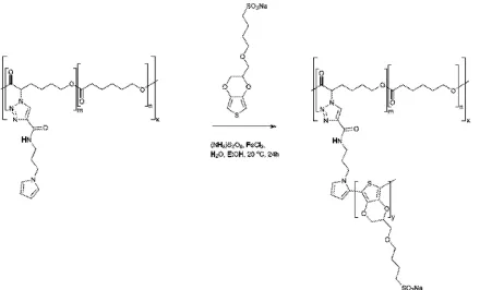
In an embodiment a sulfonate-displaying polycaprolactone derivative can be prepared
The solvent, temperature, time, and other reactant variables can be modified accordingly to
form the same polymer.
Asmentioned above, embodiments of the present disclosure provide for
differentiation of human mesenchymal stemcells. An embodiment of the method includes
introducing human mesenchymal stem cells to the structure including the electroactive
scaffold. The structure and the human mesenchymal stem cells are cultured in an osteogenic
medium. The human mesenchymal stem cells can be periodically provided electrical
stimulation to the human mesenchymal stem cells to cause differentiation of human
mesenchymal stem cells towards osteogenic outcomes.
Electrical stimulation can include direct contact of the material with a power source
via a wire, wireless energy transfer, magnetic force, and the like. The term "periodically"
refers to applying the electrical stimulation at established time frames that may be at regular
or irregular time intervals on the time frames of seconds, hours, days, weeks, or months (e.g.,
about 1 s to 2 months, about 1 hour to 1 day, about 1 day to 1 month, or other the like)
depending upon the specific circumstances. In an embodiment, the impulses of the electrical
stimulation can last on the time frame of seconds, hours, or days (e.g., about 1 second to 1
day, about 10 seconds to 1 hour, about 1 minute to12 hours, about 1 hour to 1 day, or the
like) depending upon the specific circumstances. In an embodiment, the electrical
stimulation can be in the range of millivolts to volts (e.g., about 10mV to 10volts, about 1
of the electrical stimulation can be designed based on particular circumstances and
requirements of a specific situation.
In a particular embodiment, the electroactive scaffold can be designed to enable the
growth of biominerals on the surface of the electroactive scaffold. In an embodiment, the
biomineral silica (Si02) can be grown on the electroactive scaffold when the R group is
. The amine functional group is conducive to growing the biomineral silica
because it is positively charged under physiological pH conditions and interacts with silicic
acid(Si(OH)4),which is negatively charged to produce a layer of neutral silica (Si02) .
In an embodiment, calcium carbonate, calcium phosphate, or a combination thereof
canbe grown on the electroactive scaffold when the R group displays a negatively charged
moiety such as a carboxylic acid or sulfonic acid. Examples of such monomers include
PEDOT-carboxylic acid (PEDOTY-C02H): ' Negatively charged functional
groups (e.g.,carboxylic acids, C02H) are conducive to growing the calcium carbonate,
calcium phosphate, or a combination thereof because they form complexes with the
positively charged calcium 2+ ions (switching the charge on the surface of the material)
enabling the deposition of negatively charged ions(e.g.,carbonate or phosphate anions), and
this process can be repeated to obtain a layer of the desired thickness (nm to mm scale).
In an embodiment, calcium carbonate, calcium phosphate, or a combination thereof
sulfonate moiety, e.g., PEDOT-sulfonate (PEDOT-S0 3Na) . The sulfonic
acidfunctional groups are conducive to growing the calcium carbonate, calcium phosphate,
or a combination thereof because they form complexes with the positively charged calcium
2+ions (switching the charge on the surface of the material) enabling the deposition of
negatively charged ions (e.g., carbonate or phosphate anions), and this process can be
repeated to obtain a layer of the desired thickness (nm to mm scale)
In an embodiment the osteogenic medium is based on standard cell culture medium
with the optional addition of other components such asserum, non-essential amino acids,
bone morphogenetic protein 2 (BMP-2), dexamethasone, β-glycerophosphate, ascorbic acid,
ascorbic acid-2-phosphate, heparin, retinoic acid, and 1,25-dihydroxycholecalciferol (for
example: high glucose Dulbecco's Modified Eagle Medium (DMEM, 425 mL); fetal bovine
serum (50 mL); antibiotic-antimycotic (5mL); non-essential amino acids (5mL),
dexamethasone (100 nM), β-glycerol phosphate (10 mM) and ascorbic acid (50 µΜ)). The
volume of medium used should be in line with the recommended guidelines of the
manufacturer of the cell culture dishes.
As statedabove, embodiments of the present disclosure provide for a structure having
anelectroactive scaffold. In an embodiment, the electroactive scaffold can include one or
more agents (e.g., a chemical or biological agent), where the agent can be disposed indirectly
or directly onthe electroactive scaffold. As described herein, the agent can include a stem
cell suchasa human mesenchymal stem cell. In particular, human mesenchymal stem cells
(e.g., oncollagen-1 coated substrates) and the differentiated products of the stem cells are
within the electroactive scaffold. Furthermore, the electroactive scaffold includes ALP,
which represents asteptowards formation of bone tissue.
In addition, an additional agent that can be disposed on the electroactive scaffold can
include, but is not limited to, a drug, a therapeutic agent, a radiological agent, a small
antibodies (monoclonal or polyclonal)), antigens, nucleic acids (both monomeric and
oligomeric), polysaccharides, haptens, sugars, fatty acids, steroids, purines, pyrimidines,
ligands, and aptamers) and combinations thereof, that can be used to image, detect, study,
monitor, evaluate, and the like, the differentiation of the stem cells. In an embodiment, the
agent is included in an effective amount to accomplish its purpose (e.g.,ALP production),
where such factors to accomplish the purpose are well known in the medical arts.
In general, the agent can be bound to the structure by a physical, biological,
biochemical, and/or chemical association directly or indirectly by a suitable means. The term
"bound" can include, but is not limited to, chemically bonded (e.g., covalently or ionically),
biologically bonded, biochemically bonded, and/or otherwise associated with the
electroactive supramolecular polymeric assembly. In an embodiment, being bound can
include, but is not limited to, a covalent bond, a non-covalent bond, an ionic bond, a chelated
bond, aswell asbeing bound through interactions suchas,but not limited to, hydrophobic
interactions, hydrophilic interactions, charge-charge interactions, π-π stacking interactions,
combinations thereof, and like interactions. In an embodiment, cell-electroactive scaffold
interactions could be controlled through the inclusion of cell-adhesive peptides (e.g.,RGD,
YIGSR, KQAGDV, KHIFSDDSSE, KRSR), protease-labile domains (e.g.,APGL,VRN, or
indeed oligoalanines suchasthose in the backbone of MTTl and MTT2 that are degraded by
elastase), osteoinduction can be enhanced (e.g.,NSPVNSKIPKACCVPTELSAI), and
directing mineralization (e.g.FHRRIKA).
While embodiments of the present disclosure are described in connection with the
Examples and the corresponding text and figures, there is no intent to limit the disclosure to
the embodiments in these descriptions. On the contrary, the intent is to cover allalternatives,
modifications, and equivalents included within the spirit and scope of embodiments of the
present disclosure.
EXAMPLE 1 :
Tissue scaffolds allowing the behavior of the cells that reside on them to be controlled
areof particular interest for tissue engineering. Herein we describe biomineralized
conducting polymer-based bone tissue scaffolds that facilitate the electrical stimulation of
human mesenchymal stem cells, resulting in enhancement of their differentiation towards
Bone conditions requiring surgical intervention are of growing importance in societies
with populations in which life expectancies are increasing, motivating the development of
pro-regenerative biomaterials.1Non-biodegradable materials (e.g. titanium), biodegradable
materials (e.g. biopolymers, calcium phosphate cements) and multifunctional materials that
combine habitats for the cells with the capability to deliver drugs, have been investigated as
potential bone tissue scaffolds.1Biomineralized materials are commonly investigatedasbone
tissue scaffolds, because the presence of the biomineral in the scaffold may promote
osteogenesis.2
Conducting polymer (CP)-based biomaterials (suchasderivatives of polyaniline,
polypyrrole or polythiophene), have potential for both long term biomedical applications (e.g.
electrodes) and short term biomedical applications (e.g. drug delivery or tissue engineering).3
CP-based scaffolds have been developed for the regeneration of bone, muscle and nerve
tissue.3Langer and coworkers first reported the use of CP-based materials for their
applicationasbone tissue scaffolds.4Theapplication of a potential difference of 20 mV mm 1
over 2-dimensional polypyrrole films encouraged bone marrow-derived stromal cells to
differentiate towards osteogenic outcomes (assayedasanincrease in alkaline phosphatase
(ALP)activity per cell relative to non-stimulated control substrates).4
A variety of research groups have reported further developments in conducting
polymer-based materials for bone tissue engineering in the absence5orpresence6of an
electrical field,commonly finding improved osteogenesis for the electrically stimulated
samples. Moreover, the success of inorganic bone substitutes in the clinic has led researchers
to develop conducting polymer-based coatings for calcium phosphate-based,7steel-based,8
andtitanium-based9biomaterials which offer a method of directly electrically stimulating
cellsresiding on the materials, or delivering a drug from such a coating upon the application
of an electrical stimulus.10
Inthis Example we describe the preparation of polycaprolactone (PCL derivatives
displaying pyrrole moieties from which conducting polymers (such aspolypyrrole or
polythiophene derivatives) can be grown. Polymers displaying amines, carboxylates or
sulfonates(Figure 1) facilitate mineralization of silica or calcium carbonates or phosphates.
Theseconducting bone tissue scaffolds enable electrical stimulation of human mesenchymal
stemcellswhich promotes their differentiation towards osteogenic outcomes.
Propiolic acid was coupled to aminopropylpyrrole1 112bycarbodiimide-mediated
moiteties by Cu(I)-mediated triazole formation (Scheme S2), after which the copper was
removed by incubation in a solution of ethylenediaminetetraacetic acid (EDTA). 4 The
material was extensively washed to remove traces of EDTA and vacuum dried yielding
pyrrole-displaying PCL derivative (depicted in Figure 1) with n =5.0kDa and w n of
1.95 (Figure 5)in the form of a light brown powder. Films of the resulting polymer were
solution cast on either commercially available tissue-culture treated Corning® Costar® tissue
culture plates (TCP) or glass. An interpenetrating network of either amine displaying
polypyrrole derivative (PPy-NH2, Figure 1) or carboxylate displaying
poly(3,4-ethylenedioxythiophene) derivative (PEDOT-CO2H ,Figure 1)were generated by incubation
of the pyrrole-functionalized PCL films in aqueous solutions of the appropriate pyrrole and
EDOT derivatives in the presence of the initiators ammonium persulfate and ferric chloride
(Scheme S3 and S4, respectively).15 Films of the amine or carboxylate derivative displaying
films were washed thoroughly with water to remove the by-products (e.g. initiators,
monomers, oligomers and polymers) and vacuum dried. The brown-black PPy-NH2films
were biomineralized with silica and those of the blue-grey PEDOT-C0 2H were
biomineralized with calcium phosphate. Energy dispersive X-ray (EDX) analysis of the films
confirms that their surface chemistry is different. Peaks in the EDX spectra of the PCL
derivatives displaying pyrrole moieties have lines at 0.277 and 0.525 keV that are the
characteristic Ka emissions of carbon and oxygen, respectively, and the very weak emission
at 0.392 keV is the Ka emission of nitrogen (Figure 2A-E). The peaks in the spectra of the
films after the polymerization reactions at2.621 and 6.398 keV are characteristic Ka
emission lines of chlorine and iron, the peak at 0.705 keV is the La emission line of iron
(Figure 2B-E), and the peak at 2.307 keV is the Ka emission line of sulphur present in the
backbone of the PEDOT-C0 2H (Figure 2D and 2E). The successful biomineralization of the
PPy- H2films (Figure 2B) with silica is clear from the appearance of the Ka emission peak
of silicon at 1.739keV (Figure 2C). Likewise, the successful biomineralization of the
PEDOT-C0 2H films (Figure 2D) with calcium phosphate is clear from the appearance of the
peaks at 2.013 and 3.690 keV, that are characteristic of the Ka emissions of phosphorous and
calcium, respectively (Figure2E). The inset SEM images show the surface morphologies of
the films (Figure 2A-E), with nanometer to micrometer scale pores present on the surface of
the biomineralized films (Figure 2B-E).
The electrical sheet resistance of the biomineralized samples was measured in
accordance with the method described by Schmidt1 1'16 and Zhang.17The PPy- H
biomineralized with silica had sheet resistances of 3 1.6± 9 .1 k , and those of PEDOT-CO2H
biomineralized with calcium phosphate had sheet resistances of 248.6 ± 71.8 k , which is of
a similar order of magnitude to interpenetrating networks of polypyrrole and
polystyrenesulfonate in PCL (68.0 ± 18.1 kQ). 6While the electrochemical stability of the
polypyrrole and PEDOT derivatives are known to decrease over long periods of time which
may be problematic for biointerfaces intended for long term use,18we and others have found
them to be acceptable for the short term stimulation of cells residing in tissue scaffolds such
asthose reported here.3'4'61 1'16 7
To investigate the potential of the biomineralized CPs to act as bone tissue scaffolds,
we seeded human mesenchymal stem cells (HMSCs) on their surfaces and cultured them in
osteogenic medium for 3 weeks. We seeded sixdifferent systems: 1) cells seeded on TCP
controls; 2) cells seeded on PCL (80 kDa); 3) cells seeded on silica-coated PPy- H2films
without electrical stimulation; 4) cells seeded on silica-coated PPy- H2films with electrical
stimulation; 5) cells seeded on silica-coated PEDOT-C0 2H films without electrical
stimulation; 6) cells seeded on silica-coated PEDOT-C0 2H films with electrical stimulation.
Those samples that were electrically stimulated were cultured for 2 days without stimulation,
followed by four periods of stimulation at 10mV mm 1for 8 hours then 40 hours without
stimulation, and no stimulation thereafter).
After 3 weeks in culture, cells were fixed with paraformaldehyde and cell nuclei and
actin filaments within cells were stained with 4',6-diamidino-2-phenylindole (DAPI) and
Alexa Fluor® 488 Phalloidin, respectively. We observed that cells were homogeneously
distributed on the TCP and PCL controls, and that cells had infiltrated the biomineral
coatings on the biomineralized CP films (Figure 3A-F) which is promising for their
integration in the body where infiltration of cells such asmacrophages and osteoclasts
facilitates remodelling of implanted biomaterials. 19The differentiation of the cell population
towards osteogenic fates was shown using a biochemical assay for alkaline phosphatase
(ALP) activity which is a characteristic marker of bone formation. To within experimental
error, ALP activity of cells cultured on the TCP and PCL control substrates was the same
(Figure 4). ALP activity for cells cultured on the conductive biomineralized scaffolds was
reduced relative to the TCP and PCL control substrates, which is likely to be because of
subtle differences in cell-matrix interactions as observed for analogous systems.20
Interestingly, ALP activity of cells cultured on the scaffolds mineralized with calcium
which is likely to be because the calcium phosphate acts as a source of calcium and
phosphate ions enabling the production of calcified extracellular matrix.2 1Furthermore, the
ALP activity of cells cultured on the conductive biomineralized scaffolds was increased after
electrical stimulation (four periods during which a potential step of 10 mV mm1was applied
acrossthe conductive substrates for 8 hours), which is in line with reports by Langer4and
others.6Therefore, our biochemical analysis reveals that while the non-conductive scaffolds
support differentiation of HMSCs towards osteogenic outcomes, the application of an
electrical stimulusto HMSCs residing in a conductive scaffold enhances levels of ALP
activity which is a hallmark of bone tissue formation.
Conclusions
Pro-regenerative biomaterials for the treatment of bone conditions and disorders that
require surgical intervention are of growing importance in modern societies in which life
expectancies are increasing. Bone tissue scaffolds that control the behaviour of cells residing
onthem are particularly interesting for such applications. We report the first examples of
biomineralized conductive bone tissue scaffolds and show that the electrical stimulation of
HMSCs residing thereon enhances levels of ALP activity, which represents an important step
towards the formation of bone tissue.
Calcium carbonateisincreasingly interesting in biomedicine asa novel scaffolds for
bone tissue engineering,22 andit is possible to biomineralize PEDOT-CO
2Hfilms with
calcium carbonate (Figure6).While it is possible to biomineralize analogous materials
incorporating interpenetrating networks of sulfonate displaying PEDOT-S0 3Na (Figure 1,
Scheme S5)23with calcium-based biominerals we found them to be mechanically unstable
during long term cell culture experiments. PEDOT-S03Na is the most hydrophilic/water
solubleof the conducting polymers tested, which is likely to increase rates of enzymatic
degradation of the PCL matrix as we have observed for interpenetrating networks of PCL
with water insoluble polyplexes of polypyrrole/polystyrenesulfonate.16Moreover, we know
that such PCL/polypyrrole/polystyrenesulfonate-based materials are stable to long term cell
culture,16 andallow the growth of calcium-based biominerals suchascalcium carbonate
(Figure7).
We believe it should be possible to prepare a variety of conductive biomineralized
tissue scaffolds by chemical modification of the scaffolds with peptides directing the
cell behaviour(e.g. RGD, YIGSRorKRSR forcell adhesion, and
NSPVNSKIPKACCVPTELSAIfor osteoinduction),24therebyallowing usto tailorthe
propertiesofthe scaffoldtospecificnicheapplications (and potentially specific patients).
1 A .Atala,J .Tissue Eng. Regen. Med., 2007, 1,83; J . O .Hollinger, S .Winn andJ .
Bonadio, Tissue Engineering, 2000, 6,341; C .T. Laurencin, A .M . A .Ambrosio, M .
D .Borden andJ .A .Cooper, Jr.,Annu. Rev. Biomed. Eng., 1999, 1, 19; J .R .Porter, T.
T. Ruckh and K .C .Popat, Biotechnol. Prog, 2009, 25, 1539; M . A .
Fernandez-Yague, S .A .Abbah, L .McNamar, D .I .Zeugolis, A .Pandit and M .J .Biggs,Adv.
DrugDeliv. Rev., 2014, http://dx.doi.Org/10.1016/j.addr.2014.09.005; D .Marolt, M .
Knezevic and G .Vunjak-Novakovic, Stem CellRes. Ther., 2010, 1, 10; W .L .
Grayson, T. P. Martens, G .M .Eng., M .Radisic and G .Vunjak-Novakovic, Semin.
Cell Dev. Biol., 2009, 20, 665; M .Frohlich, W .L .Grayson, L . Q .Wan, D .Marolt, M .
Drobnic and G .Vunjak-Novakovic, Curr. Stem CellRes. Ther., 2008, 3,254.
2 N . M .Alves, I .B .Leonor, H .S .Azevedo, R . L .Reis andJ .F. Mano, J .Mater. Chem.,
2010, 20, 2911; S .V . Dorozhkin and M .Epple,Angew. Chem. Int. Ed., 2002, 41,
3130; A .R .Boccaccini, M .Erol, W .J . Stark, D .Mohn, Z .Hong and J .F . Mano,
Comp. Sci. Technol, 2010, 70, 1764; F.C .Meldrum and H .Colfen, Chem. Rev.,
2008, 108, 4332; S .Heinemann, T. Coradin and M .F. Desimone, Biomater. Sci.,
2013, 1,688; C .Liand D .L .KALPan, Curr. Opin. Solid State Mater. Sci., 2003, 7,
265;N . M .Alves, I .B .Leonor, H .S .Azevedo, R . L .Reis and J .F . Mano,J .Mater.
Chem., 2010, 20, 2911;A .Dey, G .de With and Nico A .J .M .Sommerdijk, Chem.
Soc. Rev., 2010, 39, 397; A .J .Salinas, P. Esbrit andM .Vallet-Regi, Biomater. Sci.,
2013, 1,40; E .Ko and S.-W. Cho,Int.J . Stem Cells, 2013, 6,87.
3 B .Guo, L .Glavas and A .C .Albertsson, Prog. Polym. Sci., 2013, 38, 1263; M .
Muskovich and C . J .Bettinger, Adv. Healthcare Mater., 2012, 1,248; M .Berggren
and A .Richter-Dahlfors, Adv. Mater., 2007, 19, 3201; R .Balint, N .J .Cassidy and S .
H .Cartmell, Acta Biomaterialia, 2014, 10, 2341; R .A .Green,N . H .Lovell, G . G .
Wallace and L .A .Poole-Warren, Biomaterials, 2008, 29, 3393; Mihai Irimia-Vladu,
Chem. Soc. Rev., 2014, 43, 588;N . K .Guimard, N .Gomez and C .E .Schmidt, Prog.
Polym. Sci., 2007, 32, 876;J .G .Hardy, J .Y. Lee andC .E .Schmidt, Curr. Opin.
Biotechnol., 2013, 24, 847; D .Svirskis, J .Travas-Sejdic, A .Rodgers and S .Garg,J .
Geissler, J .K .Chow, L .Nguy, J .M .Kim and C .E .Schmidt,J .Mater. Chem. B,
2014, 2,6809; T . F. Otero andJ .G .Martinez, J .Mater. Chem. B, 2013, 1,26; R .
Gracia and D .Mecerreyes, Polym. Chem., 2013, 4,2206; T.H .Qazi, R .Rai and A .R .
Boccaccini, Biomaterials , 2014, 35, 9068; J .Zimmerman, R .Parameswaran and B .
Tian,Biomater. Sci., 2014, 2, 619.
V . P. Shastri,N .Rahman, I .Martin andR .Langer,Mat. Res. Soc. Symp. Proc, 1999,
550, 215.
C .Rincon andJ .C .Meredith, MacromoI. Biosci., 2010, 10, 258; C .Rincon, C.-C.
Chen andJ . C .Meredith, Macromol. Biosci., 2010, 10, 1536; E .De Giglio, S .
Cometa, C.-D. Calvano, L .Sabbatini, P.G .Zambonin, S .Colucci, A .DiBenedetto
and G .Colaianni, J .Mater. Sci.Mater. Med., 2007, 18, 1781; B .Lakard, L .Ploux, K .
Anselme, F. Lallemand, S .Lakard, M .Nardin and J.Y. Hihn, Bioelectrochemistry ,
2009, 75 148; J .S .Moreno, S .Panero, S .Materazzi, A .Martinelli, M .G .Sabbieti, D .
Agas and G Materazzi, J . Biomed.Mater. Res., 2009, 88A, 832; H .Castano, E .A .
O'Rear, P.S .McFetridge and V.I .Sikavitsas,Macromol. Biosci. 2004, 4,785.
J .Cao, Y. Man and L .Li,Biomedical Reports, 2013, 1,428; L .Liu, P. Li, G .Zhou,
M .Wang, X .Jia, M .Liu, X .Niu, W . Song, H .Liu and Y. Fan,J .Biomed.
Nanotechnol, 2013, 9, 1532;W.-W. Hu, Y.-T. Hsu, Y.-C. Cheng, C .Li,R.-C. Ruaan,
C.-C. Chien, C.-A. Chung and C.-W. Tsao,Mater. Sci. Eng. C,2014, 37, 28; G .Jin
and G .Kim,J .Mater. Chem. B , 2013, 1, 1439; J .Zhang, K .G .Neoh, X .Hu, E.-T.
Kang and W . Wang, Biotechnol. Bioeng, 2013, 110, 1466; Y. Liu, H .Cui,X .Zhuang,
Y. Wei and X .Chen,Acta Biomaterialia, 2014, 10, 5074; H .Cui, Y. Liu, M .Deng, X .
Pang, P . Zhang, X .Wang, X .Chen and Yen Wei, Biomacromolecules, 2012, 13,
2881; H .Cui, Y. Wang, L .Cui, P. Zhang, X .Wang, Y. Wei and X .Chen,
Biomacromolecules, 2014, 15, 3146; S .Meng, M .Rouabhia andZ .
Bioelectromagnetics , 2013, 34, 189; S .Meng, Z . and M .Rouabhia, J .Bone
Miner. Metab., 2011,29, 535.
S .Yala, H .Khireddine, D .Sidane, S .Ziani and F. Bir,J .Mater. Sci., 2013, 48, 7215;
Y. Liu, H .Cui, X .Zhuang, P . Zhang, Y. Cui, X .Wang, Y. Wei andX .Chen;
Macromol. Biosci., 2013, 13, 356.
D .Gopi, S .Ramya, D .Rajeswari, M .Surendiran andL .Kavitha, Coll. Surf. B :
J .Liao,H .Pan, C .Ning, G .Tan,Z .Zhou, J .Chen and S .Huang,Macromol. Rapid
Commun., 2014, 35, 574; J .Liao, Y. Zhu, Z .Yin,G .Tan, C .Ning andC .Mao,J .
Mater. Chem. B, 2014, 2,7872; E .De Giglio, M .R .Guascito, L .Sabbatini and G .
Zambonin, Biomaterials, 2001, 22, 2609; E .De Giglio, L .Sabbatini and P.G .
Zambonin, J .Biomater. Sci. Polymer Edn., 1999, 10, 845; E .De Giglio, L .Sabbatini,
S .Colucci and G .Zambonin, J .Biomater. Sci. Polymer Edn., 1999, 10, 1073; E .De
Giglio, L .De Genarro, L .Sabbatini and G .Zambonin, J .Biomater. Sci. Polymer Edn.,
2001, 12, 63;E .De Giglio, S .Cometa, C.-D. Calvano, L .Sabbatini, P.G .Zambonin,
S .Colucci, A .Benedetto and G .Colaianni, J .Mater. Sci.Mater. Med., 2007, 18,
1781.
0S .Sirivisoot, R .A .Pareta and T.J .Webster, Solid State Phenomena, 2009, 151, 197;
S .Sirivisoot, R .A .Pareta and T.J .Webster, Nanotechnology, 2011,22, 085101.
. Y. Lee, C .A .Bashur, A .S .Goldstein and C .E .Schmidt, Biomaterials, 2009, 30,
4325
J .Y. Lee and C .E .Schmidt, J .Biomed. Mater. Res. Part A , 2014, DOI:
10.1002/jbm.a.35344.
3S .Lenoir, R .Riva, X .Lou, C .Detrembleur, R .Jerome and P. Lecomte,
Macromolecules , 2004, 37, 4055; R .Riva, P . Lussis, S .Lenoir, C .Jerome, R .Jerome
and P. Lecomte, Polymer, 2008, 49, 2023; R .Riva, S .Schmeits, F. Stoffelbach, C .
Jerome, R .Jerome and P. Lecomte, Chem. Commun., 2005, 5334.
.Malkoch, R .Vestberg, N .Gupta, L .Mespouille, P . Dubois, A .F. Mason, J .L .
Hedrick, Q .Liao, C .W . Frank, K .Kingsbury and C .J .Hawker, Chem. Commun.,
2006, 2774.
5R .H .Karlsson, A .Herland, M .Hamedi, J .A .Wigenius, A .Aslund, X .Liu, M .
Fahlman, O .Inganas and P . Konradsson, Chem.Mater., 2009, 21, 1815.
6J .G .Hardy, R . C .Cornelison, R . C .Sukhavasi, R . J .Saballos, P . Vu,D .L .Kaplan
and C .E .Schmidt, Bioengineering, 2015, 2, 15.
7X. P. Jiang, D .Tessier, L .H .Dao andZ .Zhang, J .Biomed. Mater. Res., 2002, 62,
507.
8A .Kros,N . A .J .M .Sommerdijk andR .J .M. Nolte, Sensors and Actuators B , 2005,
106,289.
9 P.M .Mountziaris and A .G .Mikos, Tissue Eng. Part B , 2008, 14, 179; L .J .Raggatt
Fajardo, H . Hagenmuller, K .Nuss, M . Arras, R .Muller, B .von Rechenberg, D .L .
Kaplan, M . P. Merkle andL .Meinel, Eur.J .Pharm. Biopharm., 2013, 85, 119; D .J .
Hadjidakis andI .I .Androulakis, Ann. N . Y.Acad. Sci., 2006, 1092, 385; J .P.
Santerre, R . S .Labow and E .L .Boynton, Can. J .Surg., 2000, 43, 173; N . A . Sims
and T .J .Martin, BoneKEy Reports, 2014, 3, 481; J .C .Crockett, M .J .Rogers, F. P.
Coxon, L .J .Hocking and M . H . Helfrich, J .Cell Sci., 2011, 124, 991.
20G . G .Wallace, M .J .Higgins, S .E .Moulton and C .Wang, Nanoscale, 2012, 4,4327;
M .J .Higgins, P.J .Molino, Z . Yue andG . G .Wallace, Chem.Mater., 2012, 24, 828;
A .Gelmi, M .K .Ljunggren, M . Rafat and E . W . H . Jager, J .Mater. Chem. B ,2014, 2,
3860.
2 1Y.-R. V . Shih, Y. Hwang, A . Phadke, H . Kang, N .S .Hwang, E .J .Caro, S .Nguyen,
M .Siu,E .A .Theodorakis, N .C .Gianneschi, K .S .Vecchio, S .Chien, O .K .Lee and
S .Varghese, PNAS, 2014, 111, 990.
22C .Combes, B .Miaoa, R .Bareilleb, C .Rey, Biomaterials, 2006, 27, 1945; W .
Schneiders, A . Reinstorf., W . Pompe, R .Grass, A . Biewener, M . Holch, H . Zwipp and
S .Rammelt, Bone, 2007, 40, 1048; K . Sariibrahimoglu, S .C .G .Leeuwenburgh, J .G .
C .Wolke, L .Yubao, J .A .Jansen; J .Biomed. Mater. Res. Part A ,2012, 100A, 712; A .
H .Dewi, I .D .Ana, J .Wolke, J .Jansen, J .Biomed. Mater. Res. PartA ,2013, 101A,
2143; H . Zhu, J . Schulz, H . Schliephake, Clin. Oral Implants Res., 2010, 21, 182.
23 O .Stephan, P. Schottland, P.-Y. Le Gall, C .Chevrot, C .Mariet and M . Carrier,
Electroanal. Chem., 1998, 443, 217; M . Yamada, N . Ohnishi, M . Watanabe and Y.
Hino, Chem. Commun., 2009, 7203.
24 K .G .Sreejalekshmi and P. D . Nair, J .Biomed. Mater. Res., Part A ,2011,96A, 477;
H .Shin, S .Jo and A .G .Mikos, Biomaterials, 2003, 24, 4353.
SUPPLEMENTARY INFORMATION FOR EXAMPLE 1:
Materials
Unless otherwise stated, allchemicalsfor synthesis and physicochemical analysis
were of ACS grade, purchased from Sigma-Aldrich and used asreceived without further
purification. Phosphate buffered saline (PBS) was at pH7.4. Reagents forcell culturewere
purchased from Invitrogen (Carlsbad, CA) unless otherwise noted. Human mesenchymal
stemcells (HMSCs) froma 24 year old drug- and disease-free male were purchased from
Experimental methods
1
H and C NMR spectra were recorded on a Varian Mercury 400 MHz MR
spectrometer, using residual solvent 1H peaks as internal references for the 1H NMR spectra.
Mass spectra were recorded on an Agilent 6530 QTOF mass spectrometer in electrospray
ionization mode. Infrared spectroscopy was carried out on a Thermo Scientific Nicolet 380
FT-IR Spectrometer (Thermo Fisher Scientific Inc., USA). Spectra were recorded in ATR
mode at2 1°C, with a 1 cm- 1resolution and 64 scans (corrected for background and
atmosphere using OMNIC software provided with the spectrometer). Gel permeation
chromatography (GPC) was performed on a Viscotek GPCmax Solvent/Sample Module. Two
fluorinated polystyrene columns (FMBHW-3078 and FMBLMW-3078) were used in series
and maintained at 22°C.DMF was used asthe eluent at a flow rate of 1 mL min . Detection
was performed using a Viscotek V E 3580 refractive index detector. Molecular weight and
dispersity data are reported relative to polystyrene standards in DMF with 0.01 M LiBr at 40
°C, and a commercially available sample of polycaprolactone (GPC: M =40.1 kDa, w
of 2.02) from Polysciences Inc., (Warrington, PA, USA) in DMF at 22°C.Differential
scanning calorimetry (DSC). DSC experiments were carried out with a DSC Q100 (TA
Instruments, USA), using airtight aluminum pans. Material was weighed into aluminum pans
(TA Instruments, USA), and analyses were carried out under a nitrogen atmosphere (flow
rate of 50mL min- 1) .The samples were treated asfollows: heated from room temperature to
125 °C (10 °Cmin 1),cooled to30 °C(10 °Cmin 1),heated from 30 °Cto 125 °C (5°C
min 1) and finally cooled to30 °C(5 °Cmin 1) .The first temperature ramp removed traces of
volatile solvents and the T from the second ramp is reported.
Synthesisof alkyne-displaying aminopropylpyrrole derivative
Aminopropylpyrrole (3.6 g,29 mmol), propiolic acid (1.4g,20 mmol),
dicyclohexylcarbodiimide (DCC, 6.0 g,29 mmol), hydroxybenzotriazole (HOBt, 3.9g,29
mmol) and triethylamine (3.1 g,4.4 ml, 29 mmol) were stirred for48hours in
dichloromethane (DCM, 100mL), after which the mixture was filtered to remove the
precipitated dicyclohexylurea. The filtrate was washed with aqueous solutions of NaHS0 4
(160 g L ),NaHC0 3(saturated), NaHS0 4, NaHC0 3, water and finally brine. The solution
was dried over MgS04 and the volatiles removed with a rotary evaporator. The crude product
was purified by silica column chromatography (eluting with a gradient of DCM 100%,to
CHC13 100%,to DCM:MeOH, 98:2) to give the product, a viscous oil,in a yield of (2.3 g,
5.87 (1H, CONH), 3.95 (2H, CH2),3.29 (2H, CH2),2.75 (2H, CH2) .ESI-MS (m/z) calculated
for C10H12N2O [M + H]+requires 177.09; found, 177.10. IR (ATR)Vmaxcm 13344(CH
alkyne), 3224 ( H , amide), 2105 (alkyne), 1636 (amideI), 1530 (amideII). See Scheme SI.
SchemeSI. Synthesis of alkyne-displaying aminopropylpyrrole derivative
Synthesisof pyrrole-displaying polycaprolactone derivative
To a solution of 2-chlorocyclohexanone (25 g, 189 mmol) in DCM (250 mL) was
added 3-chloroperbenzoic acid, (MCPBA, 50 g,290 mmol) and the reaction mixture was
stirred at room temperature under an atmosphere of nitrogen for96hours. The reaction
mixture was cooled to -20 °C, resulting in the precipitation of residual 3-chlorobenzoic acid.
After filtration, the solution was washed three times with a saturated aqueous solution of
NaHS0 3, three times with an aqueous solution of NaHC0 3, and once with water. The organic
phase was dried over MgS04, filtered, and the solvent was removed on a rotary evaporator.
The residue was distilled under reduced pressure, yielding a-chloro-s-caprolactone (22g,
boiling point 75-77°C at0.1 mm Hg). ε-caprolactone (4.0g), a-chloro-s-caprolactone (1.0
g), stannous stearate (0.212 g) and ethanol (1.8 ) were dissolved in toluene (15 mL) and
stirred under nitrogen for 24 hours at room temperature, after which the product was
precipitated in cold hexanes, dissolved in DCM and reprecipitated in cold hexanes and dried
under high vacuum for48hours, affording poly(s-caprolactone-co-a-chloro-s-caprolactone)
asa white powder in a yield of 2.26g .Poly(s-caprolactone-co-a-chloro-s-caprolactone) (2.26
g)and sodium azide (3.045 g) were dissolved in dimethylformamide (DMF, 15mL) and
stirred under nitrogen for 24 hours at room temperature, after which the product was
precipitated in water. The polymer was redissolved in DMF and reprecipitated in water,
isolated by filtration under vacuum, and dried under high vacuum for48hours, affording
poly(s-caprolactone-co-a-N 3-s-caprolactone) asa white powder in a yield of 2.86g .
Poly(s-caprolactone-co-a-N3-s-caprolactone) (1.14 g), alkyne-displaying aminopropylpyrrole
0.03g) were stirred under nitrogen for 24 hours at35 °C. The polymer was precipitated in
water and the solids were incubated in 50mL of an aqueous solution of
ethylenediaminetetraacetic acid (EDTA, 5gin 100 mL water, pH 8)for 24 hours at 4°C, after
which the EDTA solution was decanted and replaced with fresh EDTA solution and
incubated for a further 24 hours at 4°C, after which the EDTA solution was decanted and
replaced with water (50 mL) in which it was incubated for 24 hours at 4°C, after which the
water was decanted and replaced with fresh water (50 mL) in which it was incubated for 24
hours at 4 °C, after which the water was decanted and the polymer dried under high vacuum
for 48hours. This process afforded the pyrrole-displaying polycaprolactone derivative in a
yield of 1.1 g in the form of a light brown powder. 1H M R (400 MHz, CDC1
3)δΗ7.52 (CH
triazole), 7.09 (CONH), 6.53 (CH -p oie),6.15 (CH -p oie),4.21, 4.04, 3.86, 3.65, 3.29, 2.32,
1.92, 1.64, 1.38, 1.10. IR (ATR) vmaxcm 13326 (ML amide), 2099 (residual azide), 1721
(ester), 1622 (amide I), 1571 (amide II), 1457 (triazole). GPC: = 5.0kDa ( w/ of
1.95).DSC: T =54.5 °C. See Scheme S2.
Preparation of films of pyrrole-displaying polycaprolactone derivative
Solutions of the pyrrole-displaying polycaprolactone derivative in chloroform (0.5g
in 20 mL) orhexalfuoroisopropanol (0.5 g in 20 mL) were cast on glass slides or in
tissue-culturetreated Corning® Costar® tissue culture plates, respectively. The solvent was allowed
to evaporate for 24 hours and the films were subsequently dried under high vacuum for48
hours.
Preparation of conductive films displaying amines
Aminopropylpyrrole (0.2 g) was dissolved in ethanol (2mL), and addedto a solution
of ammonium persulfate (0.29g), andferric chloride (0.005g) in water (20 mL). 0.2mL of
this solution was coated on the surface of films of the pyrrole-displaying polycaprolactone
derivative and allowed to polymerize for 24 hours, after which the brown-black films were
washed extensively with water, followed by rinsing with aqueous ethanol (70%) and drying
under high vacuum for48hours. See Scheme S3.
SchemeS3. Synthesisof amine-di splaying polycaprolactone derivative
Preparation of conductive films displaying carboxylic acids
EDOT carboxylic acid (0.2 g), ammonium persulfate (0.29 g), andferric chloride
(0.005g) inwater (20mL). 0.2mL of this solution was coated on the surface of films of the
which the blue-grey films were washed extensively with water, followed by rinsing with
aqueous ethanol (70%) and drying under high vacuum for48hours. See SchemeS4.
SchemeS4. Synthesis of carboxylic acid-displaying polycaprolactone derivative
Preparation of conductive films displaying sulfonates
Hydroxymethyl EDOT (3 g, 17.8 mmol, Sarchem laboratories, Inc., Farmingdale, NJ,
USA) and sodium hydride (0.47g, 19.6 mmol, 1.1 eq.) and were stirred and heated at reflux
in toluene (50 mL) for 2 hours. Butane sultone (2.42g, 17.8mmol, 1 eq.) was added and the
mixture was heated at reflux for a further 2 hours. After cooling to room temperature, the
product was precipitated in acetone, isolated by filtration and subsequently dried under high
vacuum for48hours. The process yielded a brown powder, EDOT-S (3.71 g, 11.4mmol). 1H
M R (400 MHz, D20 )δΗ6.36, 4.70, 4.26, 4.12, 3.94, 3.59, 3.44, 2.78, 1.64, 1.55. EDOT-S
(0.2g), ammonium persulfate (0.29g), and ferric chloride (0.005g) in water (20 mL). 0.2mL
of this solution was coated on the surface of films of the pyrrole-displaying polycaprolactone
derivative and allowed to polymerize for 24 hours, after which the blue-black films were
washed extensively with water, followed by rinsing with aqueous ethanol (70%) and drying
under high vacuum for48hours. See Scheme S5.
Preparation of silica-coated conductive films
0.2mL of phosphate buffer (pH 5.5) was added to conductive films displaying
amines. Tetraethylorthosilicate (2.33 mL), water (3.85 mL), ethanol (3.85 mL) and 1N HC1
(0.1 mL)were mixed and incubated for 10 minutes at room temperature, and aliquots of this
solution (20 L) were added to the phosphate buffer covered the amine-displaying conductive
films. Thereaction mixture was incubated for 1 hour after which it was removed and the
films were washed thoroughly with Millipore water, rinsed with aqueous ethanol (70%) and
dried under high vacuum for48hours.
Preparation of calcium phosphate-coated conductive films
Conductive films displaying carboxylic or sulfonic acids were incubated in an
aqueous solution( 1mL) of calcium chloride (200 mM) for 20 minutes, after which the
solution was removed and the samples were washed with water (3x 1 mL). Thereafter,
sampleswere incubated in an aqueous solution ( 1mL)of sodium phosphate (120 mM) for 20
minutes, after which the solution was removed and the samples were washed with water (3 x
1mL). The cycle of incubation with calcium chloride and sodium phosphate was repeated a
further sixtimes (i.e. a total of 7 cycles), after which the samples were washed with water,
Preparation of calcium carbonate-coated conductive films
Conductive films displaying carboxylic or sulfonic acids were incubated in an
aqueous solution( 1mL) of calcium chloride (10 mM). The films were placed in an airtight
container with a beaker containing ammonium carbonate for 24 hours. The samples were
subsequently washed with water until the pH was neutral, after which the samples were
washed with water, rinsed with aqueous ethanol (70%) and dried under high vacuum for48
hours.
Electrical properties
Resistance (R inΩ) was measured between the two silver electrodes using a digital
multimeter (DM-8A, Sperry Instrument, Milwaulkee, WI). Sheet resistance (Rs)inΩ/square
was calculated asfollows:
= i
where W is the width of the electrode and L is the distance between the two silver electrodes.
The electrodes were moved to different positions after each measurement, and the resistance
R was recorded in at least ten different positions on the materials.
Scanningelectron microscopy (SEM) and energy dispersive X-ray (EDX) spectroscopy
Samples were mounted on a Scanning Electron Microscopy (SEM) stuband sputter
coated with carbon. All samples were imaged using a Hitachi S5500 SEM field emission
scanning electron microscope equipped with an energy dispersive spectroscopy probe located
atthe Texas Materials Institute.
Invitro culture of human Mesenchymal stem cells without electrical stimulation
HMSCs were supplied by Lonza (Walkersville, MD). Samples were inserted in
untreated polystyrene tissue culture plates and sterilized by incubation in 70% ethanol
followed by exposure to UV for 60min. After sterilization, the samples were incubated for
30minutes in 24 well plates containing HMSC growth medium that was composed of: high
glucose Dulbecco's Modified Eagle Medium (DMEM, 440 mL); fetal bovine serum (50 mL);
antibiotic-antimycotic (5mL); non-essential amino acids (5mL), and2 ng mL 1basic
fibroblast growth factor. Medium was aspirated and replaced prior to HMSC seeding. Cell
viability before starting the experiment was determined by the Trypan Blue exclusion
method, and the measured viability exceeded 95% in allcases. HMSCs were seeded at
10,000 cellsper cm2, and incubated at37 °C, 95% humidity, and a C0
2content of 5% .After
3 days the medium was aspirated, the materials were washed gently with PBS and transferred
to a fresh 24 well plate containing osteogenic medium that was composed of: high glucose
antibiotic-antimycotic (5mL); non-essential amino acids (5mL), dexamethasone (100 nM),
β-glycerol phosphate (10 mM) and ascorbic acid (50 µΜ) .Thereafter the osteogenic medium
was aspirated and replaced every 2 days until the samples were analysed.
Invitro culture of human Mesenchymal stem cells with electrical stimulation
Electrical stimulation of Human Mesenchymal Stem cells was achieved employing a
custombuilt setup. Non-conductive glass slides, polycarbonate wells (square polycarbonate
blocks, thickness of 1cm, sides of2.5 cm, with square holes with sides of0.9 cm cut out),
Dow Corning®high vacuum grease, and medium binder clips (Staples®, Framingham, MA)
were sterilized by autoclaving. Holes were drilled into the sides of 10 cmpolystyrene Petri
dishes using a Dremel saw (Lowes, Mooresfield, NC, USA), and the plates were sterilized by
exposure to UV for 60min. Adhesive-backed copper tape (5mm width, Ted Pella, Inc.),
waterproof Kapton®tape ( 1cmwidth, Fisher Scientific, Waltham, MA, USA), wires and
alligator clips were sterilized by exposure to UV for60min.
Electroactive PCL-based tissue scaffolds were placed on glass slides and secured in
position with two thin strips of adhesive-backed copper tape that were attached to the
scaffolds, parallel to one another and separated by a distance of ca. 4cm. One face of the
polycarbonate wells was coated with vacuum grease and placed on the electroactive tissue
scaffolds, greased side down, in contact with the glass slide. A binder clip on either side of
the well was used to secure this in position and render it water tight. A strip of copper tape
was run between the parallel copper strips attached to the scaffolds and the ends of the slides
aspoints of contact for the alligator clip-terminated wires attached to the multipotentiostat
(CH Instruments, Austin, TX, USA). The counter and reference electrodes were connected
together and clipped to copper tape on one side of the slide, andthe working electrode was
clipped to copper tape on the other side of the slide.
The setup was sterilized by exposure to UV (30 minutes), the samples were incubated
for 30minutes in HMSC growth medium that was composed of: high glucose Dulbecco's
Modified Eagle Medium (DMEM, 440 mL); fetal bovine serum (50 mL);
antibiotic-antimycotic (5mL); non-essential amino acids (5mL), and2 ng mL 1basic fibroblast growth
factor. Medium was aspirated and replaced prior to HMSC seeding. Cell viability before
starting the experiment was determined by the Trypan Blue exclusion method, and the
measured viability exceeded 95% in allcases.HMSCs were seeded at 10,000 cells per
scaffold, and incubated at37 °C, 95% humidity, and a C02content of 5% .After 2 days the
24well plate containing osteogenic medium that was composed of: high glucose Dulbecco's
Modified Eagle Medium (DMEM, 425 mL); fetal bovine serum (50 mL);
antibiotic-antimycotic (5mL); non-essential amino acids (5mL), dexamethasone (100 nM), β-glycerol
phosphate (10 mM) and ascorbic acid (50 µΜ) .
The tips of the wires attached to the samples were wound around alligator
clip-terminated wires attached to the multipotentiostat (CH Instruments, Austin, TX, USA). The
counter and reference electrodes were connected together and clipped to the wire protruding
from one end of the sample, and the working electrode was clipped to the wire protruding
from the other side of the sample. Wires and alligator clips were secured in position with
adhesive copper tape (Ted Pella, Inc., Reading, CA, USA) and wrapped in Parafilm® to
render them electrically insulating and waterproof (i.e. suitable for use inside an incubator).
The electrical stimulation paradigm was asfollows: a potential step of 10mV mm 1was
placed across the samples for the duration of 8 hours followed by 40 hours without
stimulation, repeated three times (32 hours of stimulation in total), after which the wires were
disconnected and the substrates cultured asnormal. Throughout the electrical stimulation
experiments the osteogenic medium was aspirated and replaced every 2 days. Thereafter the
osteogenic medium was aspirated and replaced every 2 days until the samples were analysed.
Biochemical assays
The DNA content and Alkaline Phosphatase (ALP) activity of samples that were
broken up in a buffer of 0.2% Triton X-100 were quantified concurrently, using the
PicoGreen® assay (Life Technologies, Thermo Fisher Scientific Inc., USA) for DNA
quantitation in accordance with the manufacturer's protocol, a SensoLyte® pNPP Alkaline
Phosphatase Assay Kit (AnaSpec, Inc., Freemont, CA,USA) for ALP quantitation in
accordance with the manufacturer's protocol, and a Synergy HT Multi-Mode Microplate
Reader (Bio-tek US, Winooski, VT).
Fluorescence staining and imaging of cells
Cells fixed with paraformaldehyde were permeabilized with 0.1% Triton X-100
(Fluka) and 2% bovine serum albumin (BSA) in PBS buffer for 5 min, followed by blocking
with 2% BSA in PBS buffer for30min at room temperature. Actin filaments and cell nuclei
within cells were stained with Alexa Fluor 488® Phalloidin (Life Technologies, USA) for30
min and 4',6-diamidino-2-phenylindole (DAPI, Invitrogen, USA) for 5 min, respectively.
The cells were thereafter washed three times with PBS and stored at 4°Cuntil images were
microscope equipped with an Olympus DP80 dual color and monochrome digital camera (a
1.4megapixel Bayer mosaic color CCD camera) that was attached to the microscope with a
0.63B-mount. Image Analysis was done using Olympus cellSens® imaging software,
Version 1.11 .
It should be noted that ratios, concentrations, amounts, and other numerical data may
be expressed herein in a range format. It is to be understood that such a range format is used
for convenience and brevity, and thus, should be interpreted in a flexible manner to include
not only the numerical values explicitly recited as the limits of the range, but also to include
allthe individual numerical values or sub-ranges encompassed within that rangeasif each
numerical value and sub-range is explicitly recited. To illustrate, a concentration range of
"about 0 .1%to about 5%" should be interpreted to include not only the explicitly recited
concentration of about 0.1 wt% to about 5 wt%, but also include individual concentrations
(e.g., 1%, 2%, 3%, and4%) andthe sub-ranges(e.g.,0.5%, 1.1%, 2.2%, 3.3%, and4.4%)
within the indicated range. In an embodiment, the term "about" can include traditional
rounding according to significant figures of the numerical value. In addition, the phrase
"about 'x' to includes "about ' 'to about 'y"\
Many variations and modifications may be made to the above-described
embodiments. All such modifications and variations are intended to be included herein
CLAIMS
We claim:
A structure, comprising:
anelectroactive scaffold comprising the following poly
wherein mis 1 to 1,000,000, n is 1 to 1,000,000, x is 1 to 1,000,000, and n for each of the R
group is 1 to 1,000,000.
2 . The structure of claim 1,wherein human mesenchymal stem cells are disposed on the
electroactive scaffold.
3 . The structure of claim2,wherein the electroactive scaffold is a polymer film.
Thestructure of claim2,wherein the R group is
Thestructure of claim2,wherein the R group is
A method of differentiation of human mesenchymal stem cells, comprising:
providing a structure having an electroactive scaffold including the following
ΡΕ∞ ΐ- ¾ P T- C I
wherein m is 1 to 1,000,000, n is 1 to 1,000,000, x is 1 to 1,000,000, and n for each of the R
group is 1 to 1,000,000;
introducing human mesenchymal stem cells to the structure, wherein the structure and