Cite this:Phys. Chem. Chem. Phys.,
2018,20, 26734
Direct solid state NMR observation of the
105
Pd
nucleus in inorganic compounds and palladium
metal systems†‡
Thomas J. N. Hooper,aThomas A. Partridge,aGregory J. Rees,aDean S. Keeble, b Nigel A. Powell, cMark E. Smith, dIryna P. Mikheenko,eLynne E. Macaskie, e Peter T. Bishopcand John V. Hanna *a
The ability to clearly relate local structure to function is desirable for many catalytically relevant Pd-containing systems. This report represents the first direct 105Pd solid state NMR measurements
of diamagnetic inorganic (K2Pd(IV)Cl6, (NH4)2Pd(IV)Cl6 and K2Pd(IV)Br6) complexes, and micron- and
nano-sized Pd metal particles at room temperature, thereby introducing effective 105Pd chemical shift
and Knight shift ranges in the solid state. The very large 105Pd quadrupole moment (Q) makes the
quadrupole parameters (CQ, ZQ) extremely sensitive to small structural distortions. Despite the
well-defined high symmetry octahedral positions describing the immediate Pd coordination environment,
105
Pd NMR measurements can detect longer range disorder and anisotropic motion in the interstitial positions. The approach adopted here combines high resolution X-ray pair distribution function (PDF) analyses with105Pd,39K and35Cl MAS NMR, and shows solid state NMR to be a very sensitive probe of short range structural perturbations. Solid state 105Pd NMR observations of B44–149 mm Pd sponge,
B20–150 nm Pd black nanoparticles, highly monodisperse 163 nm PVP-stabilised Pd nanoparticles, and highly polydisperseB2–1100 nm biomineralized Pd nanoparticles (bio-Pd) on pyrolysed amorphous carbon detect physical differences between these systems based on relative bulk:surface ratios and monodispersity/size homogeneity. This introduces the possibility of utilizing solid state NMR to help elucidate the structure–function properties of commercial Pd-based catalyst systems.
Introduction
The accessible oxidation states, flexibility and relatively lower cost in comparison to other precious metals makes Pd a viable option in many industrial, technological and catalytic processes.1–3 The most prominent uses of Pd involve its application to hydro-genation reactions of fatty acids and the cracking of petrochemical systems, dental applications, hydrogen fuel cells, and important automotive catalytic applications which realise NOx/SOxreduction
and the abatement of other greenhouse gas emissions.1When alloyed with Pt, it is key component of car exhaust catalytic
converters due to its resistance to both oxidation and high temperature corrosion.
The excessive functional demands on modern materials require highly accurate refinements of their proposed crystal structures. Most catalytic applications of Pd involve the use of organometallic Pd systems, or the deployment of pure or alloyed Pd metal nanoparticles (and sometimes sub-micron/micron particles) on various substrates and which often assume layered arrangements. This renders the characterisation by conven-tional techniques such as X-ray diffraction (XRD) somewhat problematic, and these systems often lack the effective long-range order that enables these measurements. The short-long-range, element specific nature of techniques such as solid state NMR provide a useful alternative that can readily probe structural aspects of materials that lack long-range order. It can be an excellent probe for characterising disordered systems, and it exhibits great complementarity with other techniques such as diffraction and vibrational spectroscopies.
No thorough NMR investigation of the 105Pd nucleus in diamagnetic materials has ever been undertaken and the field of 105Pd solid state NMR is undeveloped and unexplored. a
Department of Physics, University of Warwick, Coventry, CV4 7AL, UK. E-mail: [email protected]
bDiamond Light Source, Harwell Science and Innovation Campus, Didcot,
OX11 0DE, UK
cJohnson Matthey Technology Centre, Reading, RG4 9NH, UK
dUniversity of Lancaster, Lancaster, LA1 4YW, UK
eSchool of Biosciences, University of Birmingham, Birmingham, B15 2TT, UK
†The experimental data for this study are provided as a supporting dataset from WRAP, the Warwick Research Archive Portal at http://wrap.warwick.ac.uk/108012.
‡Electronic supplementary information (ESI) available. See DOI: 10.1039/c8cp02594k Received 24th April 2018,
Accepted 31st August 2018
DOI: 10.1039/c8cp02594k
rsc.li/pccp
PAPER
Open Access Article. Published on 16 October 2018. Downloaded on 4/5/2019 2:07:28 PM.
This article is licensed under a
Creative Commons Attribution 3.0 Unported Licence.
The dearth of105Pd solid state NMR studies has largely been a consequence of the extremely large quadrupole moment (Q = 66 fm2, I = 5/2),4 low gyromagnetic ratio (g = 1.23 107 rad s 1 T 1), and modest natural abundance of the 105Pd nucleus at 22%.5 To date, these combined factors have
hampered 105Pd measurements to the point where only very high-symmetry (cubic) metallic palladium environments, mostly at low temperatures, have been reported.6–9However, the large quadrupole moment makes this nucleus extremely sensitive to small structural variations at the Pd position. This phenomenon allows the detection of extremely small structural variations, and when coupled with techniques such as X-ray pair distribution function (PDF) analyses, an unprecedented level of structural accuracy can be achieved. This study is the first report of 105Pd solid state NMR of diamagnetic materials, and this is
demonstrated on Pd(IV)-hexahalo complexes with supposedly
high octahedral point symmetry.
The105Pd static NMR technique is then adopted to study Pd metal particles of very different size and morphology. Micron-sized Pd metal sponge, Micron-sized Pd metal black and nano-sized poly(N-vinyl-2-pyrrolidone) (PVP) stabilised Pd nano-particles are all relevant functional materials in industrial processes such as hydrogen storage and catalysis of fine chemical synthesis.10–12The105Pd Knight shift is very sensitive to physical differences and it can highlight some important aspects of the catalytic interface and the different electronic properties of the system.
Results and discussion
A first step in the development of a methodology for studying an NMR-active nucleus is to determine an appropriate isotropic chemical shift reference to standardize all105Pd chemical shift measurements. The IUPAC recommended reference K2PdCl6 (in D2O) is of limited use as the low solubility of this complex in aqueous solutions and the broad 105Pd linewidth limit its general applicability. Fedotov et al.13 previously observed the 105Pd solution NMR resonance of H
2Pd(IV)Cl6 (octahedral Pd point symmetry), however their synthetic route facilitates the fast reduction to H2Pd(II)Cl4 (planar Pd point symmetry), an observation supported by many industrial processes and extrac-tions at low pH.14 This species yields a very broad 105Pd resonance in solution which offers very limited resolution and accuracy as a chemical shift standard. This study proposes a 0.33 M solution of H2PdCl6(aq)inaqua regia(conc. HCl(aq): conc. HNO3(aq) = 3 : 1) as a chemical shift standard, as the higher symmetry Pd(IV) species is maintained under these conditions; this synthetic route is described in the Experimental section. As demonstrated in the Supplementary Section (SI1, ESI‡), the 105Pd chemical shift from H
2PdCl6exhibits a minor concentra-tion dependence with the shift variaconcentra-tion inaqua regiadecreasing at low concentrations ofr0.15 M.
Furthermore, the proximity to a convenient external solid state reference KCl(s)is also proposed to be very effective, thus eliminating the repetitive handling ofaqua regia solutions in
solid state NMR probes. The utility of both H2Pd(IV)Cl6(aq)and KCl(s)referencing approaches is shown in Fig. 1(a) which demon-strates the consistency of these shifts across allB0field strengths.
Fig. 1 (a) The105Pd NMR spectra of the primary reference H2PdCl6(aq)
(black) atdiso= 0.0 ppm, the secondary reference KCl(s)(orange) and three
octahedral palladium compounds: (NH4)2PdCl6(s)(red), K2PdCl6(s)(blue) and
K2PdBr6(s)(green). The spectra were acquired at 14.1 T (u0= 27.49 MHz),
11.7 T (u0= 22.93 MHz), 9.40 T (u0= 18.30 MHz) and 7.05 T (u0= 13.75 MHz)
with an MAS frequency ofu0= 3 kHz for the solid samples. (b) The105Pd
centre-of-gravity shifts (dCG) from (NH4)2PdCl6(red), K2PdCl6(blue), K2PdBr6
(green), Pd sponge/black and PVP stabilised Pd nanoparticles (black) against the inverse square of the Larmor frequency (u0) at 14.1 T, 11.7 T, 9.40 T and 7.05 T.
Open Access Article. Published on 16 October 2018. Downloaded on 4/5/2019 2:07:28 PM.
This article is licensed under a
[image:2.595.309.545.50.561.2]The cubic point symmetry of K in the KCl lattice rigorously restricts the39K quadrupolar coupling constant (CQ)15,16to be zero thus creating a field independent reference in the solid state; this has been verified against KCl(aq).17The39K nucleus is conveniently close to105Pd in frequency with a gyromagnetic ratio of 1.23107rad s 1T 1, thereby requiring only minimal retuning of the NMR probe upon switching observation. The H2PdCl6(aq) solution reference shift was set to 0.0 ppm and the39K resonance of KCl(s)was recorded at a shift position of 17 647 ppm at four fields (14.1 T, 11.7 T, 9.40 T and 7.05 T). This field independence of the105Pd chemical shift confirms that the H2PdCl6species does not have a measurable105Pd quadrupolar coupling constantCQ, as observed in some solutions containing species which possess large quadrupole momentsQ.18
Solid state 105Pd NMR and X-ray PDF analyses have been utilised to investigate three model diamagnetic inorganic Pd(IV)
complexes to establish the observable chemical shift range, and to investigate the small reduction in symmetry that occurs in the notionally high-symmetry octahedral Pd coordination. According to previously reported powder XRD studies, all three systems are characterised by a face centred cubic (FCC) structure (space groupFm3m) in which the Pd(% IV) centre occupies a rigidly
octahedral environment.19Despite the quadrupolar nature of the I= 5/2105Pd nucleus there should be no electric field gradient (EFG) at these Pd(IV) positions, and hence no measurable
2nd order quadrupolar contribution augmenting the isotropic chemical shift (diso). A multi-field MAS NMR study of the solid (NH4)2Pd(IV)Cl6, K2Pd(IV)Cl6 and K2Pd(IV)Br6 systems is also shown in Fig. 1(a). In contrast to the chemical shift references proposed above, the 105Pd MAS NMR data from the three complexes exhibits distinct variable field behaviour as clearly shown in Fig. 1(b). For any quadrupolar nucleus the apparent/ measurable chemical shift (dcg) is comprised of both field independent isotropic chemical shift (diso) and field dependent second-order quadrupolar shift (d(2)
Q,iso) components:20–23
dcg=diso+d(2)
Q,iso(I,m) (1)
where
d(2)Q,iso(I,m) = (3CQ2/(40no2I2(2I 1)2))
[I(I+ 1) 9m(m 1) 3](1 +ZQ2/3) (2)
When considering the central transition (i.e. m=1
2) for a spin
I= 5/2 nucleus such as105Pd, eqn (2) reduces to:
d(2)Q,iso= (6CQ2/(1000no2)(1 +ZQ2/3)) (3)
From the variable field data of Fig. 1(a) further analysis enables disoto be determined graphically from the y-intercept of this behaviour, and the quadrupole product (PQ) defined as:
PQ=CQO(1 +ZQ2/3) (4)
can be determined from the slope. This treatment of the data shown in Fig. 1(b) yields isotropic chemical shifts diso of
2092 ppm, 201 2 ppm and 5853 ppm measured for
(NH4)2PdCl6, K2PdCl6 and K2PdBr6, respectively. A change of anion from Cl to Br in the immediate coordination sphere causes an upfield chemical shift of B 800 ppm due to the increased shielding caused by greater electron donation from the Br atoms to the Pd(IV) position. Changing the cations in the second coordination sphere also has an effect on the 105Pd isotropic chemical shift position, as demonstrated by the com-paratively smallerdisochange ofB+10 ppm measured between K2PdCl6and (NH4)2PdCl6. These distinct shift changes of up to B2–3 orders of magnitude difference reflect an extreme sensi-tivity of the105Pddisovalues to the speciation occupying both the nearest-neighbour and next-nearest-neighbour positions upon substitution in these Pd(IV) systems.
[image:3.595.41.553.613.686.2]Of great interest are the observable slopes that the data in Fig. 1(a and b) describe; the small but measurable PQ/CQ determined from these data are summarised in Table 1. This suite of complexes displays smallPQvalues in the range of 0.47 and 0.81 MHz (CQ=PQ, assuming anZQof 0.0) which conflicts with the original reported crystal structures which depict octa-hedral Pd(IV) centres in perfect face centred cubic structures. Other octahedral inorganic compounds, containing 16-fold symmetrical centres, have been shown to exhibit a PQof zero due to the lack of measureable EFGs.20,21 Hence, these data indicate a departure from perfect cubic point symmetry at the Pd positions. The very large105Pd quadrupole moment (Q) renders these positions very sensitive to the influence of structural disorder, as indicated by the MAS NMR resonances in Fig. 1(a) which exhibit a 2nd order quadrupole contribution in the centre-of-gravity shift of the central transition. This contribution is
Table 1 The105Pd and39K isotropic shift (diso) and quadrupolar product (PQ) NMR parameters as determined by the variable field graphical approach
exploiting the centre-of-gravity/2nd order quadrupole shiftB0dependence of the MAS NMR data, the simulation of the disorder indisoandPQparameters
influencing the central transition lineshape (assuming Gaussian distributions in these parameters), and the simulation of spinning sideband manifolds, for the three cubic Pd(IV) compounds
Sample
105Pd MAS NMR 39K MAS NMR
diso(exp)a/
(ppm)
PQ(exp)a/
(MHz)
diso(sim)b/
(ppm)
PQcentre (sim)b,d/
(MHz)
PQwidth (sim)b,d/
(MHz)
diso(exp)a/
(ppm)
PQ(sim)c,d/
(MHz) (NH4)2PdCl6 2092 0.470.05 211.10.6 0.520 0.519 — —
K2PdCl6 2012 0.500.02 201.30.3 0.520 0.519 10.20.5 0.007
K2PdBr6 5853 0.810.05 584.81.2 0.810 0.809 16.20.5 0.029 aVariableB
0field graphical method utilising the 2nd order quadrupole shift.bVariableB0field simulation of the central transition spectrum
using QUADFIT.cSimulation of the spinning sideband manifold using the TopSpin SOLA utility.dThe simulation of the105Pd central transition and39K satellite transition simulation utilised anZQof 0 (i.e. PQ=CQ) and 1 (i.e. PQ= 1.15CQ), respectively.
Open Access Article. Published on 16 October 2018. Downloaded on 4/5/2019 2:07:28 PM.
This article is licensed under a
more clearly demonstrated in Fig. 2(a–c), where the 105Pd central transition lineshapes for each complex exhibits an asymmetric tail to higher field (lower frequency), indicative of
a second order quadrupolar distribution due to residual struc-tural disorder.20–23The quadrupolar105Pd MAS NMR lineshapes for the three disordered complexes were accurately simulated using the QUADFIT programme (see Fig. 2(a–c)), with the central values and statistical distributions governing these NMR para-meters also summarised in Table 1. These values have been constrainedviasimulation at three different magnetic fields and
Fig. 2 The105Pd MAS NMR spectra of (a) (NH
4)2PdCl6(s)at 14.1 T, 9.40 T
and 7.05 T, (b) K2PdCl6(s)at 14.1 T, 9.40 T and 7.05 T, and (c) K2PdBr6(s)at
20.0 T, 14.1 T and 9.40 T (black); each of the disordered quadrupolar central transition lineshapes has been simulated using QUADFIT (red). (d) Expanded views of the spinning sideband manifolds of each complex at 7.05 T, 7.05 T and 20.0 T, respectively (black). The quadrupolar spinning sideband manifolds have been simulated using TopSpin (red).
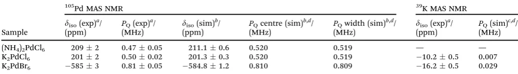
Fig. 3 (a) The39K MAS NMR data (u
r = 3 kHz) for K2PdCl6 (blue) and
K2PdBr6(green) acquired at 14.1 T (u0= 27.97 MHz), 9.40 T (u0= 18.61 MHz)
and 7.05 T (u0= 13.98 MHz) referenced to KCl(s)(atdiso= 47.8 ppm). The
asterisks mark the spinning sideband positions. The quadrupolar spinning sideband manifolds of both complexes at 7.05 T have been simulated (red) using Topspin. (b) The39K centre-of-gravity shifts (dCG) of K2PdCl6(blue)
and K2PdBr6 (green) plotted against the inverse square of the Larmor
frequency (u0) at 14.1 T, 9.40 T and 7.05 T.
Open Access Article. Published on 16 October 2018. Downloaded on 4/5/2019 2:07:28 PM.
This article is licensed under a
[image:4.595.51.287.85.642.2] [image:4.595.313.547.144.632.2]exhibit excellent agreement with those determined by the graphical approach shown in Fig. 1(b). The spinning sideband manifold of the105Pd MAS NMR data presented in Fig. 2(d) also supports this observation. An accurate simulation of each sideband manifold was achieved using the parameters given in Table 1. To ensure that these observations were not influ-enced by an impurity Pd(II) species, the complex K2PdCl6was
studied before and after washing withaqua regia; no contribu-tion was detected as evidenced by the105Pd MAS NMR data in Supplementary Section (SI2, ESI‡).
Further evidence of this phenomenon is demonstrated by the 39K and 35Cl MAS NMR data presented in Fig. 3 and 4, which also indicate the existence of non-zero EFGs influencing these data. These 39K MAS NMR spectra show single narrow resonances from K2PdCl6and K2PdBr6atdiso= 10.20.5 ppm anddiso = 16.2 0.5 ppm (calibrated against KCl(s)(diso = 47.8 ppm)), respectively. The lack of a field dependent shift (Fig. 3(b)), is contradicted by the associated low intensity spinning sideband manifolds which indicate the presence of non-zero EFGs influencing the K centres which occupy the (1
4, 1 4,
1 4)
positions in theFm3%munit cell. By simulation of the spinning sideband manifolds of the 7.05 T MAS NMR data from both com-plexes (see Fig. 3(a)), very smallPQvalues of 0.007 and 0.029 MHz can be determined for K2PdCl6and K2PdBr6, respectively (see Table 1). In contrast to the105Pd results presented above which specifies that a 2nd order quadrupole interaction is influencing the MAS NMR data (by virtue of the very largeQ), the39K MAS NMR data in Fig. 3(b) shows that there is no observable field dependent 2nd order quadrupole contribution to the observed centre-of-gravity shift position, and that the quadrupole influ-ence upon the39K data is to 1st order only. Nevertheless, in an
analogous fashion to the 105Pd studies, the quadrupole con-tribution to the39K MAS NMR data reflects a structural disorder phenomenon in these systems. Similarly, the35Cl MAS NMR data from K2PdCl6and (NH4)2PdCl6(Cl occupying the (0.24,0,0) positions), as observed in Fig. 4, yields narrow resonances at diso= 3.60.8 ppm anddiso= 742 ppm, respectively which are also accompanied by low intensity spinning sideband manifolds. These 35Cl MAS NMR data are reported against a cubic NaCl internal reference (diso= 46.1 ppm).
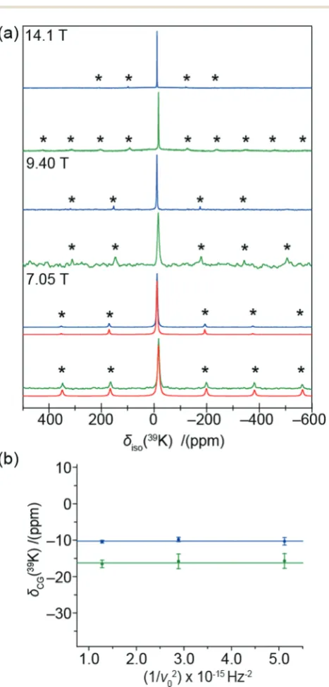
The non-zero EFGs detected by the NMR data imply a deviation from the perfect cubic symmetry reported by powder XRD.19However, X-ray pair distribution function (PDF) analyses provide descriptions of the localized structural disorder that can produce these phenomena. The real space fits of the X-ray PDF data from each Pd(IV) complex exhibits a very narrow peak
at approximately 2 Å which represents a rigid and well-defined PdX6(X = Cl, Br) bond (see Fig. 5(a–c)). The widths of peaks from the PDF experiment represent a statistical measure of the distribution of the bond lengths, hence a very narrow peak indicates that there is no displacement of the Pd position from
Fig. 4 The35Cl MAS NMR data (ur= 12 kHz) for (NH4)2PdCl6(red) and
K2PdCl6(blue) acquired at 11.7 T (u0= 49.00 MHz), and internally
refer-enced to NaCl(s)(atdiso= 46.1 ppm). The asterisks mark the spinning
sideband positions.
Fig. 5 Analysis of the X-ray PDF data forr= 0–20 Å with the experimental data (black), the PDF fittings (red), the offset difference curves (blue) and the refined structures for (a) (NH4)2PdCl6, (b) K2PdCl6and (c) K2PdBr6. The
dashed black line highlights the K–Pd correlation length which is present in K2PdCl6, but absent in K2PdBr6due to disorder.
Open Access Article. Published on 16 October 2018. Downloaded on 4/5/2019 2:07:28 PM.
This article is licensed under a
[image:5.595.310.550.301.662.2] [image:5.595.49.286.432.671.2]each octahedral PdX6 centre, as suggested from the average structure. Each octahedral hexahalopalladate unit is so well defined that the peak is at the resolution limit of the X-ray PDF measurement; this rare occurrence is responsible for the truncation artefacts observed around each 2 Å peak. Further modelling of these data also reveals that there is no measure-able distortion of the Cl and Br anions from their octahedral positions. Therefore, it is highly unlikely that the deviation from cubic symmetry observed by the105Pd and35Cl MAS NMR is induced from within the octahedral PdX6units.
The modelling of the real space X-ray PDF date for K2PdBr6 (see Fig. 5(c)) requires the additional refinement of a large atomic displacement parameter for the K site exceeding what would be expected for a normal thermal parameter range. This is clear when comparing the PDF data for K2PdCl6and K2PdBr6; the K–Pd bond lengths determined from the literature struc-tures of both complexes are 4.2 and 4.6 Å, respectively. The accompanying peak from this length is present in the PDF data for K2PdCl6(see Fig. 5(b)), but not for K2PdBr6. Hence, there is a negligible probability of instantaneously finding the K+cation precisely at the (1
4, 1 4,
1
4) position in K2PdBr6. This is due to the larger Br anions inducing an increased unit cell volume, thus leaving a larger residence volume for the K+cation to occupy. The bond valence sums (BVS) for the K+ cations reflect this, with the K+ position in K2PdBr6 showing significant under-bonding (BVS of 0.845,cf.1.006 for the K2PdCl6).24This devia-tion from cubic symmetry at the cadevia-tion posidevia-tion explains the largest PQ determined for K2PdBr6 (see Table 1), but it does not rationalise why smaller but non-zero EFGs are observed in (NH4)2PdCl6 or K2PdCl6(the former exhibits only a small peak at theB4 Å region because of the relative lower sensitivity of X-rays to (NH4)+than K+). These structures refined against the PDF data reveal no other local departures from cubic symmetry. It is therefore postulated that the hexachloride complexes are characterised by reduced degrees of disorder at their respective NH4+and K+cation positions (compared to that observed in K2PdBr6), and that the extreme sensitivity of the 105Pd NMR to variations in the Pd environment are enhanced
by the very large 105Pd quadrupole moment (Q). Such small degrees of disorder can be comprised of surface vacancies, dislocations, point defects and defect migration induced by many effects including particle size; this phenomenon has been previously observed in many MAS NMR studies of other inorganic solids including Li2O,25 KBr,26 NaBr,26 KMnO4,26 AgBr,27MgO28and NaCl.29,30
This established 105Pd solid state NMR methodology has also been applied to four Pd metal particle systems; Pd sponge, Pd black, poly(N-vinyl-2-pyrrolidone) (PVP) stabilised Pd, and bio-Pd nanoparticles. The various size distributions have been summarised in Table 2, with the nanoparticle size distributions determined viaTEM (see Fig. 7). The 105Pd static solid state NMR data of the face-centered cubic fcc Pd metal nanoparticles shown in Fig. 6 depicts conventional Knight shifted resonances, which is an interaction between the105Pd nuclei and the Pauli susceptibility of the delocalized conduction band electrons.31 This results in an enormous shift in the resonance frequency and the total shift (Ktot) is often expressed as a percentage of the isotropic reference frequency. The total shift is a combination of the Knight shift (K) and a conventional shielding term (s) as shown in eqn (5):
Ktot=K+s=Ks+Korbd +Kcp+s (5)
The Knight shift can be split into an s-electron contribution (Ks) and a d-electron contribution (Korbd ), both of which yield positive shifts. If the hyperfine fields induce polarization of the inner shell electrons a negative shift known as the core polar-ization (Kcp) can also contribute toKandKtot. The data in Fig. 6 and Table 2 clearly demonstrates that core polarization is the dominant contribution toKtotfor the measured Pd metal/metal nanoparticle shifts.
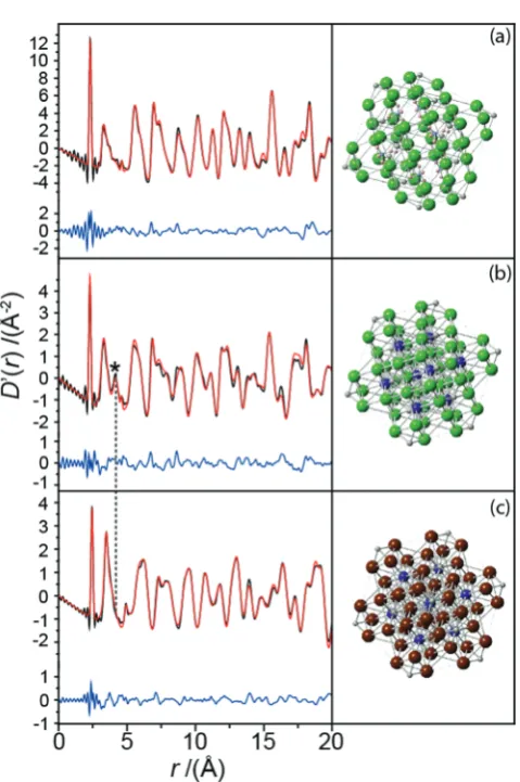
[image:6.595.315.540.46.303.2]From Fig. 6 and Table 2, theB44–149mm Pd metal (sponge) particle and the B20–150 nm Pd black nanoparticle systems yield similar observable resonances atKtot= 32 05060 ppm andKtot= 32 11090 ppm, respectively. Within experimental
Table 2 Particle size distributions of the Pd metal systems determinedvia
TEM statistical analysis and the experimental 105Pd NMR values of the Knight shift and the full-width at half-maximum. The particle size range quoted for the bio-Pd system is a coarse estimation
Sample Particle size
105Pd
Ktot(exp)/(ppm) FWHM (exp)/(kHz)
Pd sponge 44–149mm 32 05060 131 Pd black 20–150 nm 32 11090 161 PVP stab. Pd 163 nm B 31 100400 B8010 Bio-Pd 2 41000 nm B 31 100500 B5515
Fig. 6 The 105Pd VOCS static NMR spectra PVP stabilised Pd
nano-particles (163 nm), Pd black nanoparticles (20–150 nm) and Pd metal sponge (44–149mm) acquired at 14.1 T, 11.7 T and 7.05 T. All data were referenced to H2PdCl6(aq)(atdiso= 0.0 ppm).
Open Access Article. Published on 16 October 2018. Downloaded on 4/5/2019 2:07:28 PM.
This article is licensed under a
[image:6.595.42.294.647.720.2]error these measurements both represent aKtot of 3.205 0.06%. This value compares favorably with a value of Ktot of 3.10.4% calculated from magnetic susceptibility measure-ments at 3001C.6It should be noted that the small discrepancy inKtot obtained from the 105Pd static NMR and the magnetic susceptibility measurements is partially attributed to differences in referencing (implicit withins, see eqn (5)), with the NMR determined Ktot value being measured against the proposed H2PdCl6(aq)reference. In contrast to the data presented for the diamagnetic Pd(IV)-hexahalo systems above, these 105Pd metal
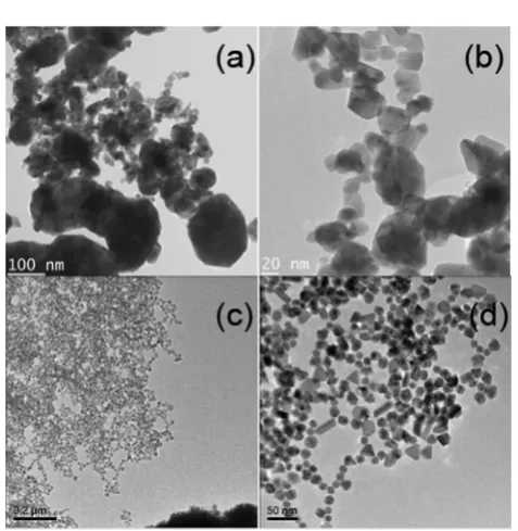
shifts for the largerB44–149mm Pd sponge particles and the B20–150 nm Pd black nanoparticles are field independent (see Fig. 6 and 1(b)) indicating that no EFG can be measured (i.e. PQ=CQ= 0), as expected for a highly symmetrical cubic fcc structure with no observable elements of intrinsic disorder. For these cases the narrower linewidths suggest that the highly ordered bulk positions of each particle system dominates these signals in comparison to that of the lower symmetry surface positions. Further evidence for these well-formed Pd black structures is shown in the TEM images shown in Fig. 7(a and b) from which the reported particle size distribution is drawn; these images confirm the occurrence of well-defined nanoparticle structures characterising the Pd black system.
These observations contrast markedly with the105Pd static NMR data from the smaller (highly monodisperse) B16 nm PVP-stabilised Pd nanoparticles, which exhibit a much broader, ill-defined resonance. Assuming that a cube-octahedral model describes the morphology of these PVP-stabilised nanoparticles, a Pd metal unit cell volume of 58.9 Å3implies that aB16 nm particle would consist ofB150 000 Pd atoms;32nevertheless, the 105Pd resonances measured from this highly monodisperse
system do not exhibit any statistically well-defined elements of
bulk character. The very high degree of monodispersity in this system is shown in the TEM images of Fig. 7(c and d) demon-strates that this observation is strictly an intra-particle phenom-enon. These smaller nanoparticles have a higher ratio of surface to bulk Pd environments. Hence, despite the much greater particle size uniformity, the increased disorder and concomitant loss of translational symmetry induced by the surface and near-surface layers is reflected in these105Pd data acquired at 14.1 and 7.05 T (see Fig. 6). As exemplified by the Pd(IV)-hexahalo
systems above, the very large 105PdQ value renders the NMR response to be extremely sensitive to structural perturbations causing deviation from perfect cubic fcc symmetry. The result in Fig. 6 implies that the wholeB16 nm PVP-stabilised Pd nano-particle experiences some degree of deviation from high cubic symmetry, ranging from a large loss of symmetry at or near the surface layers, to smaller perturbations towards sub-surface and bulk regions. It is important to realise that, although the105Pd static NMR data for this system shown in Fig. 6 and 1(b) exhibits no real field dependence, this does not infer that on average PQ = CQ = 0 for the Pd positions within these particles. In contrast, this behavior will be underpinned by a complex relationship between the distributions of EFGs generated by the variable departure from cubic fcc symmetry depending on proximity to the particle surface, and the distributions of Knight shifts emanating from the layers comprising each particle (e.g. see the core–shell cubo-octahedral model in ref. 33). These assertions are supported by the absence of bulk structural components represented by a narrow resonance at KtotB 32 100 ppm (see the105Pd static NMR data for the Pd black and Pd sponge in Fig. 6). The centre-of-gravity shift now occurs at an increased (more downfield) Knight shift of Ktot B 31 100 400 ppm demonstrating that this shift correlates with less bulk metal character (and more surface character) being realised. These results clearly demonstrate that the 105Pd Knight shift is sensitive to changes in particle size and local particle symmetry.
A similar scenario is observed with the105Pd static NMR study of the highly polydisperse bio-Pd system which is reported in Fig. SI3 within the ESI.‡ In this system, the polydispersity describes a very large range of particle sizes ranging from small nanoparticles up to micron sized particles; the size range quoted above and in Table 2 represents a coarse estimate for these particles to help describe the heterogeneity in this system. This polydispersity is accompanied by a similarly large range of random particle shapes. The 105Pd static NMR shows a clearly defined ordered bulk metal component to the structure within this very inhomogeneous system, as evidenced by a narrower resonance atKtot B 32 100 ppm. This component is accom-panied by broader downfield elements similar to those observed in the PVP-stabilised Pd system, which are attributed to smaller particles with structures that are perturbed from cubic fcc sym-metry;i.e.those components more proximate to surface and sub-surface environments. The observation of highly variable static NMR resonance linewidths emanating from particle size effects in metal particles and nanoparticle systems has been previously described in studies on several metal species.33,34
Fig. 7 The TEM images of (a), (b)B20–150 nm Pd black nanoparticles, and (c), (d) highly monodisperse 163 nm PVP stabilised Pd nanoparticles.
Open Access Article. Published on 16 October 2018. Downloaded on 4/5/2019 2:07:28 PM.
This article is licensed under a
[image:7.595.48.287.448.693.2]Conclusions
This study illustrates the feasibility of105Pd solid state NMR on diamagnetic and metallic systems. The large105Pd quadrupole moment gives rise to measureable EFGs that can act as a very sensitive probe of small structural distortions which are not observable by laboratory source X-ray diffractometers. X-ray PDF refinements confirm the nature of these secondary shell distortions. Future solid state105Pd NMR analyses will be aided by the proposal of standardised direct and indirect 105Pd chemical shift references such as H2PdCl6(aq)and KCl respec-tively. The105Pd Knight shift is also shown to be a sensitive probe of particle size effects in metallic systems. It has been demonstrated that the room temperature confirmation of the Knight shift of Pd metal provides an excellent basis for further 105Pd studies of intermetallic (PdZn,35PdGa36and PdCu37) and
alloyed catalysts (PtPdRh38) used in fuel cell, electrocatalysts and auto-catalyst technologies, and capped/stabilised nano-particles systems used in hydrogenation reactions and fine chemical syntheses.1–3,10–12
Experimental
All three Pd(IV) complexes (potassium hexachloropalladate, 99%; ammonium hexachloropalladate, 99.9%; potassium hexabromo-palladate, 99.9%) are commercially produced and were purchased from Alfa Aesar. Pd metal particles in the form of B44–149 mm Pd sponge, B20–150 nm Pd black and highly monodisperse 163 nm PVP-stabilised Pd nanoparticles were synthesized and provided by Johnson Matthey.11,39A sample of highly polydisperse bio-Pd nanoparticles of average size B2–41000 nm (coarse approximation) was synthesized via the biomineralized reduction of Pd(II) to Pd(0) using E. coli
cells and pyrolysed in vacuum.40The particle sizes of these Pd metal particle was determined by sampling statistics performed under transmission electron microscopy (TEM) analyses.
For the calibration of the 105Pd NMR measurements the synthesis of the 0.33 M H2PdCl6(aq)chemical shift reference was undertaken by the stepwise dissolution of 0.125 g of Pd metal sponge in 2 mL of concentrated (70%) HNO3. This was followed with the addition of 4–6 mL of concentrated (36%) HCl to produce a dark red colour solution in a strongly oxidizing acid/aqua regiaenvironment. The chemical equation for this reaction is proposed to be:
Pd(s)+ 4NHO3(aq)+ 6HCl(aq)-H2PdCl6(aq)+ 4NO2(g)+ 4H2O (6)
An overall excess ofaqua regiais required to impede the reduc-tion of H2Pd(IV)Cl6(octahedral Pd coordination) to H2Pd(II)Cl4 (square planar Pd coordination); the lower symmetry of the latter species induces a large quadrupolar broadening making it unsuitable as a105Pd chemical shift reference. To test for the possibility of Pd(II) impurities within the Alfa Aesar products,
K2PdCl6 was added to an aqua regia solution to remove any potential species. This solution was allowed to stand for 60 minutes,
then K2PdCl6powder was then filtered from the solution, washed with distilled water and left to dry.
All room temperature105Pd MAS and static NMR measure-ments in this study were performed at the magnetic field (B0) strengths of 20.0, 14.1, 11.7, 9.4 and 7.05 T using Bruker Avance III-850 (u0= 39.00 MHz), Bruker Avance II+-600 (u0= 27.49 MHz), Bruker Avance III-500 (u0= 22.93 MHz), Bruker Avance HD-400 (u0= 18.30 MHz) and Bruker Avance HD-300 (u0= 13.75 MHz) spectrometers, respectively. For the hexahalo-Pd(IV) systems,
both MAS and static measurements were undertaken using Bruker 7 mm HX MAS (20.0 T), Varian 9.5 mm MAS (14.1, 11.7 and 9.4 T) and Otsuka 9.5 mm MAS (7.1 T) probes, which enabled MAS frequencies ofB3–5 kHz at all fields for the acquisition of the MAS NMR data. At eachB0field a (p/4)–t–(p/2)–tHahn echo sequence was utilised to diminish ringing effects present at low frequencies, with rotor synchronizedtdelays ofB330ms and a recycle delay of 1 s being implemented throughout. For the I= 5/2105Pd nucleus, non-selective (solution)p/2 pulse lengths of 30, 60, 36, 30 and 24ms were calibrated on H2PdCl6(aq)which corresponded to selective (solid)p/4 pulse lengths of 5, 10, 6, 5 and 4ms at 20.0, 14.1, 11.7, 9.4 and 7.1 T, respectively. All 105Pd chemical shifts were referenced to the proposed standard
of 0.33 M H2PdCl6(aq) (diso = 0.0 ppm) and the neighbouring KCl(s)standard (diso= 17 647 ppm).
The solid state NMR study of metals has several associated complications due to the conductivity of the samples. For the 105Pd NMR analyses of the metal systems RF penetration (skin
depth) effects must be considered, and the sample conductivity can markedly de-tune the probe. The latter aspect is alleviated by diluting each sample with NaCl to a level of 75 wt% of the salt. In addition, the build-up of eddy currents in a conductive sample will resist MAS averaging and induce heating. These details have been treated in detail elsewhere.33 Therefore, all 105Pd measurements on the metal systems were performed
under static conditions. The ultra-wide linewidths exhibited by these systems necessitated the use of Variable Offset Cumu-lative Spectroscopy (VOCS) to achieve uniform excitation of the 105Pd lineshape. This involved the acquisition of several NMR
experiments over a uniformly stepped frequency range (30 kHz steps), and the summation of these individual sub-spectra to reconstitute the full spectrum.22,41 The 105Pd static NMR measurements on metallic systems were performed at 14.1, 11.7 and 7.05 T and utilized a (p/2)–t–(p/2) solid echo experiment to improve wide line excitation; this was stepped in 30 kHz increments across the frequency range of each total spectrum. Non-selectivep/2 pulse widths of 6ms, recycle delays of 0.01 s and tdelays of 50–150ms were common to all experiments at each field. All data were referenced to H2PdCl6(aq)(atdiso= 0.0 ppm).
The 39K MAS NMR data were measured at 20.0 T (u 0 = 39.68 MHz), 14.1 T (u0 = 27.97 MHz), 9.40 T (u0= 18.61 MHz) and 7.05 T (u0 = 13.98 MHz) using Bruker 7 mm HX MAS (20.0 T), Varian 9.5 mm MAS (14.1 and 9.4 T) and Otsuka 9.5 mm MAS (7.1 T) probes, which enabled MAS frequencies of B3–5 kHz at all fields. A single pulse experiment was used for all data acquisition, with a non-selective p/2 pulse length of 18ms being calibrated on KCl(s). For the measurements at each
Open Access Article. Published on 16 October 2018. Downloaded on 4/5/2019 2:07:28 PM.
This article is licensed under a
field a selectivep/6 of 3ms, and a recycle delay of 5 s were used throughout. All39K chemical shifts are reported with respect to the primary IUPAC recommended shift reference of 0.1 M KCl(aq) (diso= 0.0 ppm)viaa secondary KCl(s)reference (diso= 47.8 ppm).5 The corresponding35Cl MAS NMR data were acquired at 11.7 T (u0= 49.00 MHz) using a Bruker 4 mm HX MAS probe which delivered a MAS frequency of 12 kHz for all measurements. A single pulse experiment was used for all data acquisition, with a non-selective p/2 pulse length of 4 ms being calibrated on NaCl(s). The measurements utilised a selective p/4 of 1 ms, and a recycle delay of 10 s. All35Cl chemical shifts are reported with respect to the primary IUPAC recommended shift reference of 0.1 M NaCl(aq)(diso= 0.0 ppm)viaa secondary NaCl(s)reference (diso= 46.1 ppm).5
The analysis of all solid state NMR central transition lineshapes was performed using the QUADFIT simulation programme,42while the simulation of the corresponding satel-lite transition spinning sideband manifolds in the MAS NMR data were performed using the SOLA utility within the Bruker TopSpin software package.
Total X-ray scattering experiments were performed on XPDF (beamline I15-1) at Diamond Light Source, Harwell Campus, UK. Samples were packed into 1 mm borosilicate capillaries and were illuminated with photons of energy 78.34 keV (l= 0.1583 Å). The scattered intensity was acquired on a Perkin Elmer 1611 CP3; and calibrated against a CeO2 standard and integrated in DAWN.43The integrated 1D scattering pattern was used directly for Rietveld refinement and were converted to pair distribution functions in GudrunX,44while the structural refinements against both the real and reciprocal space data were performed in TOPAS Academic.45
Conflicts of interest
There are no conflicts to declare.
Acknowledgements
JVH thanks the EPSRC for funding of project EP/P511432/1, and JVH and PTB gratefully acknowledge the EPSRC and Johnson Matthey for the CASE studentships that funded TJNH and TAP. JVH thanks the EPSRC, the University of Warwick and the Birmingham Science City Programme for partial funding of the solid state NMR infrastructure at Warwick. The latter program accessed the Birmingham Science City Advanced Materials Project 1: Creating and Characterising Next Generation Advanced Materials, which derived support from Advantage West Midlands (AWM) and the European Regional Develop-ment Fund (ERDF). JVH also thanks UK National 850 MHz Solid State NMR Facility used in this research which is funded by the EPSRC, BBSRC, the University of Warwick and the Birmingham Science City Advanced Materials Projects 1 and 2, supported by Advantage West Midlands (AWM) and the European Regional Development Fund (ERDF). Collaborative assistance from the 850 MHz Facility Manager (Dinu Iuga, University of Warwick)
is acknowledged. We thank beamline I15 at Diamond Light Source (Harwell, UK) for in-house research time.
Notes and references
1 D. Jollie, Platinum 2007, Johnson Matthey, Royston, 2007, pp. 30–37.
2 G. G. Ertl, H. Kno¨zinger and J. Weitkamp, Environmental Catalysis, Wiley-VCH, Weinheim, 1999, p. 236.
3 S. Srinivasan,Fuel Cells: From Fundamentals to Applications, Springer, New York, 2006, p. 691.
4 P. Raghavan,At. Data Nucl. Data Tables, 1989,42, 189–291. 5 R. K. Harris, E. D. Becker, S. M. Cabral de Menezes, R. Goodfellow and P. Granger,Ann. Magn. Reson., 2002, 1, 43–64.
6 J. A. G. Seitchik, A. C. Gossard and V. Jaccarino,Phys. Rev., 1964,136, 1119–1125.
7 A. Narath, A. T. Fromhold and E. D. Jones,Phys. Rev., 1966,
144, 428–435.
8 P. Brill and J. Voitla¨nder,Ber. Bunsen-Ges., 1973,77, 1097–1103. 9 K. Matsuda, Y. Kohori and T. Kohara,Phys. Rev. B: Condens.
Matter Mater. Phys., 1997,55, 15223–15227. 10 F. A. Lewis,Platinum Met. Rev., 1961,5, 21–25. 11 J. Cookson,Platinum Met. Rev., 2012,56, 83–98.
12 N. Zelinsky and N. Glinka,Ber. Dtsch. Chem. Ges., 1911,44, 2305–2311.
13 M. A. Fedotov. and V. A. Likholobov,Russ. Chem. Bull., 1984,
33, 1751–1751.
14 F. E. Beamish and J. C. van Loon, Recent Advances in the Analytical Chemistry of Noble Metals, Pergamon, Oxford, 1972, pp. 7–19.
15 P. P. Man, in NMR of Quadrupolar Nuclei in Solid State Materials, ed. R. E. Wasylishen, S. E. Ashbrook and S. Wimperis, John Wiley & Sons, Ltd, 2012, pp. 3–16. 16 M. E. Smith and E. R. H. van Eck,Prog. Nucl. Magn. Reson.
Spectrosc., 1999,34, 159–201.
17 I. L. Moudrakovski and J. A. Ripmeester,J. Phys. Chem. B, 2007,111, 491–495.
18 A. Butler and H. Eckert, J. Am. Chem. Soc., 1989, 111, 2802–2809.
19 B. Douglas and S. Ho,Structure and Chemistry of Crystalline Solids, Springer, 2007, pp. 127–129.
20 A. Samoson,Chem. Phys. Lett., 1985,119, 29–32.
21 C. Ja¨ger,NMR Basic Principles and Progress, Springer-Verlag, Berlin, 1994, vol. 31, p. 135.
22 J. V. Hanna, K. J. Pike, T. Charpentier, T. F. Kemp, M. E. Smith, B. E. G. Lucier, R. W. Schurko and L. S. Cahill, Chem. – Eur. J., 2010,16, 3222–3239.
23 C. S. Griffith, V. Luca, J. V. Hanna, K. J. Pike, M. E. Smith and G. S. Thorogood,Inorg. Chem., 2009,48, 5648–5662. 24 I. D. Brown, The Chemical Bond in Inorganic Chemistry:
The Bond Valence Model, Oxford University Press, Oxford, New York, 2nd edn, 2016.
25 Z. H. Xie, M. E. Smith, J. H. Strange and C. Ja¨ger,J. Phys.: Condens. Matter, 1995,7, 2479–2487.
Open Access Article. Published on 16 October 2018. Downloaded on 4/5/2019 2:07:28 PM.
This article is licensed under a
26 J. S. Frye and G. E. Maciel, J. Magn. Reson., 1982, 48, 125–131.
27 N. Zumbulyadis and A. P. Marchetti,Colloids Surf., 1990,45, 335–346.
28 A. V. Chadwick, I. J. F. Poplett, D. T. S. Maitland and M. E. Smith,Chem. Mater., 1998,10, 864–870.
29 T. Yamanishi, T. Kanashiro, Y. Michihiro, Y. Kishimoto and T. Ohno,J. Phys. Soc. Jpn., 1995,64, 643–650.
30 Y. Michihiro, T. Yamanishi, T. Kanashiro and Y. Kishimoto, Solid State Ionics, 1995,79, 40–44.
31 J. J. van der Klink and H. B. Brom,Prog. Nucl. Magn. Reson. Spectrosc., 2000,36, 89–201.
32 R. W. G. Wyckoff,Crystal Structures, Interscience Publishers, New York, 2nd edn, 1963, vol. 1.
33 G. J. Rees, S. T. Orr, L. O. Barrett, J. M. Fisher, J. Houghton, G. H. Spikes, B. R. C. Theobald, D. Thompsett, M. E. Smith and J. V. Hanna, Phys. Chem. Chem. Phys., 2013, 15, 17195–17207.
34 S. Cadars, B. J. Smith, J. D. Epping, S. Acharya, N. Belman, Y. Golan and B. F. Chmelka, Phys. Rev. Lett., 2009, 103, 136802.
35 J. R. Gallagher, D. J. Childers, H. Zhao, R. E. Winans, R. J. Meyer and J. T. Miller,Phys. Chem. Chem. Phys., 2015,
17, 28144–28153.
36 M. Armbru¨ster, K. Kovnir, M. Behrens, D. Teschner, Y. Grin and R. Schlo¨gl,J. Am. Chem. Soc., 2010,132, 14745–14747. 37 H. S. Chang, K. C. Hsieh, T. Martens and A. Yang,J. Electron.
Mater., 2003,32, 1182–1187.
38 C. M. Hung,Int. J. Hydrogen Energy, 2012,37, 13815–13821. 39 A. F. S. Gouldsmith and B. Wilson,Platinum Met. Rev., 1963,
7, 136–143.
40 P. Yong, I. P. Mikheenko, K. Deplanche, D. F. Sargent and L. E. Macaskie,Adv. Mater. Res., 2009,71–73, 729–732. 41 D. Massiot, I. Farnan, N. Gautier, D. Trumeau, A. Trokiner
and J. P. Coutures,Solid State Nucl. Magn. Reson., 1995,4, 241–248.
42 T. F. Kemp and M. E. Smith,Solid State Nucl. Magn. Reson., 2009,35, 243–252.
43 M. Basham, J. Filik, M. T. Wharmby, P. C. Y. Chang, B. El Kassaby, M. Gerring, J. Aishima, K. Leyik, B. C. A. Pulford, I. Sikharulidze, D. Sneddon, M. Webber, S. S. Dhesi, F. Maccherozzi, O. Svensson, S. Brockhauser, G. Na’rayc and A. W. Ashtona,J. Synchrotron Radiat., 2015,
22, 853–858.
44 A. K. Soper and E. R. Barney,J. Appl. Crystallogr., 2011,44, 714–726.
45 A. A. Coelho, P. A. Chater and A. Kern,J. Appl. Crystallogr., 2015,48, 869–875.
Open Access Article. Published on 16 October 2018. Downloaded on 4/5/2019 2:07:28 PM.
This article is licensed under a