warwick.ac.uk/lib-publications Manuscript version: Author’s Accepted Manuscript
The version presented in WRAP is the author’s accepted manuscript and may differ from the published version or Version of Record.
Persistent WRAP URL:
http://wrap.warwick.ac.uk/109018
How to cite:
Please refer to published version for the most recent bibliographic citation information. If a published version is known of, the repository item page linked to above, will contain details on accessing it.
Copyright and reuse:
The Warwick Research Archive Portal (WRAP) makes this work by researchers of the University of Warwick available open access under the following conditions.
© 2018 Elsevier. Licensed under the Creative Commons Attribution-NonCommercial-NoDerivatives 4.0 International http://creativecommons.org/licenses/by-nc-nd/4.0/.
Publisher’s statement:
Please refer to the repository item page, publisher’s statement section, for further information.
Spatial representations in the primate hippocampus, and their functions in memory and navigation
Edmund T Rolls
Oxford Centre for Computational Neuroscience, Oxford, UK and University of Warwick, Department of Computer Science, Coventry, UK
and
Sylvia Wirth
Institut des Sciences Cognitives Marc Jeannerod, UMR 5229, CNRS and University of Lyon, Bron, France
Email: [email protected] Url: www.oxcns.org
Running title: Primate hippocampus
Keywords: hippocampus; spatial view cells; place cells; episodic memory; navigation; fovea
Abstract
Contents
1. Introduction
1.1. The great evolution of visual cortical and related areas in primates
1.2 Effects of hippocampal lesions in primates help to define the functions to be investigated neurophysiologically
2. On the nature of spatial representations in primates
2.1. An allocentric representation of space in the primate hippocampus 2.2. Spatial View Cells in the primate hippocampus
2.2.1 Responses when the primate is moved to different places in a cue-controlled environment. 2.2.2 Responses of hippocampal spatial view neurons during active locomotion.
2.2.3 The testing of hypotheses for spatial view vs place vs head direction vs eye position encoding.
2.2.4 Spatial view cells, and the representation of place in the primate hippocampus.
2.2.5 Comparison of primate hippocampal spatial view cells with other types of neuronal response. 2.3. Visually supported spatial representations in primates navigating in a virtual environment 2.4. Idiothetic (self-motion) update of spatial view cells
2.5. Responses of neurons in the primate hippocampus to whole body motion 2.6. Grid cells in rodents and spatial view grid cells in the primate entorhinal cortex
3. Properties of primate hippocampal spatial representations related to object and reward memory associations
3.1. Object-place neurons in the primate hippocampus
3.2. One-trial, object-place, recall-related neurons in the primate hippocampus 3.3. Reward-place neurons in the primate hippocampus
3.4. Neurons involved in learning associations between visual stimuli and spatial responses
4. Discussion: functions of hippocampal spatial representations in primates, and comparison with rodents
4.1. Self-organization by learning of primate spatial view cells related to foveal vision, and of rodent place cells related to a wide field of view
4.2. Comparison of representations in primates and rodents
4.3. Hippocampal computational similarities between primates and rodents
4.4. Functions performed by the primate hippocampus in memory, and thereby in action and navigation
4.5. Building episodic memories from associations between objects and places 4.6. The human hippocampus and the art of memory
1. Introduction
There has been great evolution of vision in primates compared to rodents. This involves great development of temporal and parietal visual cortical areas that provide inputs to the hippocampus via the perirhinal cortex and parahippocampal gyrus. It is therefore a fundamental issue to investigate whether hippocampal spatial processing, which utilizes visual inputs, in primates is identical to that in rats. In rodents, place cells are found (Hartley et al., 2014; Markus et al., 1995; McNaughton et al., 1983; Muller et al., 1991; O'Keefe, 1979, 1984). There is evidence that place is represented in the primate hippocampus (Rolls and O'Mara, 1995; Wirth et al., 2017) as in the rodent hippocampus, but the major difference is that hippocampal spatial representation in primates stems from the elements of a scene that the animal foveates rather than where the animal is, and that is the focus of this paper. We propose that the difference between primates and rodents in the spatial representations in the hippocampus is related to the development in evolution of the fovea in primates, and of the cortical areas in the temporal lobe that are specialized for processing what is represented near the fovea in complex natural scenes (Rolls et al., 2003), and of areas in the parietal lobes that guide eye movements to fixate parts of scenes (Bisley and Goldberg, 2003). We consider the implications of this difference in spatial representations for the functions of the hippocampus, and argue, inter alia, that this enables a primate located in one place to look at many locations in the environment, and associate in memory the objects or rewards present at each of those viewed locations, even though the primate is not at those locations, and remains in one place. We argue that this goes beyond the capabilities of rodent place cells that respond to places rather than locations being viewed. This is a fundamental difference important to understanding many aspects of primate including human episodic memory, which does implement this functionality of remembering objects or rewards present at viewed locations. We show that to analyse the nature of these representations,, it is necessary to perform testing while eye position is recorded when the subject is in different places, and is looking at different parts of a scene. The available evidence on this comes from neuronal recordings in macaques, with no assessment of this type performed yet in humans, and this is identified as an important area for future investigation in humans. However, we do refer to recordings and neuroimaging and lesion data for the human hippocampus and related areas, to assess to what extent they fit with the overall understanding of the functioning of the primate hippocampus described here.
We note that, consistent with the direct evidence on neuronal spatial representations in primates described here, it has recently been argued from neuropsychological and evolutionary perspectives across a range of vertebrates, that the primate hippocampus is involved in scene processing rather than place processing, related to the development of the primate fovea (Murray et al., 2017). For example, lesions to the macaque hippocampal system impair remembering the place in a scene “out there” where an object was located (Gaffan, 1994; Murray et al., 1998; Murray et al., 2017).
However, we then build on the neurophysiological evidence to show that the same underlying computational principles may be involved in setting up and using the spatial representations in both the primate and rodent hippocampus (Rolls, 2016a). Understanding the differences between the spatial representations in primates vs rodents is very timely, because it is now becoming possible to record from neurons in the human hippocampus and related structures (Ekstrom, 2015; Ekstrom et al., 2003; Ekstrom and Ranganath, 2017; Fried et al., 1997; Kreiman et al., 2000; Quiroga, 2012; Rey et al., 2015). It is likely that what is found in the human hippocampus will reflect the evolution of foveally dominated vision in primates, and therefore how the human hippocampal system operates, and how it functions in human memory and navigation.
1.1. The great evolution of visual cortical and related areas in primates
single neurons in the inferotemporal visual cortex respond to an object near to the fovea, and not to other objects in the surround (Rolls et al., 2003), and this helps the interface to action, which can then be directed to the object at the fovea (Rolls, 2016a; Rolls et al., 2003; Rolls and Deco, 2002). The presence of a fovea in primates and the cortical systems for object identification of what is at the fovea, and for directing eye movements to foveate objects, can been conceptualized as an adaptation of vision useful for finding and then reaching to obtain and grasp fruit in sylvian environments (Kaas, 2013). This primate visual capability is associated with a large system of highly developed visual cortical areas in the ventral visual stream, which provide input via the parahippocampal gyrus and retrosplenial / posterior cingulate areas to the hippocampus, and of a highly developed dorsal visual system, which includes mechanisms useful for the generation of the eye movements necessary to saccade to and then fixate objects in a scene (Fig. 1) (Bisley and Goldberg, 2010; Bremmer et al., 2002a; Bremmer et al., 2002b; Galletti and Fattori, 2017; Goodale, 2014; Rolls, 2012; Rolls, 2016a). All these specializations of visual processing in primates when compared to rodents are, we propose, necessary for understanding differences in the representations in the primate vs rodent hippocampus.
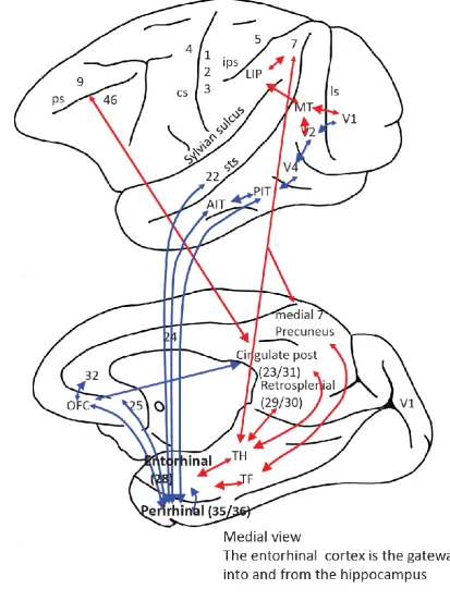
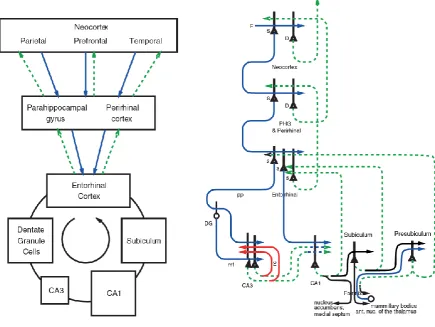
In the context of the evolutionary development of the primate cortex, which include hierarchical organization, the development of a fovea, and mechanisms for object recognition for what is at the fovea and for moving the fovea to fixate objects (Rolls, 2016a; Rolls and Webb, 2014), we describe some of the cortical connections of the primate hippocampus. These connections provide a foundation for understanding the neurons recorded in the primate including human hippocampus. Inputs reach the primate hippocampus as shown in Figs. 1 and 2 from the entorhinal cortex (area 28) which in turn receives from the considerably evolved parahippocampal gyrus (areas TH and TF) and from the perirhinal cortex. These two areas receive from the high levels of the hierarchically organised functional streams of the primate (including human) association neocortex, including the auditory and visual temporal cortical areas, the parietal cortical areas, and the prefrontal cortex (Amaral et al., 1992; Burwell et al., 1995; Suzuki and Amaral, 1994b; Van Hoesen, 1982). The hippocampus therefore has the potential to associate together object and spatial representations, including inputs from both the ventral and dorsal visual cortical streams, as shown in Fig. 1. In addition, the orbitofrontal cortex and amygdala send inputs to the entorhinal cortex, which provides reward- and punishment-related information to the hippocampus (Carmichael and Price, 1995; Pitkanen et al., 2002; Rolls, 2015; Stefanacci et al., 1996; Suzuki and Amaral, 1994a). Interestingly, reward-related information from the orbitofrontal cortex also reaches the posterior cingulate cortex (Vogt and Laureys, 2009; Vogt and Pandya, 1987), introducing reward information into the dorsal stream entry into the hippocampal system (Fig. 1). There are in addition subcortical inputs from e.g. the septum, and from the mammillary bodies which convey information about self-motion from the vestibular system. The hippocampus projects back to the neocortical areas from which it receives inputs (Van Hoesen, 1982) via the subiculum, entorhinal cortex and parahippocampal gyrus (see Figs. 1 and 2). The connectivity of the rat hippocampus is described elsewhere (Amaral and Lavenex, 2007; van Strien et al., 2009). Some differences are a relatively larger olfactory input to the entorhinal cortex in the rodent compared to the primate (Steward, 1976), and a parietal cortex which projects to the parahippocampal regions less devoted to visual space processing in the rodent than in the primate (Whitlock, 2017).
However, the computational principles of the rodent and primate hippocampus are very similar, the difference being in the nature of the representations being processed in the rodent and primate hippocampus (Kesner and Rolls, 2015). Another possible difference is that the ventral and dorsal parts of the hippocampus have somewhat different connectivity, even within CA3, in rodents (Strange et al., 2014). However, in primates the CA3 connectivity is much more widespread, allowing posterior and anterior parts of CA3 to be connected (Kondo et al., 2009), providing a basis for object, location, and reward information to be brought together within the effectively single CA3 network in the primate hippocampal system (Kesner and Rolls, 2015; Rolls, 2016a) (Fig. 2).
1.2 Effects of hippocampal lesions in primates help to define the functions to be investigated neurophysiologically
seen “out there” in space (Gaffan, 1994; Parkinson et al., 1988; Smith and Milner, 1981). In macaques, parahippocampal cortex damage even impairs object-place associations with just one pair of trial-unique stimuli to be remembered (Malkova and Mishkin, 2003). Further, neurotoxic lesions of the primate hippocampus impair spatial scene memory (Murray et al., 1998). Monkeys with fornix section also are impaired to use a viewed spatial location to learn which object to choose (Gaffan and Harrison, 1989). Hippocampal damage in macaques impairs the ability to remember the locations in an open field of rewarded objects (Hampton et al., 2004). Also, in a foraging task, monkeys with hippocampal lesions could not use allocentric, room-based, spatial cues to find food (Banta Lavenex and Lavenex, 2009). Thus, lesion evidence implicates the primate hippocampus in spatial scene memory, and provides part of the background to neuronal recordings made in primates to analyze the actual information processing being performed by the primate hippocampus.
As described here, the tasks in which primate hippocampal neurons have been recorded include those impaired by hippocampal damage (Kesner and Rolls, 2015; Murray et al., 2017) as described in section 3, and also other spatial situations described in section 2, because of what is known about spatial neurons in rodents.
2. On the nature of spatial representations in primates
Given the evidence from the effects of lesions in primates and the evidence for spatial representations in rodents (see McNaughton et al., 1983; Morris et al., 1982; Muller et al., 1991; O'Keefe, 1984), Rolls and colleagues investigated the nature of spatial representations in primates, and how hippocampal neurons activity might be related to memory tasks including object-place memory. In this section (2) we describe the evidence on the representation of locations in space in the primate hippocampus. In section 3 we consider how these spatial representations can be associated with objects and rewards. In section 4 we discuss the implications for understanding hippocampal function of the properties of spatial cells in the primate hippocampus. We also discuss the similarities (of which there are many) and the differences in the spatial representations in the primate and rodent hippocampus. We argue that these advances in understanding neuronal representations in primates provide a foundation for understating neurons in the human hippocampus, and the functions being performed by the primate, including human, hippocampus in memory, action, and navigation.
2.1. An allocentric representation of space in the primate hippocampus
The reference frames of spatial representations are important to define, in order to understand functions. An egocentric frame of reference (relative to the body or head) is useful for actions made in nearby space. An allocentric frame of reference (i.e. world-based coordinates) is useful for remembering the location of objects and rewards in the world, independently of where one is located or one’s body or head orientation. The discovery that some hippocampal neurons respond to the location on a video screen in front of the macaque (Rolls et al., 1989), and can even reflect the object shown in a location (Cahusac et al., 1989), raised the issue of which coordinate frame was used by the primate hippocampus. Feigenbaum and Rolls (1991) analysed whether the neurons utilize allocentric or egocentric spatial coordinates. They moved the video screen and the macaque relative to each other, and to different places in the room. 46% of the spatial neurons had firing that occurred in the same position on the display, or in the laboratory, when the macaque was rotated or moved to a different place in the room. Thus these hippocampal cells had spatial representations in allocentric (i.e. world-based) and not in egocentric (relative to the body or head) coordinates. 10% of the spatial neurons had firing that stayed in the same place relative to the monkey’s body/head axis when the video monitor was displaced, or the macaque was rotated, or was displaced to a different place in the room. Thus 10% of the neurons represented space in egocentric coordinates, that is, relative to the head.
fields were defined by the place in the laboratory that the animal foveated (Feigenbaum and Rolls, 1991). They were the first evidence for what Rolls and colleagues described as spatial view neurons. It would be of considerable interest to repeat these experiments with human hippocampal system recordings.
Allocentric encoding is also a property of rodent hippocampal place cells, but the encoding is of the place where the rat is, not of where in space the rat is looking. However, the parallel is that in both cases allocentric encoding is found, and this allocentric representation is important for hippocampal computation (Kesner and Rolls, 2015).
2.2. Spatial View Cells in the primate hippocampus
2.2.1 Responses when the primate is moved in a cue controlled environment to different places. In rodents, place cells are found that fire based on the place where the rodent is located in the spatial environment (see McNaughton et al., 1983; Muller et al., 1991; O'Keefe, 1984). In order to analyse whether this was the type of representation found in the primate hippocampus, Rolls and O'Mara (1993; 1995) made recordings of the firing of single hippocampal neurons when macaques were in a small chair or robot on wheels moved to different places in a cue-controlled environment (a 2m x 2m x 2m chamber with matt black walls and floors, and four cue cards that could be moved to different locations on the walls to define the spatial environment and to enable testing of whether the neurons responded to or were influenced by the cues on the walls). This environment enabled systematic tests for several different places each with several different views of the walls, and each with several different head directions, so that place vs spatial view vs head direction encoding could be distinguished. The most frequent type of neuron found responded to part of the space when the monkey looked at that part of the space, independently of the place where the monkey was located. These were termed “view” neurons, and in some cases it could be shown that the responses moved if the wall cues moved (Rolls and O'Mara, 1995). As far as we know, nothing like this has been performed in humans. Some testing of this type, with several places from each of which the same parts of a scene are visible, might be practicable in humans, and would be of great interest for understanding human hippocampal function. Some other hippocampal neurons reflected place encoding, responding for example to the place where the macaque was located, to movement to a place, or to spatial view depending on the place where the monkey was located (Rolls and O'Mara, 1995).
2.2.2 Responses of hippocampal spatial view neurons during active locomotion.
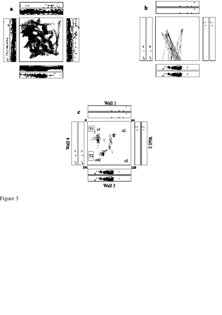
In rats, place cells respond best during active locomotion (Foster et al., 1989; Terrazas et al., 2005). To test whether place cells might be more apparent in macaques during active locomotion, as active locomotion was thought to be a key issue in rodents (Foster et al., 1989) and may be relevant in primates (Thome et al., 2017), single hippocampal neurons were recorded while monkeys actively walked on all four legs around the test environment (Georges-François et al., 1999; Robertson et al., 1998; Rolls et al., 1997a; Rolls et al., 1998). Also, to provide a good opportunity for primate hippocampal spatial neurons to reveal how they encoded space, the simple cue-controlled environment (Rolls and O'Mara, 1995) was changed to a much richer open laboratory environment approximately 5x5 m (illustrated in Fig. 4, and with for example windows on walls 1 and 2) within which the macaque had a 2.5x2.5 m area in which to walk and forage for food. The place of the monkey and the head direction were tracked continuously while the monkey walked round the environment, and the eye position (which refers to the horizontal and vertical eye directions with respect to the head), were recorded continuously to enable measurement of where the monkey was looking in the environment at all times. The monkey walked round the test area, foraging for food, to enable measurements of neuronal firing for a wide range of places, head directions, and spatial views in a very wide range of different combinations to allow analysis of the relative importance of these factors in what was encoded by the neurons.
the monkey was located, and of head direction and eye position. Moreover, the spatial view fields of the neuron were similar when the monkey was actively walking, and also when he was stationary but actively exploring with eye movements different parts of the spatial environment (Georges-François et al., 1999).
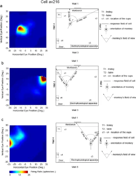
The firing of a different hippocampal cell is provided in Fig. 4, to show with a different type of analysis, how the firing is related to spatial view, and not to place, or to head direction, or to eye position. The highest firing of the cell, with the macaque at the place and with the head direction shown in Fig. 4a, occurred when the macaque looked 10° left. With the monkey in another place and with a different head direction, the highest firing was when the macaque was looking 30° right, but at the same spatial view (Fig. 4b). Fig. 4c shows the firing with the macaque at a different place (but the same head direction as in 4b), and the firing was now when the monkey looked approximately 30° left. The spatial view field was at the same place on Wall one as in Figs 4a and 4b. Previously unpublished examples of video animations to illustrate the firing of macaque hippocampal spatial view neurons are provided in the Supplementary Material.
These experiments show that it is the spatial view towards which the monkey looks that determines the neuronal responses, and not a particular place where the monkey was located, or head direction, or eye position, and this was confirmed with analyses of variance (Georges-François et al., 1999). It was found that on average the spatial view cells encoded considerably more information about spatial view (0.47 bits) than about eye position (0.017 bits), head direction (0.005 bits), or place in the room (0.033 bits) (Georges-François et al., 1999). This shows that the encoding by these primate hippocampal neurons may reflect some information about place etc but is primarily about spatial view.
The spatial view fields of these neurons typically occupy a region of space that is approximately as large as 1/16 of all the four walls of the laboratory (Rolls et al., 1998). Each neuron responds to a different view, and the partly overlapping view fields thus provide precise information about the region of space being looked at. Information theoretic measures showed that the information about spatial view increases almost linearly as the number of neurons in the sample increases, thus showing that each of the neurons makes an independent contribution within the population to representing allocentric space (Rolls et al., 1998). Given that Shannon information is a logarithmic measure, this evidence indicates that the number of spatial views (or the accuracy of the spatial representation) increases exponentially as the number of neurons in the ensemble increases. This is an important result in terms of how information is encoded by hippocampal as well as by neurons in other brain areas (Rolls, 2016a; Rolls and Treves, 2011). Moreover, this is a firing rate code, with much information present in the number of spikes from a single neuron (Panzeri et al., 1999; Rolls, 2016a; Rolls and Treves, 2011).
Many hippocampal spatial view (or “space” or “view”) cells were found in these experiments (Georges-François et al., 1999; Robertson et al., 1998; Rolls et al., 1997a; Rolls et al., 1998). In the initial sample of 352 cells recorded under these conditions, the number of spatial view cells was 40, or 11.4% (Rolls et al., 1997a). This was in a single environment (Georges-François et al., 1999; Robertson et al., 1998; Rolls et al., 1997a; Rolls et al., 1998), and of course the proportion of neurons would be expected to be higher if testing included several different environments. The spontaneous firing rate of these neurons was low (mean 0.5 spikes/s), and their mean peak firing rate was 17 spikes/s (interquartile range 11-20 spikes/s), consistent with these being hippocampal pyramidal cells, which were in both the CA1 and CA3 regions.
The finding from rodents that place cells respond better during active locomotion than passive motion (Foster et al., 1989) made it important to investigate primate hippocampal neurons during active locomotion (see also Thome et al., 2017). Having said this, Rolls and colleagues found that primate hippocampal spatial view cells have similar responses during active locomotion as when the monkey is not locomoting, but is looking around and actively exploring the spatial environment with eye movements. This is shown by the fact that spatial view fields are present when the monkey is stationary as illustrated in Fig. 4, or are walking as in Fig. 3 (Georges-François et al., 1999; Robertson et al., 1998; Rolls et al., 1997a); are found when the monkey is tested only when stationary (Rolls and O'Mara, 1995; Rolls and Xiang, 2005, 2006; Rolls et al., 2005); and as illustrated in the videos provided in the Supplementary Material. Indeed, it is an interesting hypothesis that this active exploration of a spatial environment, by moving the eyes from location to location in a scene in primates, is analogous to the active exploration performed by a rodent when it is locomoting from one place to another.
To assess whether a neuron in the primate, including human hippocampal system, responds to the place where the individual is rather than spatial view, or head direction, or eye position, extensive testing with contrasts of these different hypotheses is needed (Georges-François et al., 1999). (Eye position refers to the horizontal and vertical angles of the eye in the orbit.) If the views visible from different places differ, showing that the firing depends on the place where the individual is located is insufficient, because so does the spatial view. To separate spatial view from place cells, neurons must be tested while the individual is in one place with all of the different spatial views visible from there. Further, the same neuron must also be tested when the individual is located in a different place, but with at least many of the same spatial views visible, as has been implemented in Rolls and colleagues’ recordings in macaques. Indeed, although hippocampal neurons in squirrel monkeys were found to respond when the monkeys were in a particular location in a 3D chamber (Ludvig et al., 2004), where the monkeys were looking was not measured, so we cannot contrast spatial view with place coding in this case. Similarly, Ono et al (1993) found that when a monkey sitting in a cab was moved, some neurons responded when the cab was in specific places in the room. However, they were not able to factor out place from spatial view encoding in the type of factorial design that is necessary. These points will need to be taken into account for future investigations of hippocampal neuronal activity in humans and other primates (cf. Ekstrom, 2015; Ekstrom et al., 2003; Fried et al., 1997; Kreiman et al., 2000; Miller et al., 2013), and recording simultaneously the eye position, head direction, and head position is needed.
Having said this, it is nevertheless of interest that for humans there is now some evidence for medial temporal lobe neurons with properties like those of spatial view cells (Ekstrom et al., 2003; Miller et al., 2013), even though direct measures of eye position were not conducted. For example, in the study by Ekstrom and colleagues, cells were found to represent the interaction between the place and the view faced by the patient. It is also of interest that in humans some medial temporal lobe neurons reflect the learning of paired associations between views of places, and people or objects (Ison et al., 2015), and this implies that views of scenes are important for human hippocampal function. Consistent with this, human functional neuroimaging studies do show hippocampal activation when scenes or parts of scenes are viewed even when the human is fixed in one place for neuroimaging (Brown et al., 2016; Burgess, 2008; Chadwick et al., 2010; Chadwick et al., 2013; Epstein and Kanwisher, 1998; Hassabis et al., 2009; Maguire, 2014; O'Keefe et al., 1998; Zeidman and Maguire, 2016). Further evidence on the functioning of the human hippocampus is considered in sections 3 and 4.4.
2.2.4 Spatial view cells, and the representation of place in the primate hippocampus.
With the discovery of spatial view cells in macaques, unlike what is found in rodents, there was a strong focus on elucidating the properties of spatial view neurons. Nevertheless, as noted above some hippocampal cells with place-related activity were found, as was modulation of some spatial view neurons by place (Rolls and O'Mara, 1995). Further evidence of place-related firing of hippocampal neurons in primates, and the situations that may facilitate place-related activity, are described in sections 2.3, 4.2 and 4.4.
An interesting associated point is what happens to a spatial view cell if it is tested in the place for which it is tuned. Robertson et al (1998) provide an example in their Fig. 1, in which a spatial view cell responded to a landmark, a table, when the monkey was distant from and looked at the table, and also responded when the macaque was at the table, provided that in both cases the macaque was looking at the table. This type of evidence shows that a primate spatial view cell can respond to a landmark or spatial view when the primate is actually at the place where the landmark is located (Robertson et al., 1998; Rolls et al., 1997a).
2.2.5 Comparison of primate hippocampal spatial view cells with other types of neuronal response. To further elucidate the properties of primate hippocampal place cells, they are compared next to object cells in the macaque inferior temporal visual cortex, and then to ‘concept’ cells in humans, and then to head direction cells.
2005; Rolls et al., 2003). In contrast, parts of a spatial scene are fixed with respect to other parts of the scene, and cannot be moved independently with respect to the other parts. Thus, although hippocampal spatial view cells can respond to stimuli that are a fixed part of a spatial scene (e.g. the table T2 in Fig. 1 of Robertson et al (1998) referred to above), the point is that this is a fixed part of a continuous spatial scene. Spatial scene representations may be learned by associating together features in a scene that have a fixed spatial relationship to each other (Rolls and Stringer, 2005; Stringer et al., 2005), and this is quite different from invariant visual object learning in which the features of a single object are associated together, because of regular association of the parts of a single object that are independent of the background scene and other objects in the scene that are not constant (Rolls, 2012; Rolls, 2016a; Stringer and Rolls, 2008; Stringer et al., 2007). The inputs to the hippocampal formation that help it to form spatial view representations may come from areas such as the occipital place area (Julian et al., 2016), and from scene processing areas in the macaque temporal cortex (Kornblith et al., 2013).
Another difference from IT neurons is that many hippocampal spatial view neurons, because they represent parts of space, can respond even when the scene is not visible but that part of space is looked at (Rolls et al., 1997b); and can be updated idiothetically, that is by self-motion, in that spatial view neurons respond when a macaque moves the eyes to a location in space even when no scene is visible, and in darkness (Rolls et al., 1997b) (see section 2.4).
Another difference is that IT neurons respond well to visual stimuli in an object-reward association task (Rolls et al., 2003; Rolls et al., 1977), but hippocampal neurons have weak responses to objects in this type of non-hippocampal-dependent task, compared to the stronger object-related responses that can occur in an object-place, hippocampus-dependent, task (Rolls and Xiang, 2005).
In primates, hippocampal neurons have been described that respond in an invariant way to the sight of individual faces (Quiroga et al., 2005; Sliwa et al., 2016). The neurons found in humans described as ‘concept cells’, an example of which is a neuron that responded to Jennifer Aniston, may respond not only to Jennifer Aniston, but also to other actors in the same movie, and the places with which they are associated (De Falco et al., 2016; Quiroga, 2012; Quiroga et al., 2005; Rey et al., 2015). Object-place cells in macaques have some similar ‘concept’ properties, in that they can be activated either by the object, or by the place, in object-place memory tasks (Rolls and Xiang, 2006; Rolls et al., 2005). Similar properties have been described for human hippocampal neurons, in a task in which a human was associated with a place (Ison et al., 2015).
It can also be emphasised that spatial view hippocampal cells are quite distinct from head direction cells, found in the primate presubiculum and parahippocampal gyrus (Robertson et al., 1999). For instance, if the head direction remains constant when the macaque is moved to different places in the environment where the spatial view differs, spatial view cells provide different responses. On the other hand, head direction cells have activity that remains constant for a particular head direction, even though the spatial view differs completely (Robertson et al., 1999).
2.3. Visually supported spatial representations in primates in a virtual environment
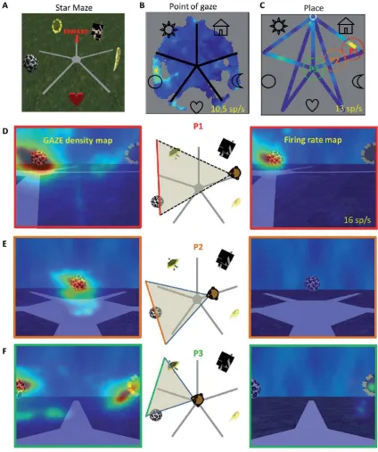
animals looked at one or sometimes more than one of the landmarks (Fig. 5B, C). Of these, the response of 83% were modulated by the position of the animal (Fig. 5D). This is illustrated by the fact that the great majority of cells responded to a landmark when it was viewed from one position only as illustrated in Fig. 5. This point is brought out by the high firing rate when the monkey looked towards the ball landmark when it is in place p1 (Fig. 5F), but no or less firing when the monkey is in place p2 or p3 (Fig 5G) (Wirth and Baraduc, 2018; Wirth et al., 2017). In addition, 17% of the total population responded when the animal was in one or more places in the virtual environment without significant modulation by where the animal was looking. Earlier results in a virtual environment had provided evidence for place-related firing, but eye position was not recorded, so it was difficult to separate view-related from place firing or head direction (Furuya et al., 2014; Hori et al., 2005).
Further, by using a task requiring navigation towards a goal, Wirth and colleagues found that some neurons responded to combinations of view, place, and task context (Wirth et al., 2017). More precisely, some cells were active in only some segments of the possible trajectories that could be followed in the environment to reach the goal. A state space representation accounts best for the activity of these cells. The state space allows separation of the activity of cells for all possible trajectories in the maze, and showed that neurons differentiated common segments between trajectories even when the view and place were identical. This suggests that the cells encoded useful information that reflects the progression along a particular trajectory during navigation rather than purely view or position information. This provides evidence that in primates in a virtual environment requiring active navigation, spatial information is anchored on what the animal foveates, but also is related to task-relevant information (Wirth et al., 2017).
This description bears similarities with spatial view cells described by Rolls and colleagues, and confirms that what the animal looks at modulates the activity. However, it suggests additionally that the modulation by cognition can have a strong influence on the expression of the firing rate. Rolls and O’Mara (1995) did find that spatial view cells could be influenced by place. What Wirth and colleagues showed is that this influence is strong in a task in which the animal needs to base its behaviour on the landmarks and where the animal is located (Wirth et al., 2017).
The development of virtual reality techniques in rodents allows a timely cross-species comparison of the types of hippocampal activity observed during virtual navigation in primates (described above) and rodents. In rats and mice it was shown that navigation to a goal could occur in a virtual environment with visual cues (Cushman et al., 2013; Holscher et al., 2005; Youngstrom and Strowbridge, 2012). These studies showed that without vestibular input, visual cues are sufficient to support a flexible route to a rewarded goal provided that the visual environment took into account the rodent’s wide-angle visual system (approximately 320 degrees (Hughes, 1979)). Further, patterns of neuronal activation were similar in virtual environments (often termed virtual reality “VR”) compared to the real world when the environments were linear tracks (Chen et al., 2013; Dombeck et al., 2010; Harvey et al., 2009). However, in one study comparing directly real world and VR, Ravassard et al (2013) showed that VR recruited fewer rat hippocampal neurons than the real world, and the firing rates were more unstable in VR. Consistently, in macaques fewer CA1 neurons were activated in a virtual environment than in real spatial environments (Thome et al., 2017).
While the first studies in VR in rodents were obtained as animals ran on 1D linear tracks, recent studies used 2D environments in which the animal, placed on a floating ball, can rotate and run in all directions. One such VR study found that hippocampal system neurons responded in VR similarly to in a real environment with place, border and head direction cells (Aronov and Tank, 2014). In another study however, in a similar 2D VR environment, place selectivity was absent when animal performed a random foraging task. However, place selectivity was reinstated when distinct landmarks at fixed locations indicated the reward position (Aghajan et al., 2015). The authors suggested that spatial selectivity can arise in 1D linear tracks and in 2D - if a visual cue shapes behaviour - because of the repeated pairing between distal visual cues and locomotion cues along systematic paths (Aghajan et al., 2015). This second condition in which landmarks are used is similar to primate studies in which spatial selectivity is found when the primates use distal room cues to plan trajectories and find a goal (Furuya et al., 2014; Hori et al., 2005; Wirth et al., 2017).
selectivity was also present in a random foraging VR task in 23% of the neurons despite no place cell responsiveness. Because this was in a VR environment (where there is reduced vestibular input), this demonstrates that visual cues alone can promote this directional selectivity (Acharya et al., 2016). It should be noted that these neurons in rodents are modulated by the direction in which the head is facing, and are different from primate hippocampal spatial view cells, which are not modulated by head direction, but respond to the location in the environment where the monkey is looking (Georges-François et al., 1999). Consistent with this, in macaques navigating a virtual environment to locate a reward, hippocampal cells code for what the animal looked at from a specific viewpoint (Wirth et al., 2017).
In summary, the comparison of findings in VR from primate and rodent studies supports the conclusion that hippocampal cells can be driven by an environment bearing visual cues only with minimal vestibular input besides optic flow. The extent to which place selectivity and head direction selectivity are modulated by the use of visual cues in a goal oriented manner is still to be determined. The fact that direction selectivity exists in VR suggests a homology between primates and rodents, in that a visual non-vestibular input may act as an external reference for the cells. However, because the findings by Acharya et al. (2016) were reported in a task in which animals performed a random foraging task, further rodent studies in which place and direction selectivity are measured in a goal oriented task would be helpful. Likewise, more studies modulating the task demand in primates would be helpful to determine what controls the emergence of target of gaze selectivity and place selectivity in hippocampal neurons.
2.4. Idiothetic (self-motion) update of spatial view cells
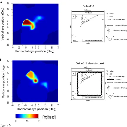
In rats, the representation of place by place cells, can be updated by self-motion, e.g. running in the dark (Jeffery et al., 1997; McNaughton et al., 1991; Quirk et al., 1990). In monkeys, the representation of the place in the scene encoded by spatial view cells can be updated by self-motion, e.g. by the monkey moving the eyes in the dark, or by the monkey turning or walking in the dark. This was shown in experiments on these spatial view cells, in which the view was obscured by black curtains, in which many of the cells could still respond when the macaque moved his eye position to look towards where the view was visible previously (Robertson et al., 1998) (see example in Fig. 6). This idiothetic update also occurs when the monkey locomotes in the dark, and then looks to a spatial view location. Some drift of the spatial view field over a few minutes when the curtains were closed was typical, consistent with the hypotheses that self-motion updating was occurring, and that the visual view details of the scene normally define the spatial view field of a neuron. These experiments (Robertson et al., 1998; Rolls et al., 1997a) show that primate hippocampal system spatial view neurons can be updated by self-motion for short periods by idiothetic information including eye position, head direction, and place movements made by the monkey, and that the drift related to the temporal integration of these signals can be corrected when the scene again becomes visible. These experiments also show that these hippocampal system spatial view cells are different from the much more visual perception-related responses of inferior temporal cortex objects and face cells, which stop responding when the object or face is removed from visibility (Rolls, 2003; Rolls and Tovee, 1994).
The neurons with only a small decrease of their response when the room was placed into darkness and/or the view details were obscured with curtains were present in CA1, the parahippocampal gyrus, and the presubiculum. On the other hand, CA3 neurons had a larger decrease (on average to 23% of their normal response) when the macaque looked towards the normally effective location in the environment but the view was not visible. There may be partial recovery of information in the CA3 network using autoassociation, and further recovery in the associative synapses from CA3 to CA1, as has been shown analytically (Schultz and Rolls, 1999) and by simulations (Rolls, 1995). Another contributory factor of the difference might be the direct perforant path input to the CA1 neurons (Rolls, 2016a; Rolls and Treves, 1998).
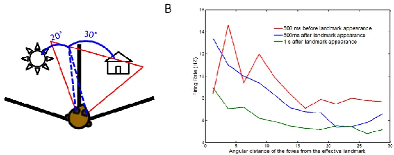
landmark had already been visible for 1 s (green in Fig 7B), providing evidence that these hippocampal neurons fire more when a landmark is first acquired, and after that tend to show a smaller response. In summary, these results indicate that hippocampal cells anticipate a landmark, using predictive recall based on knowledge of the spatial environment.
The relation between eye movements and memory has been highlighted by Buffalo and colleagues (Meister and Buffalo, 2016). Interestingly, it has been reported that some hippocampal neurons respond when eye position must be maintained in the dark (Nowicka and Ringo, 2000), and there is additional evidence about how macaque hippocampal neurons may alter their activity when the eyes are moved (Ringo et al., 1994; Sobotka et al., 1997; Sobotka and Ringo, 1997), with consistent evidence in humans (Hannula and Ranganath, 2009; Liu et al., 2017; Shen et al., 2016). Two additional phenomena relevant to anticipatory effects have been reported previously. First, it was observed that sharp wave ripples were enhanced near remembered visual objects (Leonard and Hoffman, 2017). Second, oscillatory activity was found to synchronize with visual exploration, through a theta phase reset at saccades (Hoffman et al., 2013; Jutras et al., 2013). Buffalo and colleagues described hippocampal neurons with spatial responses at different positions on a screen, that responded at different times primarily after a saccade to the effective location on the screen (Meister and Buffalo, 2016). Together, these results suggest that there is a strong relationship between visual exploration and information uptake and recall in the hippocampus. Indeed, attention has been drawn to the role of visual exploration of the world “out there” using eye movement as an important feature of primate behaviour that has implications for understanding the functioning of the hippocampus in primates (Buffalo, 2015) including humans (Ekstrom, 2015), and how this may serve memory functions of the hippocampus (Rueckemann and Buffalo, 2017).
2.5. Responses of neurons in the primate hippocampus to whole body motion
To perform idiothetic update of a spatial representation (such as that provided by spatial view or place cells), a self-motion signal is needed to update the spatial representation. The idiothetic signal might usefully be a velocity of movement signal. This velocity signal might have its origin in vestibular signals about motion, in optic flow, and/or in corollary motor discharge (Bremmer et al., 2002a; Bremmer et al., 2002b). Neurons that do respond to self-motion signals have been discovered in the primate hippocampus, in an investigation in which the monkey was moved while sitting on a robot with defined axial rotations and linear translations, and in a test situation in which optic flow visual motion cues could also be produced by rotating the whole environment round the monkey (O'Mara et al., 1994). The neurons respond to the velocity of whole body motion (O'Mara et al., 1994), which is idiothetic information. For instance, some neurons have larger responses for clockwise than for anti-clockwise whole body rotation. Occlusion of the visual field showed that some of these neurons depend on visual input. For other neurons there was no requirement for visual input, and these neurons probably responded to vestibular input. Other neurons responded to a combination of whole-body motion and view or place. Of the 45 neurons with responses related to whole body motion (9.8% of the population of hippocampal neurons recorded), 13 responded to axial rotation only, 9 to linear translation only, and 20 neurons to axial rotation or to linear translation. The sign of the motion was important for some of the neurons, with different responses for clockwise vs anticlockwise rotation, or for forward vs backward linear translation, which are different velocities. Some neurons responded to a combination of whole body motion and either a local view (n=2) or a place towards which the macaque was moving (n=1). Whole-body motion neurons are likely to be a useful component of a memory system for memorising spatial trajectories through environments for path integration that is useful in short range spatial navigation (O'Mara et al., 1994). They may provide self-motion information useful to provide the idiothetic update of spatial view cells. Consistent with this discovery (O'Mara et al., 1994), neurons have more recently been found in the rat entorhinal cortex that have a linear response with linear running speed, and have been termed ‘speed cells’ (Hinman et al., 2016; Kropff et al., 2015).
2.6. Grid cells in rodents and spatial view grid cells in the primate entorhinal cortex
the idiothetic (eye movement-related) update of spatial view cells (Robertson et al., 1998). The existence of spatial view grid cells in the entorhinal cortex of primates is predicted from the presence of spatial view cells in the primate CA3 and CA1 regions (Kesner and Rolls, 2015; Rolls, 2013; Rueckemann and Buffalo, 2017). Moreover, some of these ‘spatial view grid cells’ have their responses aligned to the visual image (Meister and Buffalo, 2018), as predicted (Kesner and Rolls, 2015).
In the human entorhinal and cingulate cortex neurons with grid-like response properties are found (Jacobs et al., 2013; Nadasdy et al., 2017), and there is neuroimaging evidence that is consistent with this (Julian et al., 2018; Nau et al., 2018). This is further evidence for the concept that representations of places being viewed in space “out there” is a key property of spatial representations in the hippocampal system of primates including humans.
3. Properties of primate hippocampal spatial representations related to object and reward memory associations
Primates have a highly developed ventral stream cortical visual system that utilises information from the fovea for object recognition, and a highly developed eye movement control system to bring the fovea to objects, using mechanisms described elsewhere (Rolls, 2012; Rolls, 2016a; Rolls et al., 2003; Rolls and Webb, 2014). These developments enable primates to explore and remember information about what is present at places seen “out there” in the spatial environment without having to visit those places. Spatial view cells would accordingly be useful as part of a memory system by providing a representation of space that does not depend on where the primate is, and that could be associated with items such as objects or rewards in those viewed spatial locations. This could enable a monkey to remember where it had seen ripe fruit, or a human to remember where in a spatial scene they had seen a person. Primate hippocampal system spatial view neurons may therefore be important in forming memories of what has been seen and where it has been seen even on a single occasion, a key component of an episodic memory. Episodic memories of this type would be useful for spatial navigation or action in space, for which according to Rolls’ hypothesis the hippocampus would implement the memory but not the spatial computation component (Kesner and Rolls, 2015), with evidence for this provided in section 4.4.
We now consider evidence that these hippocampal spatial view neurons have activity that is involved in memory-related spatial functions.
3.1. Object-place neurons in the primate hippocampus
A key issue is whether the primate including human hippocampus is for memory, or for navigation. There is emphasis on navigation for place cell function in rodents (Burgess et al., 2000; Burgess and O'Keefe, 1996; Hartley et al., 2014; O'Keefe, 1979, 1991). However, the hippocampus is implicated in episodic memory in which the place, or temporal place in a sequence of a single episodic memory is associated with for example the associated objects or rewards (Dere et al., 2008; Eichenbaum et al., 2012; Kesner and Rolls, 2015; Rolls, 1990; Treves and Rolls, 1994; Zeidman and Maguire, 2016). If the hippocampus helps to implement episodic memory, then object information would need to reach the hippocampus, where it might be combined with spatial view information to form for example, an episodic memory of a person or object seen in a viewed location.
To investigate the fundamental question of whether object information, as well as spatial information, is provided in the primate hippocampus, single hippocampal neurons were recorded during an object-place memory task in which the monkeys had to learn associations between objects and where they were shown in an open laboratory (Rolls et al., 2005). Some neurons (10%) responded to an object independently of its location; other neurons (13%) responded to spatial view independently of the object shown; and some neurons (12%) fired to a combination of a particular object and the particular place where it was shown in the laboratory. Thus in the primate hippocampus, there are separate as well as combined representations of objects and of their locations in space. These properties are needed in an episodic memory system, for associations between objects and where they are seen are prototypical for episodic memory. These discoveries provide evidence that a key requirement for a human episodic memory system, both separate and combined neuronal representations of objects and their locations “out there”, are present in the primate hippocampus (Rolls et al., 2005). These neurons might also be termed object-spatial view neurons, to emphasize the difference from what is found in rodents. Neurons that correspond have now been described in rodents, but they, as expected, encode item-place, not item-spatial view, combinations (Komorowski et al., 2009). In the rodent investigation, the items were odors.
cortical areas specialized for objects or faces, and neurons responsive for particular individuals were found both in the human medial temporal lobe (Kreiman et al., 2000; Quiroga, 2012) and the monkey hippocampus (Sliwa et al., 2016).
3.2. One-trial, object-place, recall-related neurons in the primate hippocampus
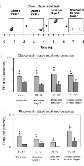
A feature of the theory of the hippocampus in episodic memory is that object and location memories should be capable of being formed in one trial, in order to be relevant to the timescale of episodic memory, and that the whole memory can be recalled from any part (Kesner and Rolls, 2015; Rolls, 1989, 1996, 2016a; Rolls and Kesner, 2006; Treves and Rolls, 1994). This has been tested in macaques in a one-trial object-place memory task. The task involved the storage of object-place information, and then the recall of the object when the place was presented as a recall cue, and the recall of the place when the object was presented as a recall cue (Fig. 8a). The design is similar to that of a one-trial odor-place recall memory task that is hippocampal-dependent in rats (Day et al., 2003), and is quite different from a long-term visual-visual associative memory task which is implemented in the perirhinal and related cortex (Fujimichi et al., 2010; Hirabayashi et al., 2013; Naya et al., 2001). Images of novel objects were used every day, and within a day the same objects were used, so that the one-trial recall task was difficult. Recordings were made from 347 hippocampal neurons during the performance of the object-place recall task (Rolls and Xiang, 2006). Some neurons performed object recall, when the recall cue was a place (Fig. 8b). Some neurons performed place recall, when the recall cue was an object (Fig. 8c). The recall-related firing is evident in stage 4, when the object or place was being recalled with no stimulus present on the screen. Details of the results are provided elsewhere (Rolls and Xiang, 2006). The findings provide evidence that the macaque hippocampus can provide for one-trial object-view association learning of the type that is prototypical for episodic memory (Rolls and Xiang, 2006). Rapid changes in neuronal response properties as a result of learning associations between items such as individuals and places have been confirmed in humans (Ison et al., 2015).
In humans, in an object-place recall task in virtual reality, some neurons during recall also reflect the recall of the place when the object recall cue is provided (Miller et al., 2013). Further, it has been shown that time as well as space is encoded in the primate hippocampus, and that some hippocampal neurons recalled the time at which a specific object was seen (Naya and Suzuki, 2009).
3.3. Reward-place neurons in the primate hippocampus
Information about where rewards are located is a key attribute of an episodic memory system. The anterior hippocampus of primates (which corresponds to the ventral hippocampus of rodents) has inputs from brain areas such as the orbitofrontal cortex and amygdala that perform reward processing (Carmichael and Price, 1995; Pitkanen et al., 2002; Stefanacci et al., 1996; Suzuki and Amaral, 1994a).
To analyse reward-related input to the primate hippocampal system, neuronal activity was recorded during a reward-place association task in monkeys in which one location in each spatial scene on a video monitor, when touched, resulted in a fruit juice reward, and a second location resulted in a less preferred juice reward. The different scenes had different locations for the two reward types (Rolls and Xiang, 2005). 18% of 312 hippocampal cells analysed responded in different scenes to the location of the preferred reward, and 5% to the place of the less preferred reward (Rolls and Xiang, 2005). Of 44 neurons tested, 60% reversed the location to which they responded when the locations of the preferred rewards were reversed in the scenes, providing evidence that the reward-place associations could be relearned in a few trials. Most (82%) of the 44 location-reward neurons in the hippocampus did not respond to object-reward associations in a visual discrimination task. Thus the macaque hippocampus represents the reward associations of places being viewed “out there”, and can store affective information as part of an episodic memory. This provides a way in which the current mood or reward/non-reward state may influence the retrieval of episodic memories, which is of interest for psychiatric disorders in which sad memories may be emphasized because of altered functional connectivity with the orbitofrontal cortex with hippocampal memory mechanisms (Cheng et al., 2016; Cheng et al., 2018a; Rolls, 2016b, 2017a, 2018a; Rolls et al., 2018).
correct reward outcomes by an increase in firing rate. These neurons did not increase their firing rates for rewards that were given randomly, but fired only after a correct response was made in the task. A second type of neuron fired more following an error (which could be the wrong bar release, but also a trial that was aborted). However, only the cells that fired for the correct outcomes were found to discriminate better between the stimuli (the object-place combinations) that were used in the set. This increase in stimulus selectivity was only found in trials and in sessions in which the animals had learned, suggesting that it is part of what helps the task to be learned (Wirth et al., 2009). These findings have been extended by showing that hippocampal neurons reflect outcome information more than prefrontal cortex neurons (Brincat and Miller, 2015).
The results indicate that the primate hippocampus can learn associations between viewed locations and objects (Rolls et al., 2005) or rewards (Rolls and Xiang, 2005), and may even shape the stimulus selective response properties to favour a better neuronal discrimination of the stimulus combination (here object-place-response) that leads to a reward (Wirth et al., 2009). Perhaps correspondingly but with a different representation of space, the responsiveness of rodent place cells can be influenced by where rewards are available (Hölscher et al., 2003; Tabuchi et al., 2003). The principle is that the hippocampus may encode information about where emotion-related (rewarding or punishing) events happened; may be involved in the recall of emotions when particular places are seen later; and may provide mechanisms by which the current mood can influence the memories that are recalled (Rolls, 2015, 2018a).
3.4. Neurons involved in learning associations between visual stimuli and spatial responses The learning of associations between visual stimuli and spatial responses may be involved in some types of navigation. This was investigated in a task for which the primate hippocampus is needed, in which monkeys learned which spatial response to make to different visual stimuli. 14% of hippocampal neurons were found to respond to combinations of visual stimuli and spatial responses (Miyashita et al., 1989). In a subsequent study by Cahusac et al (1993) to investigate the learning, 22% of such neurons in the hippocampus and parahippocampal gyrus modified their responses to become progressively different to the two stimuli while the macaque learned to make different spatial responses to the two visual stimuli (Cahusac et al., 1993). For different neurons this occurred just before, at, or just after the time when the macaque monkey learned the correct responses to make to the stimuli. The hypothesis is that, when new associations between objects and places are learned (in this case the places for responses), some hippocampal neurons learn the new object-to-spatial response associations that are required. Learning of this type could be involved in navigation in which behavioral responses such as a left turn might need to be made when a visual stimulus was seen.
In line with this interpretation, Wirth and colleagues (2003) found that when the monkeys had to learn which of four targets to reach by an eye movement within each of four visual images, the cells became more selective by gradually discriminating between the four stimulus-response associations while they were being learned. The implication that these neurons were involved in the learning was strengthened by findings that well-learned associations were represented by a sharper selectivity in these hippocampal neurons than new associations (Yanike et al., 2009).
These neurons are related to actions made in space to the locations of objects and are another example of object-spatial associative memories supported by the hippocampus.
We have now described the types of neuron found in the primate hippocampus, and move next to compare these primate hippocampal neurons with rodent hippocampal neurons. We show that there are many similarities, though the representation of space provided is very different.
4. Discussion: functions of hippocampal spatial representations in primates, and comparison with rodents
4.1. Self-organization by learning of primate spatial view cells related to foveal vision, and of rodent place cells related to a wide field of view
associated with objects or rewards present in those viewed spatial locations. This would enable humans for example to remember the viewed location where a person had been seen. These primate spatial representations would also be useful in remembering trajectories through gazing at landmarks, of use for example in spatial navigation (Wirth et al., 2017).
The spatial representation in the rodent hippocampus, of the place where the rodent is, may be related to their large visual field of view compared to the primate. A hypothesis on how this difference could be produced by a similar computational process in rodents and primates follows (de Araujo et al., 2001).
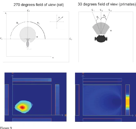
We start with the concept that in both primates and rodents, the dentate granule cells and the CA3 and CA1 neurons respond to combinations of their inputs. In primates the fovea provides high spatial resolution over a typical viewing angle of 5-10 degrees in a complex natural scene as shown by the responses of macaque inferior temporal visual cortex (IT) neurons (with a mean receptive field size of 9 degrees) (Aggelopoulos and Rolls, 2005; Rolls et al., 2003). As a result, a combination of visual features in the spatial environment will produce a spatial view cell, the effective trigger for which will be a combination of visual features within a small part of space. This is illustrated in Fig. 9 top right, where a primate hippocampal neuron responding to C1, C2 and C3 will effectively define a spatial view field. In rodents, in contrast, given the very wide visual field subtended by the retina, which may extend more than 270 degrees, and the absence of a fovea, a combination of visual features learned over such a wide visual angle would define a position in space that is a place. This is illustrated in Fig. 9 top left, where a rodent hippocampal neuron responding to C1, C2 and C3 with large angles between these cues will effectively define a place field. The computational processes by which the hippocampal neurons would learn to respond to feature combinations in rats and primates could be similar, and include competitive learning in the dentate granule cells, autoassociation learning in CA3 cells, and competitive learning in CA1 cells (Rolls, 2016a; Treves and Rolls, 1994). Thus the properties of primate spatial view cells and rodent place cells might arise by a similar computational learning process, but produce spatially different representations because primates are foveate and view a small part of the visual field at any one time, whereas rodents have a very wide visual field (see de Araujo et al (2001)).
This was tested in a simulation in which the animal explored its spatial environment, and hippocampal cells are activated by particular visual cues currently within the field of view, and learn by synaptic modification about these conjunctions of cues. If the field of view is 270°, then place cells were produced as a result of this exploration and learning, as shown in Fig. 9. If the field of view was 30° in the simulations, then spatial view cells were produced as a result of this exploration and learning (Fig. 9). Thus the same computational learning process can lead to place cells with a large field of view as in rodents, and to spatial view cells in foveate animals such as primates including humans. This is Rolls’ hypothesis about how spatial view cells are formed in primates as a result of foveate vision (de Araujo et al., 2001).
There is a hint of something similar in rodents to what is produced by the fovea in primates, in that, when a rat is running along a linear track in which it can see primarily in one direction, then place cells can show more directional properties, in that they respond at a place when the rodent can see one view, but not if the rodent is in the same place and sees the other view (Acharya et al., 2016; McNaughton et al., 1983; Muller et al., 1994). We predict that if a rat’s visual field was restricted, by for example a cone over the head, or in a virtual reality environment, then during learning new environments, or during development, the rat hippocampal neurons might become more like macaque spatial view neurons.
This model thus shows that spatial representations may be produced by similar mechanisms in rodents and primates, but become different because of the primate fovea. Spatial view representations open up the issue of memory functions of the hippocampus involved in remembering where objects and rewards are in spatial scenes, an episodic memory function. The difference in spatial representations in the rodent and primate hippocampus does have implications for understanding how the hippocampus operates in spatial function and memory in primates including humans.
maze, then the place cells would be predicted to respond primarily to the head direction in which the rodent was running, and the place cell shape would be predicted to be elongated in the direction of travel. The statistics of the spatial inputs when rodents reach a boundary and have to stop may also contribute to the formation of boundary cells in rodents.
4.2. Comparison of representations in primates and rodents
Despite the major differences in the spatial representations in the primate and rodent hippocampus, there are important similarities between the operation of the rodent and primate hippocampus, which indicate that the operation of these neural systems is comparable in rodents and primates, even though what is represented is different, as shown in this paper. Some of the similarities of the hippocampal system in primates and rodents are as follows.
First, the spatial representations are in both cases by most spatial view and place cells primarily allocentric. In monkeys, hippocampal spatial view cells during active locomotion in an open environment respond allocentrically to the view of a position in a spatial scene, relatively independently of the place where the monkey is in the open environment, of head direction, of eye position, and of where the spatial view field is relative to the monkey (Georges-François et al., 1999; Rolls and O'Mara, 1995). In a virtual environment, many of the neurons still respond to landmarks and have an allocentric representation (Wirth et al., 2017).
Second, the spatial representations can be updated idiothetically, by one’s body or eye motion in primates, as in the rat.
Third, in both cases the firing rates are low: in primates with a typical mean rate of 0.5 spikes/s, and a typical peak response rate of 17-20 spikes/s (Rolls et al., 1997a; Wirth et al., 2017). This matches numerous accounts of firing properties in rat hippocampus.
Fourth, spatial view cells may fire just before the eyes reach the centre of the spatial view field, and may have their maximal response soon after the eyes reach the spatial view field, and decrease somewhat after that, i.e. show some adaptation (Wirth et al., 2017). Analogous findings have been described for rodent hippocampal cells which generate spike sequences lasting about 2 seconds as rats traverse a place field. The firing rate in the place field shows an asymmetry which changes with experience: as rats become familiar with an environment, cells show an increase in rate before animals reach the place field, followed by a gradual decrease as rats leave the field (Mehta et al., 2000).
Fifth, in macaques, there is evidence for independent representation about spatial view by hippocampal neurons, in that the information rises linearly with the number of neurons (Rolls et al., 1998). This independence arises when the response profiles of the neurons are uncorrelated (Rolls and Treves, 2011). This is a powerful encoding, because the number of stimuli (e.g. spatial views) rises exponentially with the number of neurons. (Of course this independence applies only in a high-dimensional environment, and saturates to the limit in lower high-dimensional environments (Rolls, 2016a; Rolls and Treves, 2011).) Ensemble encoding by populations of neurons is found in rodents (Wilson and McNaughton, 1993), and it would be interesting to know whether the coding by different neurons is independent.
Sixth, rodent place cells may respond differently on the trajectory to approach a goal depending on the state of the animal (Ferbinteanu et al., 2011; Fyhn et al., 2002; Wood et al., 2000), as in primates (Wirth et al., 2017). This implies that place cells support cognition in both species.
Seventh, in macaques, object-spatial view neurons are found (Rolls et al., 2005), and one-trial object-place learning and recall can occur (Rolls and Xiang, 2006). In rodents object-place or odor-place neurons have been described (Kim et al., 2011; Komorowski et al., 2009). The presence of a barrier, which might be thought of as an object, in a place, may also be encoded by rodent hippocampal neurons (Rivard et al., 2004).
Eighth, in macaques, reward-spatial view neurons are found (Rolls and Xiang, 2005), and cells are found to encode reward outcomes (Brincat and Miller, 2015; Wirth et al., 2009). In rodents reward-place neurons have been described (Tabuchi et al., 2003), and it was found that place cells are more active after the receipt of a reward (Singer and Frank, 2009).
Ninth, in rodents, distal room cues can influence place cells (Acharya et al., 2016; Aronov and Tank, 2014; Knierim and Rao, 2003; Shapiro et al., 1997). However, this is different to the encoding of a location in a scene that is provided by primate spatial view cells, in that in rodents the distal room cues are used to encode the place where the rodent is located.