warwick.ac.uk/lib-publications
Original citation:
Ratamero, Erick Martins, Bellini, Dom, Dowson, Christopher G. and Roemer, Rudolf A. (2018) Touching proteins with virtual bare hands : visualizing protein–drug complexes and their dynamics in self-made virtual reality using gaming hardware. Journal of Computer-Aided Molecular Design. doi:10.1007/s10822-018-0123-0
Permanent WRAP URL:
http://wrap.warwick.ac.uk/103398
Copyright and reuse:
The Warwick Research Archive Portal (WRAP) makes this work of researchers of the University of Warwick available open access under the following conditions.
This article is made available under the Creative Commons Attribution 4.0 International license (CC BY 4.0) and may be reused according to the conditions of the license. For more details see: http://creativecommons.org/licenses/by/4.0/
A note on versions:
The version presented in WRAP is the published version, or, version of record, and may be cited as it appears here.
https://doi.org/10.1007/s10822-018-0123-0
Touching proteins with virtual bare hands
Visualizing protein–drug complexes and their dynamics in self-made virtual reality using
gaming hardware
Erick Martins Ratamero1 · Dom Bellini2 · Christopher G. Dowson2 · Rudolf A. Römer3
Received: 20 February 2018 / Accepted: 18 May 2018 © The Author(s) 2018
Abstract
The ability to precisely visualize the atomic geometry of the interactions between a drug and its protein target in structural models is critical in predicting the correct modifications in previously identified inhibitors to create more effective next generation drugs. It is currently common practice among medicinal chemists while attempting the above to access the infor-mation contained in three-dimensional structures by using two-dimensional projections, which can preclude disclosure of useful features. A more accessible and intuitive visualization of the three-dimensional configuration of the atomic geometry in the models can be achieved through the implementation of immersive virtual reality (VR). While bespoke commercial VR suites are available, in this work, we present a freely available software pipeline for visualising protein structures through VR. New consumer hardware, such as the HTC ViVe and the OCulus RifT utilized in this study, are available at reasonable
prices. As an instructive example, we have combined VR visualization with fast algorithms for simulating intramolecular motions of protein flexibility, in an effort to further improve structure-led drug design by exposing molecular interactions that might be hidden in the less informative static models. This is a paradigmatic test case scenario for many similar applications in computer-aided molecular studies and design.
Keywords Virtual reality · Molecular design · Protein dynamics · Anti-microbial resistance
Introduction
Proteins are three-dimensional (3D) objects [1] and, for the last century, spatially-resolved structural models of pro-teins and other biologically relevant molecules have been provided by various experimental techniques. X-ray crys-tallography was particularly instrumental in this revolution [2] with the very first structure of a protein resolved by this method in the 1950s [3]. Since then, X-ray crystallography has led to the building of detailed protein models and was
instrumental for a number of important advances, with Wat-son and Crick’s accurate 3D model of the DNA structure as a prominent example [4]. The 3D characteristics of protein molecules are important in aiding our comprehension of many biological processes. Furthermore, beyond the clas-sical “structure implies function” approach, it is now also becoming increasingly clear that protein dynamics is key to understanding protein function [5]. One of the ways we could potentially access this information is by interacting with, manipulating and visualising static and dynamic mod-els of such proteins in 3D. These might be constructed as real objects or exist in a virtual reality (VR) environment. A large part of scientific and medicinal research on drugs, such as, e.g., understanding antimicrobial resistance (AMR), revolves around clarifying the ways drugs bind to proteins and vice versa. In the, e.g., AMR context, viewing protein structures and their dynamics and understanding how muta-tions can lead to conformational changes and, thus, changes in binding regions that are relevant to AMR, is essential.
* Rudolf A. Römer [email protected]
1 Department of Physics and School of Life Sciences,
University of Warwick, Coventry CV4 7AL, UK
2 School of Life Sciences, University of Warwick,
Coventry CV4 7AL, UK
3 Department of Physics, University of Warwick,
Journal of Computer-Aided Molecular Design
1 3
Since computers became ubiquitous, the development of
computer models for proteins, as opposed to physical ones, in turn became progressively easier and cheaper. With that came the possibility to show proteins in stereoscopic 3D view. Many such projects were developed, as TAMS, for example, which used polarized slide projectors to display stereoscopic 3D images [6]. Many tools exist to make view-ing protein structures possible, such as PyMOl [7], VMD
[8], RasMOl [9], CHiMeRa [10] and isOlDe [11]. In recent
years, there was a further push to develop systems based on web browsers, such as iView [12] and JMOl [13]. To some
degree, every one of those tools can produce stereoscopic 3D images, be it through passive 3D (using chromatic distor-tion glasses), active 3D (with shutter glasses synchronised to the image displaying device) and autostereoscopic 3D (no headgear required) [14]. Though their method changes significantly, they all aim for the same near-3D effect for the end user. Besides technical drawbacks that either limit resolution or require expensive equipment, these attempts at providing 3D perception when analysing protein structures lack immersion into a different environment and do not lead to a feeling of true 3D presence.
VR allows us to address this lack of immersion, and intro-duce a level of interaction between user and visualisation tool that was not possible before. While the usage of VR in research is not new, the current levels of performance with affordable cost definitely are. Implementing a VR can be achieved through many different techniques. On one side of the complexity scale, we could point towards whole-room arrangements like cave automatic virtual environments [15], while extremely simple solutions like Google Cardboard [16] would lie at the other end of that scale. Somewhere in between we find modern head-mounted devices (HMDs), like the OCulus RifT [17] and the HTC ViVe [18]. These
create a stereoscopic 3D effect through the usage of LCD displays and lenses that allow the system to display different images for each eye, with slightly different points of view that mimic the position of the eyes of a virtual observer. These HMDs are relatively easy to use and to program, with excellent display quality and affordable prices. The promi-nence of such HMDs amongst the gaming community is particularly useful for researchers. Since VR gaming has gained popularity, the tools for programming software to use HMD capabilities have become better, more streamlined and easier to use for people even without a background in graphics programming. Initiatives such as sTeaMVR [19]
and VR addons for the uniTy3D game engine [20] decrease
the amount of work necessary to build a VR application con-siderably. Thus, VR emerges as an ideal solution for visual-ising protein dynamics. It allows us to clearly communicate the large-scale motions taking place on the protein structure, while making interaction possible for looking closer at spe-cific areas of interest, such as binding sites.
VR is now beginning to emerge as an add-on option to molecular visualizers. Projects such as RealiTyCOnVeRT [21]
and the web-based tool auTODeskMOleCuleVieweR [22]
already provide “blackbox” VR viewers, while the nanO
siMbOx [23] pioneers ambitious real-time molecular
dynam-ics simulation visualized in VR. This paper reports on a pro-tocol for introducing protein structures into VR programs, using a combination of widely and freely available software and custom-built scripts and programs. The protocol is eas-ily adapted to other molecules rather than just proteins, such as, e.g., DNA, RNA and whole viruses. Besides being a use-ful tool for researchers to visualise conformational changes in proteins, VR also provides a great outreach opportunity to motivate the general public to understand proteins and their relationship to biochemical research and its applica-tions in drug discovery better. We aim with this paper to give our readers the necessary tools to start their own VR applications. Our freely available VR setups for HTC ViVe
and OCulus RifT can be downloaded from Ref. [24].
Materials and methods
We choose the HTC ViVe [18] as a convenient HMD for VR.
It uses two infrared emitters (“lighthouses”) in the corners of the play area, which generate infrared beams in sweep-ing patterns. The headset possess multiple infrared photo diodes, which will detect these beams, and the position of the headset in the room can be reconstructed from the time differences between signals received at each diode [25]. The user can move around the room freely, with the only incon-venience being the cables attached to the headset. Further-more, the HTC ViVe can also track controllers to grab and
manipulate objects in virtual environments, and can be pre-cisely tracked and displayed inside the virtual environment as well. This allows for an immersive, interactive experience, where the limits are only the HMD cable extension and the corners of the delimited play area defined by the position of the lighthouses.
Our software pipeline uses uniTy3D, a programming
and execution environment [20]. This is a game engine, which allows us to implement ideas quickly and easily. Since it provides standard VR features such as graphics, physics (gravity and rigid-body modelling) and lighting, people without programming experience in these areas can also develop their ideas with minimal training involved. Whenever the standard features are not enough, uniTy3D
use the sTeaMVR abstraction layer. By using such a tool,
it is possible to create a single project that works seam-lessly on the two most popular HMDs (OCulus RifT and
HTC ViVe), without any code duplication. However, using
such a tool for creating our applications brings downsides as well. Not every file format can be easily imported into uniTy3D; specifically, importing 3D models with
cor-rect colours can be tricky. Since protein structures will be represented as 3D models, this requires a specific set up to allow us to visualise these structures inside uniTy3D.
Furthermore, the sTeaMVR environment is sometimes not
easily accessible from inside centrally managed networks due to restrictions on gaming.
We present our current workflow for displaying protein structures in VR in Fig. 2. The initial inputs to our appli-cation are Protein Data Bank (PDB) [26] files. These are text files that describe the geometric structure of molecules. It allows for description of atomic coordinates, rotamers, secondary structures and connections between atoms. In our case, we are mainly concerned with the 3D position of each atom in the protein at each simulation time step; these positions need to be translated to an animated 3D protein structure.
Representing individual atoms in a protein does not indi-cate clearly 3D features such as twist and fold. Therefore, we will use the ribbon diagram representation to visualise secondary structures. These are representations where the polypeptide backbone is interpolated by a smooth curve, generating helices, sheets and loops. The result is a sim-ple, yet informative diagram where 3D information about the protein can be easily displayed. Other representations such as the full “sphere” atom representation can of course be chosen as well if desired. We use the Visual Molecular Dynamics (VMD) program [8] for creating representations. The next step is generating 3D models for the ribbon rep-resentations. VMD allows us to create (waVefROnT [27])
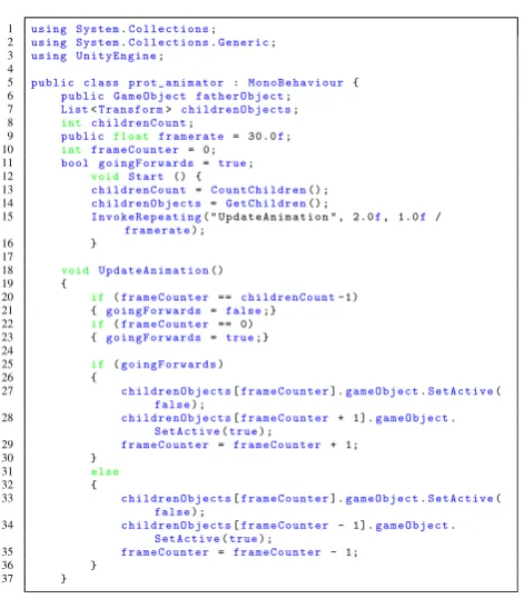
OBJ files; this is an open file format for 3D geometry, stor-ing information about individual vertices, vertex normals, faces of polygons and textures associated with them. This way, we can export the 3D ribbon representations (and any other 3D structure, for that matter) from VMD while keeping any visual information such as colouring associated with it. Let us emphasize that upon exporting the structures from VMD, they become pure geometrical objects, and a user will not be able to query them for names of amino acids, ligands, hydrogen bonds, clashes, distances between atoms, etc. The user must therefore take care to label areas of est in advance. We expect that future improvements in inter-facing game engines and molecular visualizers will make Fig. 1 C# code for uniTy3D, a part of the script written to animate
proteins. There is a father object with multiple children objects con-taining the 3D models of consecutive frames of animation, and this script replaces the object being displayed multiple times per second
[image:4.595.53.289.50.320.2] [image:4.595.233.532.553.722.2]Journal of Computer-Aided Molecular Design
1 3
interactive queries possible. Finally, it is necessary to import the generated OBJ files into uniTy3D. Fortunately, uniTy3D
imports OBJ files natively, and integrates textures without any extra effort being necessary. Therefore, the VMD-gen-erated structures can immediately become physical objects in the VR environment. uniTy3D allows us to attach collider
objects and rigid-body physics to the protein objects, i.e. giving them a virtual mass and spatial extent by integrating them seamlessly into the physics framework of the applica-tion. We find that this deepens a sense of physical reality
for the user, which also translates in an enhanced, almost playful, engagement with the protein structures.
In addition, it is also possible to introduce dynamic struc-tures into VR. We have used data generated by our coarse-graining molecular simulation method [28] in the form of multiple PDB files acting as snapshots of the protein con-formation over time. Next, we load each individual PDB file into our pipeline, obtaining 3D models for each frame. We then use a script in uniTy3D to flip through the models
over time (cp. Fig. 2), creating an animation that shows the conformational changes calculated by our simulations.
Results and discussion
A “template” room was constructed for visualising proteins structures in uniTy3D. This saves time when a quick
visuali-sation is necessary by importing the relevant OBJ files into an existing “template” project where lighting and physics
of the virtual room have previously been defined, and into which VR structures have already been hooked. Use of a “template” project reduces the tasks to simply importing OBJ files to create “father” objects for the 3D models and to define rigid body physics and collider boxes. If necessary, a script can be attached to that object for animation. Depend-ing on the number of snapshots in PDB files, the process from simulation outputs to ready-to-use VR application can be as short as 5 min. A dataset containing all source code used in this project is available in a public repository for download [24].
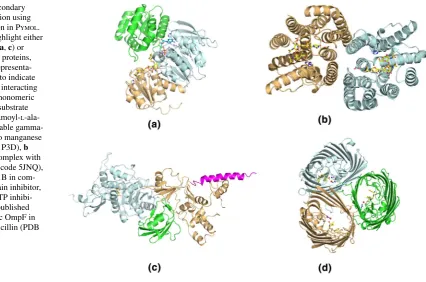
The structural models used in this study as examples for the implementation of 3D visualization in VR are those of four bacterial proteins from different cellular compartments: cytoplasm, inner membrane, periplasm and outer membrane. MurC is a cytoplasmic ligase involved in the construction of the pentapeptide stem, which is a central component of the peptidoglycan (Fig. 3a). MraY is an integral cytoplasmic membrane enzyme that catalyzes the formation of lipid II by transferring the pentapeptide to the lipid carrier, which is another essential step in peptidoglycan biosynthesis (Fig. 3b). Penicillin-binding protein 1b (PBP1b) is a bifunc-tional enzyme containing both a transglycosilation (TG) and a transpeptidation (TP) domain, which is able to polymerize lipid II to form the peptidoglycan mesh (Fig. 3c). OmpF is an integral outer membrane channel exploited by most antimicrobial drugs to enter the organism on their way to the target (Fig. 3d). All structures are in complex with either the substrate or antimicrobial drugs that inhibit bacterial cell
Fig. 3 Standard secondary structure visualization using the “cartoon” option in PyMOl. Different colors highlight either different domains (a, c) or chains (b, d) of the proteins, while the “stick” representa-tion has been used to indicate ligands and nearby interacting side-chains for: a monomeric MurC with bound substrate UDP-N-acetylmuramoyl-l -ala-nine, non-hydrolyzable gamma-imino-ATP and two manganese atoms (PDB code 1P3D), b
dimeric MraY in complex with tunicamycin (PDB code 5JNQ),
[image:5.595.116.543.442.724.2]wall (peptidoglycan) synthesis. As such, they are currently studied targets for understanding AMR resistance mecha-nisms [29].
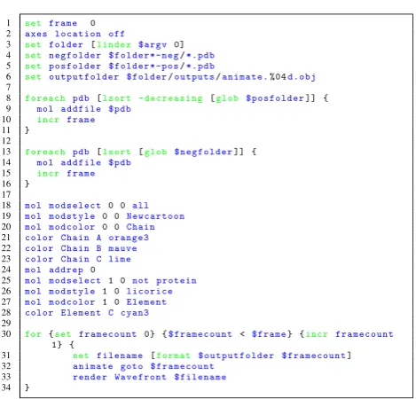
Intrinsic motions of these proteins were simulated by ana-lysing protein flexibility, defining movement modes through elastic network modeling and generating conformers based on that information and steric interactions [28]. These simu-lations produced a series of PDB files detailing the state of protein structures at certain “snapshot” moments (e.g., every 100th conformer out of 5000 overall for each mode of motion). The output of these simulations are PDB files, each containing the protein atomic coordinations at a specific time step. The PDB format is not directly supported by the VR software uniTy3D. Therefore, VMD was subsequently
used to transform PDB files into OBJ files. Clearly, any other molecular dynamics package should also be able to result in the relevant OBJ files—perhaps after using a suitable “con-verter” such as VMD. Our specific choice of modelling [28] represents simply a generic test case scenario. We automated the process by writing TCL scripts [30] for VMD to examine output folders from the simulations and generate OBJ files of each conformer. An example of such script for a specific protein is presented as Fig. 4. Here, every PDB file for pos-sible directions of protein movement gets loaded onto VMD, specific colouring and representation options for that protein are chosen and finally each frame is rendered and stored as an OBJ file [27]. Through this process, a series of OBJ files are obtained that act as 3D “frames” of animation. Upon importing these files into uniTy3D, the only task left is to
ensure that these frames are shown sequentially to impart upon the user the illusion of an animated protein. In this study we do so by updating the displayed object every 1/30 of a second. Though theoretically possible, it was decided against updating the collision structure of the object at each frame, as this would consume significant computational resources, even if it would allow for “correct” physics in VR at all times. Instead, we have chosen to define a bounding box spanning the whole range of movement for the protein as a constant collision structure.
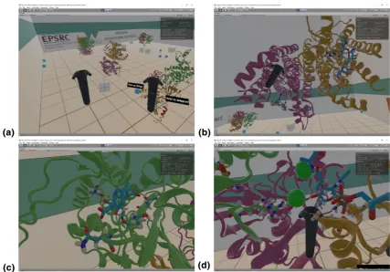
Snapshots of the VR application being used and presented here are shown in Fig. 5a–d, where a user was interacting with the four proteins discussed above in a VR environment. Con-veying a VR experience in pictures is a difficult task; readers with access to the HTC ViCe or the OCulus RifT can download
our preconfigured VR rooms from [24]. Figure 5a shows the template room containing the four protein structures. Some common physical objects such as cubes and balls have been added to emphasize the sense of “reality” and physicality around the protein models. We find that initial engagement with these familiar objects helps first-time users in their later interactions with the protein structures. This figure shows how is also possible to see both controllers being employed by the user in the VR room, with tool tips added for introducing new
players to the controls. Figure 5b shows a snapshot of a protein structure being held by an user. In this case, the user is hold-ing up dimeric MraY, with the same structure and colorhold-ing as in Fig. 3a. A close look to the drug and its interactions with protein residues can be taken as desired by “grabbing” the protein with the controller and moving it towards the viewer; tilting the controller allows to rotate the protein structure in an easy and intuitive way.
Further detail on specific residues and drug-binding pock-ets can be seen by simply approaching the areas of interest, or through clipping the protein structure through the view point. Figure 5c shows a detail of ampicillin bound to the TP domain of PBP1b. This is the same region presented in light blue in Fig. 3d. The figure shows how much and how simple VR allows for in-depth analysis of specific areas of a protein structures. Finally, in Fig. 5d, we present a detail of an area of contact between MurC, the substrate and the cofactor, includ-ing the two catalytic manganese ions. In the VR environment, investigation of protein–ligand interactions is straightforward, since complete control of the 3D position and rotation of the protein model is mapped to movement of the controllers.
Conclusion
[image:6.595.308.543.50.275.2]Journal of Computer-Aided Molecular Design
1 3
researchers to straightforwardly embed protein structures into VR programs using a combination of widely available software, standard hardware and only few custom-built codes. The immersion and interactivity that VR brings can significantly change the level of details accessible to a researcher when analysing protein–ligand interactions or conformational changes. While it is difficult to convey this with static figures as in Fig. 5, it is interesting to report our personal experiences with immersive 3D VR. For exam-ple, despite having previously worked with these protein structures for a while and having observed the drug-bound catalytic pockets of these proteins for many times on 2D rendering softwares such as PyMOl, it was still only after
we observed the same in VR that we realized that the 3D configuration of the catalytic pocket was significantly dif-ferent to the picture conveyed by the 2D projections and that we had built in our minds. These differences could translate into designing more effective modifications into the drug.
With the decreasing costs for customer VR hardware and an established workflow for importing structures into VR, immersive 3D visualization should become viable for an increasing number of research groups. It is a first step for
developing VR-based ways of interacting with proteins and probing their properties. At the moment, there is still the limitation in both 2D and 3D protein visualization software packages that pre-computed conformers are necessary and the interaction between user and structure is only at a non-interactive visualisation level. In the future, physical interac-tions may become possible, where the user could bend the protein structure or add new functional groups to the ligand while background simulations calculate in real-time whether such changes are mechanically stable or energetically favora-ble. In such a scenario, immersive VR environments can aid much better than other 2D or 3D visualizations in guiding molecule manipulation.
As well as VR being a very useful tool for visualisation of both protein conformational changes and drug design, there is also the element of fantastic potential for outreach and engagement with the general public. Indeed, at our institu-tion, the protein viewer VR project is now regularly show-cased at such events.
Acknowledgements We gratefully acknowledge funding via the EPSRC’s Cross-scale prediction of Antimicrobial Resistance: from
Fig. 5 a Template VR room containing the four protein structures discussed above. Both controllers are visible, with tool tips for teach-ing the controls. b MraY structure in VR. Details of specific residues and drug can be seen in both monomers. c Detail of PBP1b interac-tions with the drug. VR allows for close inspection of specific areas
[image:7.595.82.510.54.352.2]molecules to populations network (EP/M027503/1). UK research data statement: data is available at [24].
Open Access This article is distributed under the terms of the Crea-tive Commons Attribution 4.0 International License (http://creat iveco
mmons .org/licen ses/by/4.0/), which permits unrestricted use,
distribu-tion, and reproduction in any medium, provided you give appropriate credit to the original author(s) and the source, provide a link to the Creative Commons license, and indicate if changes were made.
References
1. Riddell FG, Robinson MJT (1974) J. H. van’t Hoff and J. A. Le Bel—their historical context. Tetrahedron 30(13):2001–2007 2. Anderson AC (2003) The process of structure-based drug design.
Chem Biol 10(9):787–797
3. Kendrew JC, Bodo G, Dintzis HM, Parrish RG, Wyckoff H, Phil-lips DC (1958) A three-dimensional model of the myoglobin mol-ecule obtained by X-ray analysis. Nature 181(4610):662–666 4. Watson JD, Crick FHC (1969) Molecular structure of nucleic
acids: a structure for deoxyribose nucleic acid. Nature 224(5218):470–471. (Reprinted from Nature, April 25, 1953) 5. Henzler-Wildman K, Kern D (2007) Dynamic personalities of
proteins. Nature 450(7172):964–972
6. Feldmann RJ, Bing DH (1980) Teaching aids for macromolecular structure. Taylor-Merchant Corp, New York
7. Pymol. www.pymol .org/pymol . Accessed 22 Aug 2017 8. Humphrey W, Dalke A, Schulten K (1996) VMD: visual
molecu-lar dynamics. J Mol Graph 14(1):33–38
9. Sayle RA (1995) RASMOL: biomoIecular graphics for all. Bio-chemistry 30:374–376
10. Pettersen EF, Goddard TD, Huang CC, Couch GS, Greenblatt DM, Meng EC, Ferrin TE (2004) UCSF Chimera—a visualiza-tion system for exploratory research and analysis. J Comput Chem 25:1605–1612
11. Croll TI (2018) ISOLDE: a physically realistic environment for model building into low-resolution electron-density maps. Acta Crystallogr D 74(6):6
12. Li H, Leung K-S, Nakane T, Wong M-H (2014) iview: an interac-tive WebGL visualizer for protein-ligand complex. BMC Bioin-formatics 15(1):56
13. Jmol: an open-source browser-based HTML5 viewer and stand-alone Java viewer for chemical structures in 3D. http://jmol.sourc
eforg e.net/. Accessed 22 Aug 2017
14. McIntire JP, Havig PR, Geiselman EE (2014) Stereoscopic 3D dis-plays and human performance: a comprehensive review. Disdis-plays 35(1):18–26
15. Creagh H. Cave Automatic Virtual Environment. In: Proceedings: electrical insulation conference and electrical manufacturing and coil winding technology conference (Cat. No.03CH37480), pp 499–504. IEEE
16. Google Cardboard Google VR. https ://vr.googl e.com/cardb oard/. Accessed 22 Aug 2017
17. Oculus Rift. https ://www.oculu s.com/rift/. Accessed 22 Aug 2017 18. VIVE. https ://www.vive.com/. Accessed 22 Aug 2017
19. SteamVR. http://www.steam vr.com/. Accessed 22 Aug 2017 20. Unity - Virtual Reality. https ://unity 3d.com/learn /tutor ials/topic
s/virtu al-reali ty. Accessed 22 Aug 2017
21. Borrel A, Fourches D (2017) RealityConvert: a tool for preparing 3D models of biochemical structures for augmented and virtual reality. Bioinformatics 33(23):3816–3818
22. Balo AR, Wang M, Ernst OP (2017) Accessible virtual reality of biomolecular structural models using the Autodesk Molecule Viewer. Nat Methods 14(12):1122–1123
23. O’Connor M, Tew, P Sage B, McIntosh-Smith S, Glowacki DR (2015) Nano Simbox: an OpenCL-accelerated framework for interactive molecular dynamics. In: Proceedings of the 3rd inter-national workshop on OpenCL-IWOCL ’15. ACM Press, New York, pp 1–1
24. Ratamero EM, Römer RA (2017) Dataset to support the article “Touching proteins with virtual bare hands: how to visualize pro-tein structures and their dynamics in virtual reality”. http://wrap.
warwi ck.ac.uk/91507 /. Accessed 22 Aug 2017
25. Dempsey P (2016) The Teardown: HTC Vive virtual reality head-set. Eng Technol 11(7):80–81
26. Bernstein FC, Koetzle TF, Williams GJB, Meyer EF, Brice MD, Rodgers JR, Kennard O, Shimanouchi T, Tasumi M (1977) The Protein Data Bank. A computer-based archival file for macromo-lecular structures. Eur J Biochem 80(2):319–324
27. Wavefront object file, from Wikipedia. https ://en.wikip edia.org/
wiki/Wavef ront_.obj_file. Accessed 29 May 2018
28. Jimenez-Roldan JE, Freedman RB, Römer RA, Wells SA (2012) Rapid simulation of protein motion: merging flexibility, rigidity and normal mode analyses. Phys Biol 9(1):016008
29. Bugg TDH, Braddick D, Dowson CG, Roper DI (2011) Bacterial cell wall assembly: still an attractive antibacterial target. Trends Biotechnol 29(4):167–173