Arthur Christopoulos
Sexton, J. Robert Lane, Meritxell Canals and
M.
David M. Thal, Shailesh N. Mistry, Patrick
Alaa Abdul-Ridha, Laura López, Peter Keov,
Acetylcholine Receptor
Muscarinic
1
Modulation at the M
Molecular Determinants of Allosteric
doi: 10.1074/jbc.M113.539080 originally published online January 17, 2014 2014, 289:6067-6079.
J. Biol. Chem.
10.1074/jbc.M113.539080 Access the most updated version of this article at doi:
. JBC Affinity Sites Find articles, minireviews, Reflections and Classics on similar topics on the
Alerts:
When a correction for this article is posted •
When this article is cited •
to choose from all of JBC's e-mail alerts Click here
http://www.jbc.org/content/289/9/6067.full.html#ref-list-1
This article cites 56 references, 28 of which can be accessed free at
at Monash University (CAUL) on March 15, 2014
http://www.jbc.org/
Downloaded from
at Monash University (CAUL) on March 15, 2014
http://www.jbc.org/
Molecular Determinants of Allosteric Modulation at the M
1
Muscarinic Acetylcholine Receptor
*
Received for publication, December 2, 2013, and in revised form, January 16, 2014 Published, JBC Papers in Press, January 17, 2014, DOI 10.1074/jbc.M113.539080
Alaa Abdul-Ridha‡1, Laura Lo´pez‡, Peter Keov‡, David M. Thal‡, Shailesh N. Mistry§, Patrick M. Sexton‡2, J. Robert Lane‡, Meritxell Canals‡3, and Arthur Christopoulos‡2,4
From‡Drug Discovery Biology and§Medicinal Chemistry, Monash Institute of Pharmaceutical Sciences and Department of Pharmacology, Monash University, Parkville, Victoria 3052, Australia
Background:BQCA is a selective allosteric modulator of the M1mAChR.
Results:Residues that govern BQCA activity were identified using mutagenesis and molecular modeling.
Conclusion:BQCA likely occupies a pocket overlapping prototypical mAChR modulators and gains selectivity through
coop-erativity with orthosteric ligands.
Significance: Understanding the structural basis of BQCA function can provide insight into the design of more tailored
allosteric ligands.
Benzylquinolone carboxylic acid (BQCA) is an unprece-dented example of a selective positive allosteric modulator of acetylcholine at the M1 muscarinic acetylcholine receptor (mAChR). To probe the structural basis underlying its selectiv-ity, we utilized site-directed mutagenesis, analytical modeling, and molecular dynamics to delineate regions of the M1mAChR that govern modulator binding and transmission of cooperativ-ity. We identified Tyr-852.64 in transmembrane domain 2 (TMII), Tyr-179 and Phe-182 in the second extracellular loop (ECL2), and Glu-3977.32and Trp-4007.35in TMVII as residues that contribute to the BQCA binding pocket at the M1mAChR, as well as to the transmission of cooperativity with the orthosteric agonist carbachol. As such, the BQCA binding pocket partially overlaps with the previously described “common” allosteric site in the extracellular vestibule of the M1mAChR, suggesting that its high subtype selectivity derives from either additional con-tacts outside this region or through a subtype-specific coopera-tivity mechanism. Mutation of amino acid residues that form the orthosteric binding pocket caused a loss of carbachol response that could be rescued by BQCA. Two of these residues (Leu-1023.29and Asp-1053.32) were also identified as indirect contrib-utors to the binding affinity of the modulator. This new insight into the structural basis of binding and function of BQCA can guide the design of new allosteric ligands with tailored pharma-cological properties.
G protein-coupled receptors (GPCRs)5mediate a multitude of biological functions in response to a variety of ligands, including hormones and neurotransmitters, and play essential roles in all physiological processes (1). As such, GPCRs are important therapeutic targets for numerous diseases (2). Given such importance, an understanding of the structural basis underlying ligand binding and activation of GPCRs is essential to design more effective therapies (3). The recent surge in high resolution family A GPCR crystal structures (4) has provided new insights into the structural and functional diversity of this protein family. This knowledge, combined with information from computational, biochemical, and mutagenesis studies, has not only mapped out the location of orthosteric binding pock-ets but is starting to unravel the molecular changes that occur upon receptor activation and the mechanisms by which differ-ent ligands stabilize distinct conformational states (5, 6).
The M1 mAChR is a family A GPCR and is one of five mAChR subtypes for which acetylcholine (ACh) is the endog-enous orthosteric agonist. The ACh binding pocket is formed by amino acids that are conserved across all five mAChR sub-types and shares structural homology with other functionally unrelated acetylcholine-binding proteins from different species (7). Along with the M4mAChR, the M1mAChR is an attractive therapeutic target for the treatment of diseases in which cogni-tion is impaired, such as Alzheimer disease and schizophrenia (8). However, because of the highly homologous ACh binding pocket across subtypes, it has been challenging to develop drugs that are sufficiently subtype-selective to avoid undesired activ-ity at other mAChRs. This has spurred intensive efforts to dis-cover allosteric ligands that act at topographically distinct regions on these receptors (9) with more potential to confer subtype selectivity. Despite the wealth of information obtained from GPCR crystal structures, challenges remain in under-standing the mode of binding and action of such small molecule *This work was supported in part by National Health and Medical Research
Council of Australia Program Grant 519461 (to A. C. and P. M. S.), Project Grant APP1011796 (to M. C.), and Grant APP1011920 (to J. R. L.) and com-putational studies were supported by Resource Allocation Scheme Grant VR0024 from the Victorian Life Sciences Computation Initiative, Peak Com-puting Facility, University of Melbourne.
1Recipient of an Australian Postgraduate Award scholarship.
2Principal Research Fellows of the National Health and Medical Research
Council of Australia.
3To whom correspondence may be addressed: Drug Discovery Biology,
Monash Institute of Pharmaceutical Sciences, Monash University, 399 Royal Parade, Parkville, Victoria 3052, Australia. Tel.: 61-3-9903-9094; Fax: 61-3-9903-9581; E-mail: meri.canals@monash.edu.
4To whom correspondence may be addressed: Drug Discovery Biology,
Monash Institute of Pharmaceutical Sciences, Monash University, 399 Royal Parade, Parkville, Victoria 3052, Australia. Tel.: 61-3-9903-9067; Fax: 61-3-9903-9581; E-mail: arthur.christopoulos@monash.edu.
5The abbreviations used are: GPCR, G protein-coupled receptor; BQCA,
ben-zylquinolone carboxylic acid; ACh, acetylcholine; mAChR, muscarinic ace-tylcholine receptor; CCh, carbachol; QNB, quinuclidinyl benzilate; NMS,
N-methylscopolamine; IP1, myoinositol 1-phosphate; TM, transmembrane
domain; ECL, extracellular loop; MD, molecular dynamics.
at Monash University (CAUL) on March 15, 2014
http://www.jbc.org/
allosteric modulators (9). High resolution structures of family A GPCRs bound to allosteric modulators are only starting to be solved (10), and even then the dynamic mechanisms contribut-ing to modulator bindcontribut-ing, receptor activation, and transmission of cooperativity between orthosteric and allosteric sites cannot be readily captured in a single structure.
The conserved ACh-binding site in mAChRs is located in the top third of the transmembrane helical bundle of the receptor with ACh contacting inward-facing residues in ECL2 and TMIII–VII (7, 11). In particular, TMIII contains a number of residues that have been implicated in both binding and activa-tion mechanisms of the mAChRs and plays a central role as a structural and functional hub of many GPCRs (1). Accumulated evidence also points toward the existence of a “common” allo-steric binding pocket utilized by structurally diverse mAChR allosteric modulators (12–14). This site is located within an extracellular “vestibule” and includes residues from both ECL2 and the extracellular regions of TMII and -VII (12, 13). Inter-estingly, we recently demonstrated that LY2033298, an allo-steric modulator originally described as being a “selective” pos-itive allosteric modulator for ACh at the M4mAChR, can also occupy this conserved allosteric pocket at the M2mAChR, where it exerts cooperative behavior with alternative orthosteric ago-nists, such as oxotremorine M, but not with ACh (14). Such probe dependence highlights the fact that selectivity of allo-steric agents can actually be attained through two mechanisms, namely the differences in the allosteric site between receptor subtypes or the differences in cooperativity upon binding to a common allosteric site.
With regard to the M1mAChR, several selective ligands have been discovered in the past few years (15). Among these, ben-zylquinolone carboxylic acid (BQCA) is a novel example of a highly selective positive allosteric modulator of ACh binding and function at the M1mAChR, displaying very low affinity but a remarkably high cooperativity with ACh (16 –18). The unprecedented subtype selectivity of BQCA thus suggests two potential scenarios as follows: (i) that BQCA binds to a com-pletely different site than other mAChR allosteric modulators or (ii) that BQCA achieves subtype-selective cooperativity upon interaction with a conserved or overlapping allosteric site. In this study, we aimed to resolve this issue by site-directed mutagenesis of residues previously shown to be important for orthosteric, allosteric, or bitopic (dual orthosteric-allosteric) ligand binding at either the M1mAChR or other mAChR family subtypes. Importantly, we also applied an analytical approach, based on the operational model of agonism (19, 20), to elucidate the effects of the introduced mutations on ligand bindingversus
signalingversustransmission of cooperativity. By doing so, we present new evidence for differential effects of distinct receptor regions on each of these molecular properties at the M1 mAChR.
EXPERIMENTAL PROCEDURES
Materials—Chinese hamster ovary (CHO) FlpIn cells and
Dulbecco’s modified Eagle’s medium (DMEM) were purchased from Invitrogen. Fetal bovine serum (FBS) was purchased from ThermoTrace (Melbourne, Australia). Hygromycin-B was pur-chased from Roche Applied Science. [3H]Quinuclidinyl
ben-zilate ([3H]QNB; specific activity, 50 Ci/mmol),N-[3 H]meth-ylscopolamine ([3H]NMS); specific activity, 85 Ci/mmol), and MicroScint scintillation liquid were purchased from Perkin-Elmer Life Sciences. IP-One assay kit and reagents were pur-chased from Cisbio (Codolet, France). All other chemicals were purchased from Sigma. BQCA was synthesized in-house at the Monash Institute of Pharmaceutical Sciences.
Cell Culture and Receptor Mutagenesis—Mutations of the
c-Myc-hM1 mAChR sequence were generated using the QuikChange site-directed mutagenesis kit (Agilent Technolo-gies, La Jolla, CA). All mutations were confirmed by DNA sequencing (AGRF, Australia). Mutant c-Myc-hM1 mAChR DNA constructs were transfected into FlpIn CHO cells (Invit-rogen) and selected using 0.2 mg/ml hygromycin for stable expression.
Whole Cell Radioligand Binding Assays—To facilitate a more
direct comparison between parameters derived from the anal-ysis of cell-based functional assays (see below), radioligand binding experiments were performed on whole cells rather than membrane preparations. Saturation binding assays were performed using cells plated at 104cells per well in 96-well Isoplates (PerkinElmer Life Sciences). The following day cells were incubated with the orthosteric antagonists [3H]QNB or [3H]NMS in a final volume of 100l of HEPES buffer (10 m
M
HEPES, 145 mMNaCl, 1 mMMgSO4䡠7H2O, 10 mMglucose, 5
mMKCl, 2 mMCaCl2, 1.5 mMNaHCO3, pH 7.4) for 2 h at room temperature. For competition binding assays, cells were plated at 2.5⫻104cells per well. The following day, cells were incu-bated in a final volume of 100l of HEPES buffer containing increasing concentrations of the competing cold ligand CCh (in the absence or presence of increasing concentrations of BQCA) for 4 h at 4 °C (to avoid potential confounding effects of com-peting agonist ligands on receptor internalization while ensur-ing reactions reach equilibrium) in the presence of 0.3 nM
[3H]QNB or [3H]NMS. Nonspecific binding was defined in the presence of 100Matropine. For all experiments, termination
of the assay was performed by rapid removal of radioligand followed by two 100-l washes with ice-cold 0.9% NaCl buffer. Radioactivity was determined by addition of 100l of Micros-cint sMicros-cintillation liquid (PerkinElmer Life Sciences) to each well and counting in a MicroBeta plate reader (PerkinElmer Life Sciences).
IP-One Accumulation Assays—The IP-One assay kit (Cisbio,
France) was used for the direct quantitative measurement of myoinositol 1-phosphate (IP1) in FlpIn CHO cells stably expressing either WT or mutant hM1mAChRs. This is a com-petitive immunoassay that measures the homogeneous time-resolved fluorescence signal transferred between a cryptate-la-beled IP1-specific monoclonal antibody andd2-labeled IP1. The fluorescence signal measured is inversely proportional to the concentration of native IP1.
Briefly, cells were seeded into 384-well proxy-plates at 7,500 cells per well and allowed to grow overnight at 37 °C, 5% CO2. The following day, cells were stimulated with CCh in IP1 stim-ulation buffer (HEPES 10 mM, CaCl21 mM, MgCl20.5 mM, KCl 4.2 mM, NaCl 146 mM, glucose 5.5 mM, LiCl 50 mM, pH 7.4) in
the absence or presence of increasing concentrations of BQCA and incubated for 1 h at 37 °C, 5% CO2. Cells were lysed by the
at Monash University (CAUL) on March 15, 2014
http://www.jbc.org/
addition of homogeneous time-resolved fluorescence reagents, the cryptate-labeled anti-IP1antibody, and thed2-labeled IP1 analog prepared in lysis buffer, followed by incubation for 1 h at room temperature. The emission signals were measured at 590 and 665 nm after excitation at 340 nm using the Envision mul-tilabel plate reader (PerkinElmer Life Sciences), and the signal was expressed as the homogeneous time-resolved fluorescence ratio: F ⫽ ((fluorescence665 nm/fluorescence590 nm) ⫻ 10
4).
Experiments using WT M1mAChR CHO FlpIn cells were per-formed in parallel each day.
Computational Methods for the Model of the Ligand-Receptor
Complex—The sequence of the hM1 mAChR was retrieved
from the Swiss-Prot database. ClustalX software (21) was used to align the hM1mAChR sequence with the crystal structure of the nanobody-stabilized active state of the human2 adreno-receptor (Protein Data Bank code 3P0G) (22). Ballesteros-Weinstein numbering was used for residues in the TMs (23).
The structural model of the receptor was built using the Modeler Version 9.12 suite of programs (24), which yielded 10 candidate models. The conserved disulfide bonds between res-idues Cys-983.25at the top of TMIII and the cysteine in the middle of the ECL2 as well as the one between Cys-3916.61and Cys-3947.29in ECL3 present in the template structure were also built and maintained as a constraint for geometric optimiza-tion. The best structure was selected from these candidates, according to the Modeler Discrete Optimized Protein Energy (DOPE) assessment score and visual inspection. The resulting receptor structure was optimized using the Duanet al.(25) force field and the general Amber force field, and HF/6 –31G*-derived restrained electrostatic potential atomic charges were used for the ligands (26).
Docking of the ligands was performed with MOE (Molecular Operating Environment, Chemical Computing Group, Inc.). CCh was docked manually into the receptor model with the protonated nitrogen interacting with Asp3.32and the carba-mate group situated toward TMVI resembling the position of the ligands described in the mAChR crystal structures (Protein Data Bank code 3UON (7) and Protein Data Bank code 4DAJ (11)). The allosteric binding site of BQCA was generated using the Alpha site finder. Dummy atoms were created from the obtained␣spheres. BQCA docking was carried out using the Induced Fit protocol, with Alpha PMI placement and Affinity dG rescoring. One main BQCA pose was obtained at an allo-steric site comprising residues from ECL2, ECL3, TMII, and TMVII at the extracellular surface of the M1mAChR. The low-est energy conformation of this pose was selected and subjected to an energy minimization using MMFF94X force field. Molec-ular dynamics (MD) simulations of the final complex was performed with NAMD2.9 (27) package using the protocol described previously (28).
Data Analysis—All data were analyzed using Prism 6.01
(GraphPad Software, San Diego). Inhibition binding curves between [3H]QNB or [3H]NMS and unlabeled ligands were fit-ted to a one-site binding model (29). Binding interaction studies with allosteric ligands were fitted to the following allosteric ternary complex model, Equation 1 (30),
Y⫽ Bmax[A]
关A兴⫹
冉
KAKB ␣⬘关B兴⫹KB冊冉
1⫹关I兴 KI⫹
关B兴 KB ⫹
␣关I兴关B兴 KIKB
冊
(Eq. 1)
where Y is percentage (vehicle control) binding;Bmaxis the total number of receptors; [A], [B], and [I] are the concentrations of radioligand, allosteric modulator, and the orthosteric ligand, respectively;KA, KB, andKI are the equilibrium dissociation constants of the radioligand, allosteric modulator, and orthosteric ligand, respectively.␣⬘and␣are the binding coop-erativities between the allosteric modulator and radioligand and the allosteric ligand and orthosteric ligand, respectively. Values of␣(or␣⬘)⬎1 denote positive cooperativity; values⬍1 (but⬎0) denote negative cooperativity, and values⫽1 denote neutral cooperativity.
Concentration-response curves for the interaction between the allosteric ligand and the orthosteric ligand in the various functional signaling assays were globally fitted to the following operational model of allosterism and agonism, Equation 2 (20),
E⫽ Em共A关A兴共KB⫹␣关B兴兲⫹B关B兴KA兲 n
共关A兴KB⫹KAKB⫹关B兴KA⫹␣关A兴关B兴兲n
⫹共A关A兴共KB⫹␣关B兴兲⫹B关B兴KA兲n
(Eq. 2)
whereEmis the maximum possible cellular response; [A] and [B] are the concentrations of orthosteric and allosteric ligands, respectively;KAandKBare the equilibrium dissociation con-stant of the orthosteric and allosteric ligands, respectively;A andBare operational measures of orthosteric and allosteric ligand efficacy, respectively; ␣ is the binding cooperativity parameter between the orthosteric and allosteric ligand, and denotes the magnitude of the allosteric effect of the modulator on the efficacy of the orthosteric agonist. In many instances, the individual model parameters of Equation 2 could not be directly estimated via the nonlinear regression algorithm by analysis of the functional data alone, due to parameter redundancy. To facilitate model convergence, we therefore fixed the equilib-rium dissociation constant of each ligand to that determined from the whole cell binding assays. This practice assumes that the affinity determined in the whole cell binding assays is not significantly different from the “functional” affinity operative at the level of the signaling assay, which may not always be the case (31), and thus may lead to a systematic error in the estimate of the operational efficacy parameter,. However, because only a single pathway (IP1) is being considered, therelativedifferences between values remain valid for statistical comparison purposes.
All affinity, potency, and cooperativity values were estimated as logarithms (32), and statistical comparisons between values were by one-way analysis of variance using a Dunnett’s multiple comparison post test to determine significant differences between mutant receptors and the WT M1mAChR. A value of
p⬍0.05 was considered statistically significant.
RESULTS
To identify the location of the binding pocket of BQCA and to gain insight into its molecular mechanism of allosteric mod-ulation at the M1 mAChR, residues from distinct locations
at Monash University (CAUL) on March 15, 2014
http://www.jbc.org/
within the receptor were mutated to alanine (Fig. 1). This includes residues previously shown to be important for orthosteric, allosteric, or bitopic ligand binding at either the M1 or other mAChR subtypes (18, 33–35).
Effects of Amino Acid Substitutions on the Binding of
Orthosteric Ligands at the M1mAChR—Whole cell [
3 H]NMS saturation binding experiments showed that the majority of the mutations led to a significant reduction in cell surface receptor expression compared with the WT (Table 1). The maximum decrease in receptor expression relative to WT was 3-fold at F3746.44A. In agreement with previous reports (36, 37), no [3H]NMS binding was detected when residues Tyr-1063.33, Trp-1574.57, Tyr-3816.51, or Val-395 were mutated to alanine. For these mutant receptors, [3H]QNB was used as the alterna-tive radioligand.
In addition to receptor expression, the equilibrium dissocia-tion constant of orthosteric antagonists [3H]NMS or [3H]QNB (pKA) or the orthosteric agonist CCh (pKI) were significantly altered for a large number of mutants (Fig. 2 and Tables 1 and 2). Most notably, and in agreement with previous studies (33, 37, 38), alanine mutation of the TMII residues Tyr-822.61or Tyr-852.64, and the conserved orthosteric site residues Trp-1013.28, Leu-1023.29, Asp-1053.32, Tyr-1063.33, Trp-1574.57, Thr-1895.39, or Thr-1925.42caused significant reduction in the equi-librium dissociation constants of both CCh and the radiolabeled antagonist used (Fig. 2 and Tables 1 and 2). Muta-tion of Leu-183 in ECL2 or Val-395 in ECL3 also led to signifi-cant decreases in the affinities of both ligands. Consistent with previous findings showing that the Tyr-3816.51residue is able to discriminate between different mAChR antagonists (39, 40), we found that Y3816.51A completely abolished [3H]NMS binding, although it showed unaltered affinity for [3H]QNB. Several mutations showed differential effects between the binding of the radioligand and CCh. F182A and E3977.32A caused signifi-cant reduction in [3H]NMS affinity but had no effect upon the affinity of CCh, whereas I180A and W4007.35A only decreased CCh affinity. Mutation of the highly conserved aromatic
resi-dues Phe-3746.44and Trp-4057.40as well as the ECL2 residue Gln-181 resulted in substantially enhanced CCh binding affin-ity, with F3746.44A also displaying reduced [3H]NMS binding. Mutation of Phe-3746.44and Trp-4057.40to alanine has been previously shown to cause constitutive receptor activity, which is likely to account for the increase in CCh affinity (38, 40 – 43). Alanine substitution of Tyr-179, Ser-1845.32, Lys-392, and Glu-4017.36did not impact the affinity of either agonist or antago-nist. Overall, the change in pKAof the radiolabeled antagonists tracks with changes in CCh pKIfor the majority of mutations FIGURE 1.Mutations and ligands investigated in this study.Asnakediagram of the human M1mAChR highlighting mutated residues and chemical structure
[image:5.603.119.474.57.267.2]of the allosteric modulator BQCA.
TABLE 1
Whole cell equilibrium saturation binding parameters for WT and mutant M1mAChRs
Values represent the mean⫾S.E. from 2 to 5 separate experiments performed in duplicate.Bmaxis the maximum density of binding sites per 10
4
cells in counts/min. pKAis the negative logarithm of the radioligand equilibrium dissociation constant.
Bmax pKA
M1WT关 3
H兴NMS 300⫾10 10.04⫾0.01 M1WT关
3
H兴QNB 290⫾13 9.85⫾0.03 Y822.61
A 225⫾7 9.76⫾0.01a
T832.62
A 302⫾5 9.95⫾0.05
Y852.64
A 398⫾10 9.82⫾0.03a
W1013.28
A 177⫾1a
9.36⫾0.06a
L1023.29
A 147⫾12a
9.40⫾0.05a
D1053.32
E 195⫾12a
9.17⫾0.07a
Y1063.33 Ab
220⫾10 9.15⫾0.20a
W1574.57 Ab
300⫾8 9.32⫾0.09a
Y179A 270⫾14 10.00⫾0.02
I180A 186⫾6a
9.95⫾0.03
Q181A 276⫾8 9.92⫾0.08
F182A 255⫾7 9.52⫾0.05a
L183A 300⫾23 9.31⫾0.05a
S1845.32
A 246⫾9a
10.04⫾0.02 T1895.39
A 390⫾13a
9.56⫾0.08a
T1925.42
A 402⫾12a
9.76⫾0.05a
F3746.44
A 90⫾8a
9.29⫾0.12a
Y3816.51 Ab
174⫾15a
9.82⫾0.02
K392A 231⫾7 9.88⫾0.09
V395Ab
315⫾24 9.30⫾0.06a
E3977.32
A 237⫾8a
9.82⫾0.02a
W4007.35
A 138⫾6a
9.95⫾0.01 E4017.36
A 236⫾18a
9.97⫾0.05 W4057.40
A 189⫾10a
9.95⫾0.02
aData are significantly different (p⬍0.05) from WT value as determined by
one-way analysis of variance with Dunnett’s post hoc test.
bExperiments and statistical comparisons are relative to WT关3
H兴QNB values.
at Monash University (CAUL) on March 15, 2014
http://www.jbc.org/
tested (Fig. 2). Those that showed the most divergent effects include Y3816.51A in the orthosteric pocket, causing a marked decrease in CCh affinity but not that of [3H]QNB and Q181A, F3746.44A, and W4057.40A that caused an increased affinity for CCh.
Effect of Amino Acid Substitution on BQCA Affinity and on
the Transmission of Binding Cooperativity with CCh at the M1
mAChR—The orthosteric binding pocket is formed by amino
acids that are fully conserved across all five mAChR subtypes (7). However, although the importance of these residues for orthosteric ligand binding has been demonstrated in numerous studies and confirmed in our results, less is known about the role of these residues in the actions of allosteric ligands. Muta-tion of Trp3.28at the M
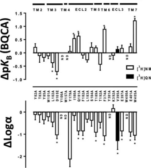
2and M4mAChRs led to a significant reduction in affinity of the allosteric modulator LY2033298 and its binding cooperativity with ACh (14, 33). To determine whether BQCA behaves in a similar manner to LY2033298 at the equivalent residue in the M1mAChR, we performed equi-librium binding studies for the interaction between CCh and BQCA at W1013.28A, as well as at other orthosteric pocket res-idues. An allosteric ternary complex model (Equation 1) was applied to the data to obtain estimates of BQCA affinity at each mutant (pKB), and its binding cooperativity with CCh (log␣) (representative examples of the analysis for different constructs are shown in Fig. 3, and all results are summarized in Fig. 4).
We found that the cooperativity of BQCA with CCh at W1013.28A, L1023.29A, or T1925.42A was not significantly dif-ferent when compared with the WT receptor estimates (Fig. 4
and Table 2). Although these residues do not form direct tacts with orthosteric ligands, they have been described to con-stitute a “second shell” that stabilizes the primary binding pocket (3, 44). Interestingly, mutation of orthosteric binding site residues substantially affected the ability of BQCA to mod-ulate CCh affinity; binding cooperativity with CCh was com-pletely abolished at Y1063.33A, W1574.57A, and Y3816.51A and was significantly reduced at D1053.32E and T1895.39A (Figs. 3D and 4 and Table 2). In addition, V395A, which displayed a reduction in affinity for [3H]QNB and CCh similar to that of orthosteric site residues, also caused a significant reduction in binding cooperativity between BQCA and CCh (Table 2 and Fig. 4 (bottom panel)). No pKBestimates for BQCA could be derived from the analysis of the binding interaction data of Y1063.33A, W1574.57A, or Y3816.51A due to the lack of allosteric modulation. The pKBof BQCA was significantly lower than WT at L1023.29A and D1053.32E but was unchanged at the remaining orthosteric site mutations (Fig. 4 (top panel) and Table 2). These results suggest that residues that form direct contacts with the orthosteric ligand (7) also play a role in the transmission of cooperativity from the allosteric binding site of BQCA.
[image:6.603.39.287.58.312.2]Equilibrium binding studies for the interaction between CCh and BQCA were also performed on residues previously described to participate in the allosteric modulation of mAChRs. Alanine FIGURE 2.Orthosteric agonist affinity estimates are differentially
modi-fied by M1 mAChR mutations.Barsrepresent the difference in pKA of
orthosteric antagonist [3H]NMS or [3H]QNB (top panel) derived from whole
cell saturation binding experiments (Table 1) or the difference in pKIof the
orthosteric agonist CCh (bottom panel) derived from whole cell competition binding experiments (Table 2), relative to the WT receptor value for each ligand at each mutant residue. Data represent the mean⫾S.E. of three exper-iments performed in duplicate. *, significantly different from WT,p⬍0.05, one-way analysis of variance, Dunnett’s post hoc test.
TABLE 2
Whole cell equilibrium competition binding parameters for the inter-action between关3H兴NMS or关3H兴QNB, CCh, and BQCA at the WT and mutant M1mAChRs
Estimated parameter values represent the mean⫾S.E. of 3– 4 experiments per-formed in duplicate and analyzed according to Equation 1.
CCh pKI a
BQCA pKB b
log␣c
M1WT关 3
H兴NMS 4.56⫾0.05 4.49⫾0.09 2.64⫾0.12 M1WT关
3
H兴QNB 4.67⫾0.20 4.18⫾0.18 2.31⫾0.36 Y822.61
A 3.89⫾0.10d
4.70⫾0.08 2.53⫾0.14 T832.62
A 4.62⫾0.12 4.39⫾0.18 2.52⫾0.24 Y852.64
A 3.84⫾0.08d
4.32⫾0.10 2.29⫾0.13 W1013.28
A 3.64⫾0.10d
4.38⫾0.06 2.43⫾0.11 L1023.29
A 3.21⫾0.06d
4.12⫾0.10d
2.19⫾0.12 D1053.32
E 3.09⫾0.08d
3.79⫾0.07d
1.61⫾0.1d
Y1063.33 Ae
1.73⫾0.10d
NDf
ND W1574.57
Ae
1.85⫾0.06d
ND ND
Y179A 4.63⫾0.07 4.55⫾0.11 0.52⫾0.20d
I180A 4.07⫾0.08d
5.03⫾0.08d
2.49⫾0.13 Q181A 5.42⫾0.05d
5.12⫾0.07d
1.79⫾0.1d
F182A 4.68⫾0.03 4.41⫾0.07 1.79⫾0.12d
L183A 3.51⫾0.03d
4.23⫾0.05 2.32⫾0.08 S1845.32
A 4.35⫾0.09 4.70⫾0.1 2.27⫾0.15 T1895.39
A 3.69⫾0.07d
4.68⫾0.04 1.73⫾0.11d
T1925.42
A 3.22⫾0.08d
4.06⫾0.13 2.18⫾0.15 F3746.44
A 5.43⫾0.08d
5.38⫾0.06d
1.57⫾0.15d
Y3816.51 Ae
2.46⫾0.06d
ND ND
K3926.62
A 4.28⫾0.05 4.40⫾0.09 2.80⫾0.12 V3957.30
Ae
2.03⫾0.06d
4.31⫾0.06 1.02⫾0.25d
E3977.32
A 4.64⫾0.07 4.66⫾0.07 1.79⫾0.13d
W4007.35
A 4.18⫾0.02d
ND ND
E4017.36
A 4.60⫾0.12 4.72⫾0.08 1.74⫾0.14d
W4057.40
A 5.40⫾0.07d
5.70⫾0.05d
1.59⫾0.12d a
Negative logarithm of the equilibrium dissociation constant of CCh.
b
Negative logarithm of the equilibrium dissociation constant of BQCA as esti-mated from Equation 1.
c
Logarithm of the binding cooperativity factor between BQCA and CCh as esti-mated from Equation 1; for this analysis, the pKAof the radiolabeled antagonist
for the WT and each of the mutant receptors was constrained to the values listed in Table 1. The cooperativity between BQCA and the radioligand was constrained to⫺2, consistent with high negative cooperativity between the two ligands.
d
Data are significantly different (p⬍0.05) from WT values as determined by one-way analysis of varianc with Dunnett’s post hoc test.
e
Experiments and statistical comparisons are relative to WT关3
H兴QNB values.
f
ND means not determined (no modulation of affinity).
at Monash University (CAUL) on March 15, 2014
http://www.jbc.org/
[image:6.603.303.553.114.338.2]substitution of the TMII residues had no affect on the binding cooperativity between BQCA and CCh (Table 2). However, ala-nine substitution of the ECL2 residues Tyr-179, Gln-181, and Phe-182 significantly reduced the binding cooperativity, with Tyr-179 having the most profound effect (Fig. 3,BandC,and Table 2). The cooperativity was unaffected at the remaining ECL2 residues I180A, L183A, and S184A (Table 2). However, although I180A did not have a significant effect on cooperativ-ity, it had significant opposing effects on the affinities of CCh and BQCA, with a decrease in the former and an increase in the latter (Figs. 2 and 4 and Table 2).
Mutation of the glutamate residues Glu-3977.32 and Glu-4017.36, which have been implicated in the binding of allosteric ligands at the mAChRs (34, 45, 46), also caused significant reduction in the binding cooperativity (Table 2). Alanine sub-stitution of the conserved Trp-4007.35residue in TMVII led to
complete loss of allosteric modulation even at the highest con-centrations of BQCA (Fig. 3E and Table 2) confirming the importance of this residue for the binding of allosteric ligands at the M1mAChR (18, 40, 47) and suggesting that this is likely to be a residue with which BQCA directly interacts.
Alanine substitution of the conserved aromatic residues Phe-3746.44or Trp-4057.40also led to substantial reductions in the binding cooperativity (Fig. 3F and Table 2). Interestingly all three mutations (Q181A, F3746.44A, and W4057.40A) that led to an increase in CCh affinity also caused an increase in BQCA affinity and a reduction in the binding cooperativity between the two ligands (Figs. 2, 3F, and 4 and Table 2). The pKB esti-mates obtained from the binding interaction studies at the remaining mutants were not significantly different from WT (Fig. 4 and Table 2). Overall, the binding interaction studies revealed a significant correlation between the changes in affin-FIGURE 3.Identification of residues that differentially govern BQCA affinity and binding cooperativity with CCh at the M1mAChR.Thecurvesrepresent competition between [3H]NMS (A–C, E,andF) or [3H]QNB (D) and increasing concentrations of CCh in the absence or presence of varying concentrations of
BQCA. All assays were performed using 0.3 nM[3H]NMS or [3H]QNB in whole cells expressing the WT or mutant c-Myc-tagged M
1mAChRs as described under
“Experimental Procedures.” Data points represent the mean⫾S.E. of three independent experiments performed in duplicate. Curves drawn through thepoints
inA–CandFrepresent the best fit of an allosteric ternary complex model (Equation 1). Parameters obtained from these experiments are listed in Table 2.
at Monash University (CAUL) on March 15, 2014
http://www.jbc.org/
[image:7.603.52.535.55.485.2]ities of CCh and BQCA (Fig. 5). Additionally, these data show that residues located in both the putative allosteric and the orthosteric pockets are conformationally linked and contribute to the transmission of binding cooperativity.
Effects of Mutations on Ligand Efficacy and on the
Transmis-sion of Functional Cooperativity between CCh and BQCA—To
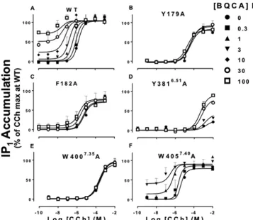
investigate the effects of the selected mutations on the ability of BQCA to modulate signaling efficacy, we determined the con-centration-response profile for CCh in the absence and
pres-ence of increasing concentrations of BQCA using IP1 accumu-lation as a canonical measure of M1 mAChR activation resulting from preferential activation of G␣qG proteins. The potency (pEC50) and maximal agonist effect (Emax) parameters for CCh in the absence of modulator are shown in Table 3. As agonist potency is determined by affinity, signaling efficacy, and receptor density, we applied an operational model of alloster-ism (Equation 2 (20)) to estimate the effect of each mutation on the operational efficacy (log) of CCh and BQCA without the confounding influence of affinity. The estimated log values were then corrected for changes in receptor expres-sion and are summarized in Fig. 6 and Table 4. Additionally, analysis of the data with Equation 2 allowed for the estima-tion of the overall funcestima-tional allosteric interacestima-tion between CCh and BQCA at each mutant (denoted by the parameter log␣).
As summarized in Fig. 6 (representative examples in Fig. 7), the majority of the M1mAChR mutations led to a significant reduction in the signaling efficacy of CCh (logA). Not surpris-ingly, the most prominent effects were seen for the orthosteric site residues in TMIII, W1574.57A in TMIV, and Y3816.51A in TMVI, consistent with reduced CCh affinity at these mutants and their importance for orthosteric ligand binding (3). No change in logAwas detected at residues for which CCh dis-played higher affinity (Q181A, F3746.44A, and W4057.40A). Interestingly, our analysis indicated that the CCh log Awas significantly larger than WT at V395A in ECL3, despite a sig-nificant reduction in CCh binding affinity (Figs. 2 and 6 and Tables 1 and 2). BQCA agonism was not detected at any of the mutants with the exception of F3746.44A (log
B0.55⫾0.10) and W4057.40A (log
[image:8.603.42.288.56.331.2]B0.25⫾0.06), where logBwas not sig-nificantly different to WT (logB0.38⫾0.05) (Fig. 7).
FIGURE 4.Effects of M1mAChR mutations on BQCA affinity and binding cooperativity estimates.Barsrepresent the difference in pKB(top panel) or
[image:8.603.302.554.106.323.2]binding cooperativity value (log␣,bottom panel) of BQCA relative to WT as derived from binding interaction experiments with CCh (Table 2). Data rep-resent the mean⫾S.E. of three experiments performed in duplicate.ND,no modulation by BQCA. *, significantly different from WT,p⬍0.05, one-way analysis of variance, Dunnett’s post hoc test.
FIGURE 5.Positive correlation between the changes in orthosteric and allosteric ligand affinities at the M1mAChR mutants.Each point repre-sents the affinity values of BQCA (pKB) and CCh (pKI) as determined from
whole cell competition binding studies as listed in Table 2.
TABLE 3
CCh pEC50and Emaxvalues for WT and mutant M1mAChRs as mea-sured from the IP1accumulation assay
Estimated parameter values represent the mean⫾S.E. of three experiments per-formed in duplicate.
pEC50
a E
max
WT 5.72⫾0.08 100
Y822.61
A 4.65⫾0.20b
66.03⫾7.35b
T832.62
A 5.27⫾0.15 20.39⫾1.35b
Y852.64
A 4.43⫾0.06b
90.56⫾5.67 W1013.28
A 2.95⫾0.50b
10.05⫾3.40b
L1023.29
A 2.46⫾0.10b
20.00⫾5.37b
D1053.32
E NAc
NA Y1063.33
A NA NA
W1574.57
A NA NA
Y179A 4.39⫾0.14b
82.38⫾4.47 I180A 4.43⫾0.27b
79.92⫾9.37 Q181A 5.120⫾0.26 80.00⫾7.60 F182A 4.85⫾0.10b
82.73⫾3.85 L183A 3.76⫾0.14b
65.32⫾5.11b
S1845.32
A 4.89⫾0.07b
85.55⫾4.00 T1895.39
A 3.94⫾0.15b
101.00⫾9.41 T1925.42
A 3.40⫾0.20b
70.82⫾9.24 F3746.44
A 4.74⫾0.09b
105.2⫾3.55 Y3816.51
A 3.10⫾0.30b
21.81⫾3.00b
K3926.62
A 5.37⫾0.12 91.19⫾5.66 V3957.30
A 4.71⫾0.11b
97.23⫾4.45 E3977.32
A 5.50⫾0.21 96.20⫾8.89 W4007.35
A 3.50⫾0.08b
71.80⫾3.70 E4017.36
A 5.12⫾0.05 99.00⫾3.39 W4057.40
A 5.21⫾0.05 84.38⫾1.98
aNegative logarithm of the EC
50value.
bSignificantly different (p⬍0.05), from WT value as determined by one-way
analysis of variance with Dunnett’s post hoc test.
cNA, not applicable (no detectable response).
at Monash University (CAUL) on March 15, 2014
http://www.jbc.org/
[image:8.603.41.289.407.582.2]A common finding was obtained from the interaction studies between BQCA and CCh at the orthosteric site mutations that substantially impaired CCh signaling (W1013.28A, L1023.29A,
D1053.32E, Y1063.33A, W1574.57A, and Y3816.51A). As opposed to the loss of cooperativity between the two ligands seen in the binding interaction studies for the majority of these mutant receptors (Fig. 3Dand Table 2), BQCA was able to rescue CCh function (Fig. 7Dand Table 4). An analogous “rescue” of ACh function by LY2033289 has been described at equivalent TMIII residues in the M4mAChR (33, 48). This finding indicates that a key part of the mechanism for the positive cooperativity medi-ated by BQCA on the orthosteric agonist involves a global drive of the receptor toward an active conformation.
The majority of mutant residues that displayed reduced binding cooperativity (log ␣) between CCh and BQCA also caused a reduction in functional cooperativity (log␣) between the two ligands (Fig. 8). These include the three residues that showed enhanced affinities for CCh and BQCA (Q181A, W4057.40A, and F3746.44A), F182A in ECL2 (Fig. 7,CandF), T1895.39A in TMV, and the two glutamate mutants E3977.32A and E4017.36A. The three residues mutated in TMII (Y822.61A, T832.62A, and Y852.64A) and L183A in ECL2 caused significant reductions in functional cooperativity despite their lack of effect on binding cooperativity between CCh and BQCA (Fig. 8 and Table 4), indicating that these residues are likely to play a role in the transmission of functional cooperativity alone. In contrast, the log ␣between BQCA and CCh at V395A was unchanged, despite significantly reduced binding cooperativity (Fig. 8 and Table 4). Consistent with the findings of Maet al.
(18), modulation of CCh efficacy was absent at Y179A and W4007.35A (Figs. 7,BandE,and 8). These results suggest that Trp-4007.35and Tyr-179 are likely to be residues with which BQCA directly interacts.
Molecular Dynamics Simulations and Ligand Docking
—Li-gand docking and molecular dynamic simulations were subse-quently performed to rationalize our findings. This resulted in one main pose of BQCA in the predicted allosteric site.
The obtained complex for BQCA and CCh bound to the modeled M1mAChR is shown in Fig. 9A. CCh forms the estab-lished salt bridge between the cationic nitrogen and Asp-1053.32and is fixed in a hydrophobic pocket formed by residues Tyr-1063.33 in TMIII, Trp-1574.57 in TMIV, Tyr-3816.51 in TMVI, and Tyr-4047.39and Tyr-4087.43 in TMVII (Fig. 9B). This is a signature network of interactions in cationic amine receptors (7, 11, 49), rhodopsins (51), and the adenosine A2A receptor (52). Moreover, the orthosteric site is further flanked by the H-bonds formed between Tyr-1063.33and Tyr-3816.51, which adds stability to the binding pocket (3), and together with Tyr-4047.39and Tyr-4087.43formed an aromatic lid separating the orthosteric and allosteric pockets. The aromatic ring of Trp-1574.57appears to form a-interaction with Tyr-1063.33 (Fig. 9B), and it has been shown to form direct contact with the aromatic ring of the antagonist QNB (7).
[image:9.603.41.291.59.244.2]The analysis of the MD trajectories shows the interaction of BQCA with residues located in the allosteric binding site (Fig. 9C). This binding site is defined by residues from TMII, TMVII, and ECL2 and is in agreement with our binding and functional studies; in particular, significant effects of the mutation of Tyr-179 in ECL2 and Trp-4007.35in TMVII can be reconciled with this pose. Tyr-179 is predicted to contribute to the stability of BQCA binding via formation of hydrophobic/edge-to-face- FIGURE 6.CCh signaling efficacy (logA) estimates are differentially
affected by M1mAChR mutations.Barsrepresent the difference in logAof
CCh at each mutant relative to the WT receptor value, as derived from appli-cation of the operational model of allosterism to the IP1interaction data at
each mutant (Equation 2). Data represent the mean⫾S.E. of three experi-ments performed in duplicate.NRindicates that CCh activity was absent.ND
indicates that Equation 2 could not be used due to loss of allosteric modula-tion by BQCA. *, significantly different to WT receptor value,p⬍0.05, one-way analysis of variance, Dunnett’s post hoc test.
TABLE 4
Operational model parameters for the functional allosteric interaction between CCh and BQCA at the WT and mutant M1mAChRs measured using IP1accumulation
Estimated parameter values represent the mean⫾S.E. of three experiments per-formed in duplicate. LogAand log␣values were obtained from analyses of
functional interaction data according to Equation 2. For this analysis, the pKIand
pKBvalues for CCh and BQCA, respectively, were fixed to those determined from
the radioligand binding assays as listed in Table 2.
logAa
log␣b
WT 1.32⫾0.07 2.03⫾0.12
Y822.61A 1.21⫾0.13 1.14⫾0.17c
T832.62
A 0.65⫾0.13c
0.73⫾0.20c
Y852.64A 0.53⫾0.07c 1.03⫾0.09c
W1013.28Ad ⫺1.11⫾0.18c 1.95⫾0.17
L1023.29
Ad ⫺
0.44⫾0.24c
0.98⫾0.15c
D1053.32Ed ⫺0.69⫾0.23c 0.97⫾0.20c
Y1063.33
Ad ⫺
0.79⫾0.21c
1.24⫾0.10c
W1574.57Ad ⫺2.01⫾0.20c 1.74⫾0.49
Y179A NDe ND
I180A 0.89⫾0.13 1.35⫾0.18 Q181A 0.24⫾0.08c 0.33⫾0.12c
F182A 0.70⫾0.07c
0.53⫾0.13c
L183A 0.52⫾0.11c 1.26⫾0.13c
S1845.32A 1.01⫾0.11 1.61⫾0.14 T1895.39
A 0.25⫾0.18c
0.71⫾0.12c
T1925.42A 0.23⫾0.11c 1.76⫾0.10
F3746.44
A 0.56⫾0.11c
0.03⫾0.02c
Y3816.51Ad ⫺0.23⫾0.11c 1.25⫾0.08c
K3926.62A 1.16⫾0.11 1.92⫾0.14 V3957.30
A 2.19⫾0.10c
1.46⫾0.14 E3977.32A 1.17⫾0.20 1.07⫾0.30c
W4007.35
A ND ND
E4017.36A 0.74⫾0.02c 0.93⫾0.04c
W4057.40A 0.38⫾0.02c 0.47⫾0.07c a
Logarithm of operational efficacy parameter for CCh (logA) was corrected for
changes in receptor expression to allow comparison with WT.
b
Logarithm of the functional cooperativity between CCh and BQCA is shown.
c
Significantly different (p⬍0.05), from WT value as determined by one-way ANOVA with Dunnett’s post hoc test.
d
pKBof BQCA was left unconstrained at W101A, L102A, D105E, Y106A, W157A,
and Y381A. The logBof BQCA was constrained to⫺2 at these mutants. e
ND, no modulation by BQCA.
at Monash University (CAUL) on March 15, 2014
http://www.jbc.org/
[image:9.603.40.289.417.623.2]interactions with both the bicyclic 4-oxoquinoline core and the benzylic pendant of BQCA. Similarly, Trp-4007.35is predicted to make a-interaction with the benzylic pendant. In this model, Glu-3977.32 also constrains this moiety of BQCA through a hydrophobic interaction, essentially forming a lid
over this part of the allosteric binding site. Tyr-852.64and Tyr-822.61are predicted to delimit the allosteric site via extra edge-to-face-/hydrophobic interactions with the 4-oxoquinoline ring system (Fig. 9C). Although the former residue only affected the functional cooperativity between BQCA and CCh, it has been found to be an important contact residue for prototypical allosteric modulators at the M2mAChR (13). Mutation of an adjacent residue at the M4mAChR (I93
2.65T) was found to be
important for the transmission of cooperativity between ACh and LY2033298 (34), suggesting a contribution of this residue to a conserved allosteric pocket within the mAChR family. Fig. 10 shows the global movements of the ECLs and TMs as well as the movements of the residues to accommodate the binding of BQCA. These include the rotation of the aromatic side chains of Trp-4007.35(Fig. 10,inset) and Trp-4057.40that may be facil-itating the accompanying shifts in the nearby TMVII residues Glu-3977.32 and Glu-4017.36 to constrain BQCA into the observed pose. The binding of BQCA also causes subtle move-ments in the ECLs; the most significant of these appear to be in ECL2 where the aromatic side chains of Tyr-179 and Phe-182 both move closer to BQCA, whereas Gln-181 adopts a horizon-tal position away from the ligand accessible cavity. These results support our finding that mutation of Glu-3977.32, Glu-4017.36, Tyr-179, and Phe-182 lead to reduced cooperativity between BQCA and CCh and that mutation of Gln-181 enhances the binding affinity of both ligands.
FIGURE 7.Identification of residues that differentially govern BQCA efficacy and functional cooperativity with CCh at the M1mAChR.Interaction between BQCA and CCh in IP1accumulation assay in CHO FlpIn cells stably expressing the WT or mutant M1mAChRs. Data points represent the mean⫾S.E.
of three independent experiments performed in duplicate.Curvesdrawn through the points inA, C, D,andFrepresent the best fit of an operational allosteric model (Equation 2 and Table 4) with the affinity of each ligand at each mutant fixed to the value determined from separate binding studies (Table 2).
FIGURE 8.BQCA functional cooperativities are differentially modified by M1mAChR mutations.Barsrepresent the difference in the functional coop-erativity value (log␣, Equation 2) relative to the WT value, as derived from application of the operational model of allosterism to the CCh and BQCA IP1
interaction data at each mutant (Equation 2) (Table 4). Data represent the mean⫾S.E. from three experiments performed in duplicate.ND,no modula-tion by BQCA. * significantly different from WT value,p⬍0.05, one-way anal-ysis of variance, Dunnett’s post test.
at Monash University (CAUL) on March 15, 2014
http://www.jbc.org/
[image:10.603.118.476.57.367.2]DISCUSSION
BQCA demonstrates a number of unique properties relative to previously described allosteric ligands of the mAChR family, including an exquisite selectivity for M1mAChRs over other subtypes and a mechanism of action that appears in strict accordance with a two-state model of receptor activity, such that it is a positive modulator of agonists but a negative modu-lator of antagonists/inverse agonists (16, 17). Moreover, the
[image:11.603.121.475.57.316.2]compound is activein vivo(18, 53), providing proof of concept for the validity of allosteric targeting of M1mAChRs in the treatment of CNS disorders, and it has been the focus of numer-ous structure-activity studies (15, 54) aimed at improving its “druggability” and affinity. However, it is now apparent that allosteric modulators can achieve selectivity by more than one mechanism,i.e.at the level of structural divergence of an allo-steric pocket across GPCR subtypes or via selective cooperativ-FIGURE 9.Structural model of the M1mAChR in complex with BQCA and CCh.A,overall view of the complex obtained using MD simulations. The ligands are shown inorange(BQCA) andyellow(CCh)spheres.B,orthosteric binding site for CCh;C,predicted allosteric binding site of BQCA. Important residues are shown byorange sticks.
FIGURE 10.Proposed rearrangement of ECLs and TMs upon BQCA binding to M1mAChR.Extracellular view of the BQC-binding site at the starting position for MD simulations (gray) or at the final position of the receptor after 20 ns of MD (orange). Important residues involved in BQCA binding or cooperativity are shown assticks. Global movements of TMs and ECLs are shown withgreen arrows,and residue shifts are indicated byblue arrows.Insetshows the movement of the side chain of Trp-4007.35.
at Monash University (CAUL) on March 15, 2014
http://www.jbc.org/
[image:11.603.130.465.359.591.2]ity at a given subtype despite acting at a “conserved” allosteric site (14). The latter paradigm is best exemplified by the mAChRs, in which are all characterized by an extracellular ves-tibule that can be recognized by structurally diverse allosteric ligands (12, 13). To better understand the basis of the selectivity of BQCA, we combined mutagenesis with mathematical and molecular modeling to identify potential structural contribu-tors to its binding pocket and its ability to allosterically modu-late the binding and signaling of the prototypical orthosteric agonist CCh.
Although no affinity values could be obtained for BQCA at residues where allosteric modulation was abolished (Tyr-1063.33, Trp-1574.57and Tyr-3816.51and Trp-4007.35), our study identified residues of the M1mAChR that contribute to the following: (i) the binding affinity of the modulator; (ii) the coop-erativity between the modulator and the orthosteric agonist CCh; (iii) the ability of the modulator to drive the receptor into an active state, and (iv) enhancement of the binding affinity of BQCA.
Alanine substitution of a number of residues from various regions in the receptor caused a decrease in the cooperativity between BQCA and CCh in binding and functional interaction studies (Figs. 4 and 8 and Tables 2 and 4). Such reductions in cooperativity resulted either from mutation of residues that form the proposed allosteric binding pocket or residues confor-mationally linked to the allosteric site and thus needed for the transmission of cooperativity or receptor activation upon ligand binding. Our MD simulations support this hypothesis and the experimental findings. As shown in Fig. 9, the proposed BQCA pocket is topographically distinct from the orthosteric binding site, and these two sites are separated by a shelf of aromatic residues. The residues that are predicted to form the BQCA binding pocket, mainly from ECL2 (Tyr-179), TMII (Tyr-852.64), and TMVII (Trp-4007.35) or those whose mutation to alanine cause significant decreases in cooperativity (Phe-182, Glu-3977.32, and Glu-4017.36), are equivalent to residues that have been implicated in the binding of several allosteric ligands at mAChRs, as follows : (i) the action of gallamine and the allo-steric antagonist MT7 at the M1mAChR (40, 47); (ii) the action of LY2033298 at the M4mAChR (34), and (iii) the action of C7/3-phth, gallamine, alcuronium, McN-A-343, alkane-bisam-monium, and caracurine V-type allosteric modulators at the M2 mAChR (13, 35, 42, 55–57).
The only residue in the proposed BQCA binding pocket that leads to complete loss of modulation in both functional and binding assays is Trp-4007.35. This suggests that the- inter-action of the benzylic pendant of BQCA and the aromatic side chain of Trp-4007.35 makes either a major contribution to BQCA binding and/or maintaining the structure of the local binding pocket of BQCA. Other residues, such as 179, Tyr-822.61, and Tyr-852.64, while predicted in our MD simulations to interact with BQCA, appear to have a predominant role in the transmission of BQCA’s modulator action on orthosteric ligands such that their mutation to alanine impairs the trans-mission of cooperativity but does not lead to a significant loss of binding affinity. Of interest, a recent molecular dynamics study identified two binding centers in the extracellular vestibule of the M2mAChR, each defined by a pair of aromatic residues
(center 1, Tyr-177ECL2 and Trp7.35; center 2, Tyr2.61 and Tyr2.64) (13). It is noteworthy that we identified these residues as key contributors for the binding and function of BQCA. Fur-thermore, Tyr2.64and Trp7.35are conserved across all mAChR receptor subtypes; Tyr2.61is conserved across all but the M
3 mAChR (where it is replaced by a similarly aromatic phenyl-alanine residue), and Tyr-179 is only present at the M1and M2 mAChRs but is a phenylalanine at the M3and M4mAChRs. This is consistent with BQCA sharing a common binding site with other prototypical mAChR allosteric modulators. Glu-3977.32and Glu-4017.36are not conserved across the mAChR family and were predicted by our modeling experiments to make minimal interaction with BQCA. Mutation of these resi-dues had no effect on BQCA binding affinity but decreased cooperativity with CCh. This suggests that such residues may govern the subtype-specific cooperative effect of BQCA upon orthosteric ligand binding from a conserved allosteric pocket.
The observation of a correlation between the change in CCh binding affinity (pKI) and BQCA binding affinity (pKB) is entirely consistent with our previous description of the mechanism of BQCA within the confines of a strict two-state model (Fig. 5) (17). Furthermore, the finding that the orthosteric site residues shown in Fig. 9Blead to complete loss (Y1063.33A, W1574.57A, and Y3816.51A), or significant reduction (D1053.32A) in the binding cooperativity between CCh and BQCA is a striking example of a conformationally linked mechanism for the transmission of cooperativity. The efficacy of CCh was severely reduced when each of these resi-dues was mutated, but this was “rescued” by BQCA. Further-more, these additional functional interaction data confirmed the ability of BQCA to bind to this set of mutant receptors, a conclusion that could not be drawn from the binding data alone. This highlights the importance of using both binding and functional assays to characterize the effect of mutations upon allosteric ligand function.
Given that an analogous observation was made for the action of the positive allosteric modulator, LY2033298, at a function-ally impaired M4 mAChR double mutant containing the Y1133.33C (48) or the D1123.32E mutation (33), our results indi-cate that BQCA may share similarities in mechanism of action and may bind to a site that is spatially conserved between the M4 the M1 mAChRs. However, the very high selectivity of BQCA for the M1mAChR (as opposed to LY2033298 that acts at both the M2and M4mAChRs) indicates that although the allosteric sites of these two ligands share some epitopes, they may engage additional distinct residues either in their mode of binding or transmission of cooperativity to the orthosteric site. In addition to regions of the receptor that were primarily important for the binding of BQCA and transmission of coop-erativity, we also identified mutations that caused a significant enhancement in the affinities of both CCh and BQCA (Q181A, F3746.44A, and W4057.40A). Previous studies have reported increases in CCh affinity at Q181A (36, 40), whereas others have reported constitutive receptor activity when residues 6.44 and 7.40 are mutated to alanine (41, 43). The movement of the side chain of Phe6.44is coupled to an outward movement of TMVI upon2-adrenergic receptor activation (58), and it has been reported to be a microswitch in GPCR activation (50). It
at Monash University (CAUL) on March 15, 2014
http://www.jbc.org/
has also been suggested that Trp-4057.40 restricts thermal motions of the extracellular domain of TMVII of mAChRs (3). In summary, we have identified key regions in the M1 mAChR that are involved in the binding and signaling of CCh and BQCA, and in the transmission of cooperativity between the orthosteric and an allosteric binding site. We propose that some of the structural determinants of these effects are analo-gous to those of other family A GPCRs, but the unique selectiv-ity of BQCA arises from the additional involvement of noncon-served residues (e.g. Glu-3977.32 and Glu-4017.36) and/or selective cooperativity with agonists at the M1mAChR. There-fore, our results provide further understanding of the structural basis of allosteric modulation that may be of general application to GPCR drug discovery and that can help guide the more ratio-nal design of allosteric ligands that target this distinct site. In particular, they challenge an important concept often associ-ated with allosteric targeting of GPCRs, namely that selective modulators gain subtype selectivity through their binding to a site that is not conserved across a receptor subfamily. Rather, as highlighted in our study, selective cooperativity via interaction with a conserved allosteric site is also possible. Given the increasing number of GPCR crystal structures now being solved, it should thus be appreciated that structure-based drug design usingin silicoscreening for novel allosteric modulators will not, in and of itself, guarantee a desired level of selectivity without complementation by additional structure-function approaches as described herein.
Acknowledgments—We are grateful to Dr. Ann Stewart for generating some of the M1mAChR mutants and Briana J. Davie for the synthesis
of BQCA.
REFERENCES
1. Venkatakrishnan, A. J., Deupi, X., Lebon, G., Tate, C. G., Schertler, G. F., and Babu, M. M. (2013) Molecular signatures of G-protein-coupled re-ceptors.Nature494,185–194
2. Lagerstro¨m, M. C., and Schio¨th, H. B. (2008) Structural diversity of G protein-coupled receptors and significance for drug discovery.Nat. Rev. Drug Discov.7,339 –357
3. Hulme, E. C. (2013) GPCR activation: a mutagenic spotlight on crystal structures.Trends Pharmacol. Sci.34,67– 84
4. Stevens, R. C., Cherezov, V., Katritch, V., Abagyan, R., Kuhn, P., Rosen, H., and Wu¨thrich, K. (2013) The GPCR Network: a large-scale collaboration to determine human GPCR structure and function.Nat. Rev. Drug Discov. 12,25–34
5. Miao, Y., Nichols, S. E., Gasper, P. M., Metzger, V. T., and McCammon, J. A. (2013) Activation and dynamic network of the M2 muscarinic recep-tor.Proc. Natl. Acad. Sci. U.S.A.110,10982–10987
6. Wacker, D., Wang, C., Katritch, V., Han, G. W., Huang, X.-P., Vardy, E., McCorvy, J. D., Jiang, Y., Chu, M., Siu, F. Y., Liu, W., Xu, H. E., Cherezov, V., Roth, B. L., and Stevens, R. C. (2013) Structural features for functional selectivity at serotonin receptors.Science340,615– 619
7. Haga, K., Kruse, A. C., Asada, H., Yurugi-Kobayashi, T., Shiroishi, M., Zhang, C., Weis, W. I., Okada, T., Kobilka, B. K., Haga, T., and Kobayashi, T. (2012) Structure of the human M2 muscarinic acetylcholine receptor bound to an antagonist.Nature482,547–551
8. Conn, P. J., Christopoulos, A., and Lindsley, C. W. (2009) Allosteric mod-ulators of GPCRs: a novel approach for the treatment of CNS disorders.
Nat. Rev. Drug Discov.8,41–54
9. Conn, P. J., Jones, C. K., and Lindsley, C. W. (2009) Subtype-selective allosteric modulators of muscarinic receptors for the treatment of CNS
disorders.Trends Pharmacol. Sci.30,148 –155
10. Tan, Q., Zhu, Y., Li, J., Chen, Z., Han, G. W., Kufareva, I., Li, T., Ma, L., Fenalti, G., Li, J., Zhang, W., Xie, X., Yang, H., Jiang, H., Cherezov, V., Liu, H., Stevens, R. C., Zhao, Q., and Wu, B. (2013) Structure of the CCR5 chemokine receptor–HIV entry inhibitor maraviroc complex.Science 341,1387–1390
11. Kruse, A. C., Hu, J., Pan, A. C., Arlow, D. H., Rosenbaum, D. M., Rose-mond, E., Green, H. F., Liu, T., Chae, P. S., Dror, R. O., Shaw, D. E., Weis, W. I., Wess, J., and Kobilka, B. K. (2012) Structure and dynamics of the M3 muscarinic acetylcholine receptor.Nature482,552–556
12. De Amici, M., Dallanoce, C., Holzgrabe, U., Tra¨nkle, C., and Mohr, K. (2010) Allosteric ligands for G protein-coupled receptors: A novel strategy with attractive therapeutic opportunities.Med. Res. Rev.30,463–549 13. Dror, R. O., Green, H. F., Valant, C., Borhani, D. W., Valcourt, J. R., Pan,
A. C., Arlow, D. H., Canals, M., Lane, J. R., Rahmani, R., Baell, J. B., Sexton, P. M., Christopoulos, A., and Shaw, D. E. (2013) Structural basis for mod-ulation of a G-protein-coupled receptor by allosteric drugs.Nature503,
295–299
14. Valant, C., Felder, C. C., Sexton, P. M., and Christopoulos, A. (2012) Probe dependence in the allosteric modulation of a g protein-coupled receptor: implications for detection and validation of allosteric ligand effects.Mol. Pharmacol.81,41–52
15. Davie, B. J., Christopoulos, A., and Scammells, P. J. (2013) Development of M1 mAChR allosteric and bitopic ligands: prospective therapeutics for the treatment of cognitive deficits.ACS Chem. Neurosci.4,1026 –1048 16. Abdul-Ridha, A., Lane, J. R., Sexton, P. M., Canals, M., and Christopoulos,
A. (2013) Allosteric modulation of a chemogenetically modified G pro-tein-coupled receptor.Mol. Pharmacol.83,521–530
17. Canals, M., Lane, J. R., Wen, A., Scammells, P. J., Sexton, P. M., and Chris-topoulos, A. (2012) A Monod-Wyman-Changeux mechanism can explain G protein-coupled receptor (GPCR) allosteric modulation.J. Biol. Chem. 287,650 – 659
18. Ma, L., Seager, M. A., Seager M, Wittmann, M., Jacobson, M., Bickel, D., Burno, M., Jones, K., Graufelds, V. K., Xu, G., Pearson, M., McCampbell, A., Gaspar, R., Shughrue, P., Danziger, A., Regan, C., Flick, R., Pascarella, D., Garson, S., Doran, S., Kreatsoulas, C., Veng, L., Lindsley, C. W., Shipe, W., Kuduk, S., Sur, C., Kinney, G., Seabrook, G. R., and Ray, W. J. (2009) Selective activation of the M1 muscarinic acetylcholine receptor achieved by allosteric potentiation.Proc. Natl. Acad. Sci. U.S.A.106,15950 –15955 19. Black, J. W., and Leff, P. (1983) Operational models of pharmacological
agonism.Proc. R. Soc. Lond. B Biol. Sci.220,141–162
20. Leach, K., Sexton, P. M., and Christopoulos, A. (2007) Allosteric GPCR modulators: taking advantage of permissive receptor pharmacology.
Trends Pharmacol. Sci.28,382–389
21. Thompson, J. D., Gibson, T. J., Plewniak, F., Jeanmougin, F., and Higgins, D. G. (1997) The CLUSTAL_X windows interface: flexible strategies for multiple sequence alignment aided by quality analysis tools.Nucleic Acids Res.25,4876 – 4882
22. Rasmussen, S. G., Choi, H. J., Fung, J. J., Pardon, E., Casarosa, P., Chae, P. S., Devree, B. T., Rosenbaum, D. M., Thian, F. S., Kobilka, T. S., Schnapp, A., Konetzki, I., Sunahara, R. K., Gellman, S. H., Pautsch, A., Steyaert, J., Weis, W. I., and Kobilka, B. K. (2011) Structure of a nanobody-stabilized active state of the2adrenoceptor.Nature469,175–180
23. Ballesteros, J. A., and Weinstein, H. (1995) inMethods in Neurosciences
(Stuart, C. S., ed) pp. 366 – 428, Academic Press, New York
24. Sali, A., and Blundell, T. L. (1993) Comparative protein modelling by sat-isfaction of spatial restraints.J. Mol. Biol.234,779 – 815
25. Duan, Y., Wu, C., Chowdhury, S., Lee, M. C., Xiong, G., Zhang, W., Yang, R., Cieplak, P., Luo, R., Lee, T., Caldwell, J., Wang, J., and Kollman, P. (2003) A point-charge force field for molecular mechanics simulations of proteins based on condensed-phase quantum mechanical calculations.
J. Comput. Chem.24,1999 –2012
26. Wang, J., Wolf, R. M., Caldwell, J. W., Kollman, P. A., and Case, D. A. (2004) Development and testing of a general amber force field.J. Comput. Chem.25,1157–1174
27. Phillips, J. C., Braun, R., Wang, W., Gumbart, J., Tajkhorshid, E., Villa, E., Chipot, C., Skeel, R. D., Kale´, L., and Schulten, K. (2005) Scalable molec-ular dynamics with NAMD.J. Comput. Chem.26,1781–1802
at Monash University (CAUL) on March 15, 2014
http://www.jbc.org/