The Role of Ets2 Transcription Factor in the Induction of
MicroRNA-155 (miR-155) by Lipopolysaccharide and Its
Targeting by Interleukin-10
*
Received for publication, September 27, 2013, and in revised form, December 19, 2013Published, JBC Papers in Press, December 20, 2013, DOI 10.1074/jbc.M113.522730
Susan R. Quinn‡1, Niamh E. Mangan§, Brian E. Caffrey¶, Michael P. Gantier§, Bryan R. G. Williams§, Paul J. Hertzog§, Claire E. McCoy‡§2, and Luke A. J. O’Neill‡2
From the‡School of Biochemistry and Immunology, Trinity Biomedical Sciences Institute, Trinity College Dublin, Dublin 2, Ireland,
the§Monash Institute of Medical Research, Monash University, Clayton, Victoria 3168, Australia, and the¶Smurfit Institute of
Genetics, Trinity College Dublin, Dublin 2, Ireland
Background:miR-155 is strongly induced by LPS, a response inhibited by IL-10.
Results:The Ets2 transcription factor is required for induction of miR-155 by LPS. IL-10 can subsequently decrease miR-155 via suppression of Ets2.
Conclusion:Ets2 is an important transcription factor for regulation of miR-155.
Significance:This study reports a detailed mechanism of induction of miR-155 and provides a new means of inhibition for IL-10 via suppression of Ets2.
MicroRNA-155 (miR-155) is highly expressed in many can-cers such as B cell lymphomas and myeloid leukemia and inflammatory disorders such as rheumatoid arthritis, atopic dermatitis, and multiple sclerosis. The role of miR-155 as both a promoter of inflammation and an oncogenic agent provides a clear need for miR-155 itself to be stringently regulated. We therefore investigated the transcriptional regulation of miR-155 in response to the respective pro- and anti-inflammatory medi-ators LPS and IL-10. Bioinformatic analysis revealed Ets binding sites on the miR-155 promoter, and we found that Ets2 is critical for miR-155 induction by LPS. Truncation and mutational anal-ysis of the miR-155 promoter confirmed the role of the Ets2 binding site proximal to the transcription start site for LPS responsiveness. We observed increased binding of Ets2 to the miR-155 promoter and Ets2 deficient mice displayed decreased induction of miR-155 in response to LPS. IL-10 inhibited the induction of Ets2 mRNA and protein by LPS, thereby decreasing Ets2 function on the pri-155 promoter. We have thus identified Ets2 as a key novel regulator in both the positive and negative control of miR-155 in the inflammatory response.
The immune response to infection involves a carefully orchestrated and timed signaling cascade resulting in up-regu-lation of an abundance of immune and inflammatory genes, followed by a stringently regulated anti-inflammatory response to ensure a timely return to homeostasis and prevent excess
inflammation (1). Toll-like receptors (TLRs)3play a central role
in this system, recognizing conserved microbial patterns and launching an efficacious and controlled immune response. These responses induced by TLRs must be meticulously regu-lated, and one such class of regulators is microRNAs (miRNAs). miRNAs are a class of non-coding molecules that bind to the 3⬘-UTR of target mRNAs, regulating their translation (2). They are highly efficient fine tuners of gene expression, exerting sub-tle yet essential effects throughout the genome.
miRNAs have been shown to dampen down immune responses, fine tune expression of inflammatory mediators, and control the differentiation and development of immune cell lineages. Certain miRNA, for example miR-146a, target key immune regulators to prevent excess inflammation (3). Others, such as miR-155, are proinflammatory. miR-155 is processed from a primary noncoding transcript B cell integration cluster (or pri-155), originally identified in lymphomas (4). miR-155 is potently induced in response to various TLR ligands (5) and targets a number of key players in the immune response, such as inhibiting the negative TLR4 regulator SHIP1 (6, 7) and SOCS1 (8). The targets of miR-155 in immunity and inflammation have been well characterized (9), but the detailed mechanism by which this miRNA is potently induced upon ligation of TLR receptors has not been extensively studied. We have previously shown that the anti-inflammatory cytokine IL-10 inhibits 155, but not the anti-inflammatory miRNAs 21 or miR-146a (10). This observation revealed a new axis of regulation for this anti-inflammatory cytokine on the TLR pathway. IL-10 is crucial for stifling the inflammatory response after infection and is vital to protect the host from excess inflammation (11). The exact mechanism of this inhibition, similar to much of the functions of IL-10, has not yet been unraveled in detail.
*This work was supported by the Science Foundation Ireland, Health
Research Board/Marie Curie CoFund, and the European Research Council. The Monash Institute of Medical Research was supported by the Australian National Health and Medical Research Council, Australian Research Coun-cil, Health Research Board/Marie Curie CoFund, and the Victorian govern-ment’s operational infrastructure support program.
1To whom correspondence should be addressed: School of Biochemistry and
Immunology, Trinity Biomedical Sciences Inst., 152-160 Pearse St., Trinity College Dublin, Dublin 2, Ireland. Tel.: 35318962449; E-mail: quinns1@ tcd.ie.
2Both authors are joint last authors of this work.
3The abbreviations used are: TLR, Toll-like receptor; miRNA, microRNA;
BMDM, bone marrow-derived macrophage; TRIF, TIR-domain-containing
adapter-inducing interferon-.
at IReL (Trinity College Dublin) on January 6, 2016
http://www.jbc.org/
In this study, we have examined in detail the mechanism of induction of miR-155 by LPS. We have found a key role for the transcription factor Ets2 in this induction. Furthermore, IL-10 suppresses Ets2 expression and thereby inhibits the induction of miR-155 by LPS. We have therefore identified a novel tran-scriptional inducer of miR-155, revealed a new downstream target of IL-10 in the form of Ets2 and an important process through which IL-10 acts to exert its anti-inflammatory properties.
EXPERIMENTAL PROCEDURES
Reagents—LPS fromEscherichia coli, serotype 0111:B4, was from Alexis. Recombinant mouse and human IL-10 were from R&D Biosystems. Oligonucleotides and SYBR primers were from Eurofins, and TaqMan probes were from Applied Biosys-tems. Ets2 antibody was from Santa Cruz Biotechnology (sc-351), and-actin antibody was from Sigma (AC-74). STAT3 inhibitor was from Merck Millipore (catalog no. 573099).
Cell Culture and Isolation—Raw264.7 and HEK293T cell lines, obtained from the European Cell Culture Collection, and immortalized WT, myD88⫺/⫺, and TIR-domain-containing
adapter-inducing interferon- (TRIF)⫺/⫺ bone marrow-de-rived macrophages, obtained from Dr. Elaine Kenny were maintained in Dulbecco’s modified Eagle’s medium. Ets2fl/fl/
LysMCre⫹/⫺and matched wild type mice were obtained from Paul Hertzog (Monash Institute of Medical Research, Monash University, Victoria, Australia). Bone marrow was isolated from the tibias and femurs of C57BL/6 mice, and cells were grown in macrophage colony stimulating factor-conditioned DMEM. Peri-toneal macrophages were isolated from Ets2fl/fl/LysMCre⫹/⫺or C57BL/6 mice. Purity of macrophages was confirmed by Facs analysis. Mice were euthanized by CO2, and warm DMEM was
injected into the peritoneal cavity and massaged for 5 min prior to removal of lavage, which was placed on ice. The lavage was centrifuged and replated in warm DMEM medium to allow for macrophage attachment. After 1 h, nonadherent cells were removed by washing, and attached cells were harvested imme-diately for RNA measurement or treated with LPS (100 ng/ml) for the specified times prior to harvesting. In all cases, DMEM medium were supplemented with 10% fetal calf serum and 1% penicillin/streptomycin solution (v/v).
Bioinformatic Analysis of pri-155 Promoter—Nucleotide sequences forHomo sapiensand eight orthologous sequences were obtained for the upstream region of the pri-155 promoter from the Ensembl database (assembly GRCH37.p8). Upstream regions were taken as 2500 bases upstream and 500 down-stream from the transcription start site. The identification of evolutionarily conserved transcription factor binding sites was performed using PhyloGibbs; a Gibbs sampling technique that utilizes phylogenetic footprinting. PhyloGibbs identifies both evolutionarily conserved and over-represented binding sites utilizing only sequence data and without the use of binding profile/experimental date. PhyloGibbs was used to analyze the orthologous upstream sequences using all possible binding sizes between 4 and 20, fixing all other parameters at the default settings. Secondary identification of transcription factor bind-ing sites was performed usbind-ing JASPAR and ConSite. JASPAR is a transcription factor binding profile database, which performs
a single sequence comparison to all high-quality transcription factor models. ConSite is a web-based tool that performs both transcription factor model comparisons in combination with phylogenetic footprinting leading to results of greater signifi-cance. A default binding threshold of 0.8 was used for the both the JASPER and ConSite analysis.
RNA Isolation and Real Time PCR—Cells (primary bone mar-row-derived macrophages (BMDMs), Raw264.7, immortalized BMDM, or primary peritoneal macrophages) were plated 1 day prior to stimulation. Cells were stimulated with LPS⫾IL-10 as indicated in the figure legends. Total RNA was extracted using the RNeasy kit (Qiagen), modified to obtain small RNA species. cDNA for miRNA and mRNA analysis was prepared from 5–100 ng/ml total RNA using the high-capacity cDNA archive kit (Applied Biosystems) according to the manufacturer’s instructions and incorporating TaqMan primers for miR-155 and RNU6B for miRNA analysis. miRNA expression was mea-sured by Taqman analysis using specific Taqman Assays for miR-155 or RNU6B (Applied Biosystems) according to the manufacturer’s instructions. mRNA expression was measured using SYBR Green-based chemistry (KAPA-Sybr) using the fol-lowing primers: Pri-mmu-155, 5⬘-aaa cca gga agg gga agt gt-3⬘ (forward) and 5⬘-caa gag tca ccc tgc tgg at-3⬘(reverse); Ets2, 5⬘-aat gca ggc acc aaa cta cc-3⬘(forward) and 5⬘-gtc ctg gct gat gga aca gt- 3⬘(reverse); Ets1, 5⬘-tcc aga cag aca cct tgc ag-3⬘ (forward) and 5⬘-ggt gag gcg gtc aca act at-3⬘(reverse); GAPDH, 5⬘-ttc acc acc atg gag aag gc-3⬘(forward) and 5⬘-ggc atg gac tgt ggt cat ga-3⬘(reverse); SHIP1, 5⬘-ggt ggt acg gtt tgg aga ga-3⬘ (forward) and 5⬘-atg ctg agc ctc tgt ggt ct-3⬘(reverse). miRNA and mRNA expression were measured on the 7900 RT-PCR system (Applied Biosystems), and fold changes in expression were calculated by the ⌬⌬CT method using RNU6B as an endogenous control for miRNA analysis and GAPDH as an endogenous control for mRNA expression. All fold changes are expressed normalized to non-stimulated control for each cell type.
Enzyme-linked Immunosorbent Assay—Murine TNF-␣ expres-sion was measured from the supernatants of stimulated cells using an enzyme-linked immunosorbent assay DuoSet kit (R&D Biosys-tems) according to the manufacturer’s instructions.
Protein Expression—Differentiated BMDM or Raw264.7 cells were seeded at 4⫻105in six-well plates and stimulated with
LPS⫾IL-10 as indicated in the figure legends. Cells were lysed in low stringency lysis buffer complete with protease inhibitors. Protein concentration was then determined using the Coomas-sie Bradford reagent (Pierce). Lysates were resolved on 10% SDS-PAGE gels and transferred onto polyvinylidene difluoride membrane. Membranes were blocked in 5% (w/v) dried milk in TBS-T (50 mMTris/HCl, pH 7.6, 150 mMNaCl, and 0.1% (v/v) Tween 20) before being immunoblotted with anti-Ets2 (Santa Cruz Biotechnology) or anti--actin (AC-74, Sigma) antibodies (1:1000 or 1:10,000, respectively) in 5% (w/v) dried milk in TBS-T at 4 °C overnight, or at room temperature for at least 2 h. Membranes were then incubated with the appropriate horserad-ish peroxidase-conjugated secondary antibody diluted 1:2000 in 5% (w/v) dried milk in TBS-T for 1 h. Blots were developed by enhanced chemiluminescence according to the manufacturer’s instructions (Cell Signaling Technology).
at IReL (Trinity College Dublin) on January 6, 2016
http://www.jbc.org/
Luciferase Assays—Pri-155 luciferase plasmid along with the Ets1 mutant (M_pri-155) were a kind gift from Eric Flemington (Tulane University, New Orleans, LA). Truncated pri-155 plas-mids were generated by PCR amplification using specific prim-ers to create plasmids of varying lengths that were recloned in to the luciferase plasmid at the NheI and HinDIII sites. The reverse primer, 5⬘-tca ccg ccg ggt taa ctg-3⬘, was used for each truncation along with specific forward primers as follows: T1_pri-155, 5⬘-ctg aga ccc atg aat gag-3⬘; T2_pri-155, 5⬘-ctc tta ggg acc tgc tgg-3⬘; T3_pri-155, 5⬘-caa agg ttg gag ccc aag-3⬘; T4_pri-155, 5⬘-cag aaa agg cgc ctg gtc-3⬘. Raw264.7 cells seeded at 2⫻105/ml in 24-well plates were transfected using 6% JetPEI
macrophage transfection reagent as per the manufacturer’s protocol, with each plasmid and TK-Renilla.Cells were rested for 24 – 48 h prior to stimulation with LPS. In all cases, cells were lysed in passive lysis buffer before being analyzed for both luciferase and TK-Renillaactivity as described previously (23). Data were normalized to TK-Renillaactivity and represented as mean⫾S.D. for triplicate determinations where fold changes are expressed normalized to non-stimulated control.
Chromatin Immunoprecipitation—Raw264.7 cells were set up at 4⫻105cell/ml in DMEM, treated with LPS⫾IL-10 for
24 h and fixed by adding a final concentration of 1% formalde-hyde to each culture dish. Flasks were incubated for 10 min at room temperature. A 1/20 volume of 2.5Mglycine was then added to each flask and allowed to set at room temperature for 5 min prior to washing in PBS and resuspension in 6 ml of ChIP lysis buffer (SDS lysis buffer with leupeptin, aprotinin, and PMSF) and immediately snap frozen in liquid nitrogen. The samples were thawed and resuspended in SDS:Triton buffer and then sonicated at 22% intensity, 10 ⫻ 30 s per sample, placing on ice in between pulses. Preblocked protein A beads were incubated with samples prior to overnight incubation with primary antibodies; anti-Ets2 (Santa Cruz Biotechnology, sc22803) and anti-HA (Sigma, H6908). Quantitative RT-PCR was carried out using primers for either the pri-155 promoter consensus Ets binding site (0/⫹9), a non-Ets binding site (⫺1003/⫺995). Data are presented as percent of input.
Affinity Purification with Biotinylated Oligonucleotides— Oligonucleotides for the terminal Ets binding site on the pri-155 promoter were annealed 90 –95 °C for 3–5 min and then heat block was allowed to cool to room temperature (forward, 5⬘BIO-gcgccggcttcctgtacgc-3⬘; reverse, 5⬘ -cgcgtacaggaagccg-gcg-3⬘). Raw264.7 cells were seeded at 4⫻ 105 cell/ml and
treated with LPS⫾IL-10. 24 h later, cells were lysed in 100l of oligonucleotide buffer (25 mMTris, 5% glycerol, 50 mMEDTA, 5 mMNaF, Nonidet P-40 1%, 1 mMDTT, 150 mMNaCl, and protease and phosphatase inhibitors), and snap-frozen. Sam-ples were then thawed on ice and diluted with a further 900l of oligonucleotide buffer containing no NaCl. 10 l sample of lysate was kept to which 40l of 5⫻SDS buffer was added. Remaining lysates were then precleared with 20 l of pre-washed streptavidin-agarose beads, rotating at 4 °C for 15 min before centrifuging at 2500 rpm for 5 min at 4 °C. Supernatants were removed to a fresh tube with 30l of prewashed strepta-vidin-agarose beads and 30g of 5⬘-biotinylated oligonucleo-tide. Binding was performed for 2 h at 4 °C, rotating. Samples were centrifuged for 5 min at 4 °C to pellet the beads, which
were washed three times before 50 l of 5⫻ SDS sample buffer was added to the beads. Samples were loaded on a 10% gel and Ets2 binding detected by Western blotting as described previously.
RESULTS
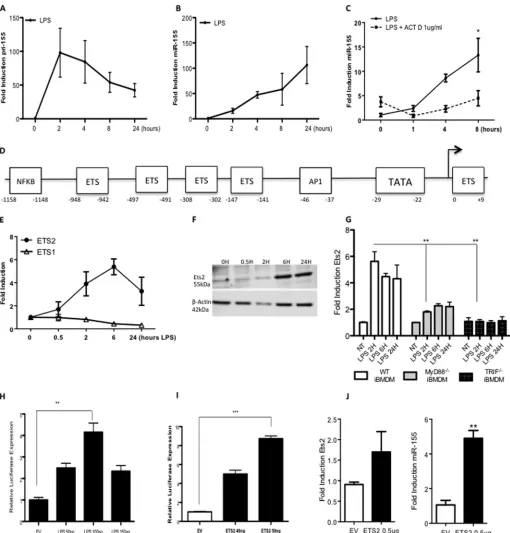
The pri-155 Promoter Is Controlled by Ets Transcription Factors—As miR-155 has previously been shown to be potently induced in response to the TLR4 ligand LPS (5), we wished to investigate the mechanism of this induction. The strong induc-tion over time of pri-155 and miR-155 by LPS in BMDM was first verified (Fig. 1,AandB). IL-6 was also measured as a pos-itive control for LPS treatment (data not shown). The potent transcriptional inhibitor, actinomycin D, blocked the induction of miR-155 by LPS as shown in Fig. 1C. The miR-155 promoter gene pri-155 was next investigated bioinformatically to deter-mine key transcription factor binding sites. Regions 2500 bases upstream and 500 downstream from the transcription start site were analyzed in the miR-155 gene. The identification of evo-lutionarily conserved transcription factor binding sites was per-formed using PhyloGibbs across eight different ,species includ-ing human and mouse. Combininclud-ing the most significant results from PhyloGibbs and JASPAR identified ETS family binding sites as stand-out candidates. Further verification was carried out using ConSite, which verified all sites in the previous tests. The analysis indicated the miR-155 promoter was heavily con-trolled by the Ets family of transcription factors. Five of the putative Ets binding sites are identified in the schematic shown in Fig. 1D. Positions are indicated relative to the transcription initiation site as described previously (12).
As we were interested in the induction of miR-155 in an inflammatory context, we next examined the induction of Ets family members in response to the TLR4 ligand LPS. Fig. 1E
shows that Ets2 is induced over time is response to LPS, whereas the closely related Ets family member Ets1 is unaf-fected by LPS stimulation. Further investigation of Ets2 revealed it is also induced strongly at the protein level (Fig. 1F) and that induction by LPS is MyD88- and TRIF-dependant (Fig. 1G), confirming it as a key modulator of downstream TLR4 responses.
Having confirmed that Ets2 is induced by LPS and that the miR-155 promoter is controlled by many Ets binding sites, we next investigated the effect of Ets2 on the pri-155 promoter. First, we confirmed our previous finding that LPS can drive the pri-155 promoter. Pri-155 promoter constructs fused to a lucif-erase reporter were transfected into Raw264.7 cells and treated with LPS (50 –150 ng), which drove the pri-155 promoter 5-fold at 100 ng (Fig. 1H), confirming that miR-155 is regulated by LPS at the transcriptional level. Subsequently, pri-155 promoter constructs fused to a luciferase reporter were cotransfected with plasmids encoding Ets2. Fig. 1Ishows that overexpression of Ets2 could drive the pri-155 promoter in a dose-dependent manner. To investigate the effect of Ets2 on mature miR-155, gain of function studies were utilized, where Ets2 was overex-pressed and mature miR-155 was measured by real-time PCR. As revealed in Fig. 1J, 0.5g Ets2 is sufficient to induce a 5-fold increase in mature miR-155, indicating Ets2 alone is sufficient
at IReL (Trinity College Dublin) on January 6, 2016
http://www.jbc.org/
FIGURE 1.The pri-155 promoter is controlled by Ets transcription factors.A–C, primary BMDMs from C57BL/6 mice (n⫽3) were treated with LPS (100
ng/ml) for 0 –24 h (AandB) or pretreated with actinomycin D (ACT D; 1g) prior to LPS stimulation for 24 h (C). RT-PCR analysis of RNA was carried out with
primers specific for pri-155 or mature miR-155 as indicated. Expression is normalized to that of GAPDH (for pri-155 expression) or RNU6B (for miR-155
expression) and is presented relative to that of untreated controls. Data are presented as the mean⫾S.E. of one representative experiment shown of three
independent experiments (n⫽3 per experiment).D, bioinformatic analysis of the pri-155 promoter revealed highly conserved Ets transcription factor binding
sites.EandF, primary BMDM from C57BL/6 mice (n⫽3) were treated with LPS (100 ng/ml) for 0 –24 h. RT-PCR analysis of RNA was carried out with primers
specific for Ets2 or Ets1 mRNA (E), and protein levels were measured by Western blot with antibodies specific for Ets2 or-actin (F). Data are representative of
three separate experiments, with each point assayed in triplicate, witherror barsrepresenting S.D.G, wild type, MyD88⫺/⫺, or TRIF⫺/⫺immortalized BMDMs
(iBMDM) were plated at 5⫻105/ml and stimulated with LPS for 0 –24 h prior to lysis for RNA extraction. Ets2 or GAPDH control was measured by real-time PCR.
Data are the mean⫾S.D. of triplicate determinations from three independent experiments.HandI, pri-155 promoter activation was assayed in Raw264.7 cells
using a pri-155 luciferase reporter construct and increasing doses of LPS (50 –100 ng) (H), or plasmid encoding Ets2 (40 –50 ng) or empty vector (EV) control (I).
Luciferase activity was determined 24 h after transfection and normalized to TKRenillacontrol. Results are presented as the mean⫾S.D. for triplicate determinations
and are representative of three separate experiments.J, Raw264.7 cells were transfected with 0.5g of Ets2 plasmids or empty vector control and lysed for RNA.
miR-155 expression was measured by real-time PCR. Statistical analysis was carried out using Student’sttest; *,p⬍0.05; **,p⬍0.001; ***,p⬍0.0001.
at IReL (Trinity College Dublin) on January 6, 2016
http://www.jbc.org/
for the transcription of mature miR-155, thus focusing our study on Ets2 regulation of miR-155 in response to LPS.
Ets2 Binds to a Highly Conserved Site on the pri-155 Promoter— Having confirmed that Ets2 and LPS can drive the pri-155 moter and that LPS could induce Ets2, the regions of the pro-moter that were most important for this interaction were next examined. Four additional pri-155 luciferase constructs were generated, each truncated to contain one less Ets binding site
[image:5.594.118.482.59.525.2]until only the terminal Ets binding site and the TATA box remained (T1-T4_PRI-155). A schematic of these promoter constructs is shown in Fig. 2A,top panel. Each construct was co-transfected with an Ets2 expression plasmid, and interest-ingly, it was evident that Ets2 could continue to drive the trun-cated pri-155 promoters (T1-T4_PRI-155) to the same extent as the full-length pri-155 promoter (Fig. 2A,lower panel). This was examined in more detail by transfecting higher amounts of
FIGURE 2.Ets2 binds to a highly conserved binding site on the pri-155 promoter.Pri-155 promoter constructs were truncated sequentially such that one
Ets binding site was deleted at a time (T1-T4_pri-155), only the terminal Ets binding site remained (T4_pri-155) or the terminal Ets binding site was mutated
(M_pri-155).A, pri-155 promoter and T1-T4_pri-155 promoter activation was assayed in HEK293T cells using an Ets2 expression plasmid.B, pri-155 promoter
and T4_pri-155 promoter activation was assayed in Raw264.7 cells using increasing doses of Ets2 expression plasmid (50 – 80 ng).CandD, pri-155 promoter and
M_pri-155 promoter activation was assayed in Raw264.7 cells with increasing doses of Ets2 expression plasmid (50 –100 ng) (C) or LPS (100 ng/ml) (D).
Luciferase activity was determined 24 – 48 h after transfection and normalized to TKRenillacontrols.E, Raw264.7 cells were treated with LPS for 0, 6, and 24 h
and lysed, and an oligonucleotide (oligo) pulldown assay was carried out with the terminal Ets binding site oligonucleotide sequence at position 0/⫹9. Samples
were probed for Ets2 by Western blot. Densitometry was carried out relative to lysates and are relative to non-treated control (NT). Bar graph represents the
mean of two independent experiments.F, Raw264.7 cells were treated with LPS for 6 or 24 h, after which a ChIP assay was performed. Primers specific for the
terminal Ets binding site on pri-155 (black bars); and additionally, a non-Ets-binding site (white bars) were designed, and binding events were measured as
percent of input by real time PCR using antibodies against HA (control antibody) or Ets2. In all cases, results are presented as the mean⫾S.D. for triplicate
determinations and are representative of at least three separate experiments. Statistical analysis was carried out using Student’sttest. *,p⬍0.05; **,p⬍0.001.
at IReL (Trinity College Dublin) on January 6, 2016
http://www.jbc.org/
plasmid encoding Ets2 with the pri-155 luciferase construct with only the terminal Ets binding site remaining. Again Ets2 could be seen to drive the T4_PRI-155 construct to an equal extent as the full-length pri-155 (Fig. 2B). This finding implied that the terminal Ets binding site might be key for Ets2 to drive transcription of the pri-155 promoter. We therefore examined the effect of overexpression of Ets2 on a pri-155 construct in which the terminal Ets binding site is mutated (12) because we could not create a truncation as this site occurs 3⬘to the TATA box. Fig. 2Creveals that Ets2 can no longer drive induction of the pri-155 promoter when this terminal Ets binding site is mutated (M_PRI-155). Additionally, induction of pri-155 by LPS was strongly decreased when the terminal Ets binding site was mutated as shown in Fig. 2D. Together, these findings indi-cate that Ets2 requires the terminal Ets binding site on the pri-155 promoter for induction by LPS.
We next examined binding interaction at this site using a number of novel approaches. First, oligonucleotide pulldown assays were employed to investigate binding at this terminal Ets site. Oligonucleotides specific to the Ets binding site were incu-bated with lysates from Raw264.7 cells treated with LPS for 6 and 24 h, and Fig. 2Ereveals Ets2 binding to this specific and highly conserved site on the pri-155 promoter in response to LPS. To investigate this finding further, and in an endogenous context, ChIP at the key terminal Ets binding site was next carried out. This technique allows us to examine the endoge-nous binding of transcription factors to chromatin in response to LPS. As shown in Fig. 2F, Ets2 binds to the pri-155 promoter at 24 h of LPS treatment (black bars). Ets2 does not bind to a non-Ets-binding site (white bars) or the-actin promoter (data not shown). These findings confirmed that LPS induces Ets2 binding to the Ets binding site at the initiation site on the miR-155 promoter, whose function we have confirmed by muta-tional analysis.
pri-155 Expression Is Ablated and miR-155 Induction Is Reduced in Response to LPS in Ets2⫺/⫺Mice—We next investi-gated miR-155 induction in cells from Ets2⫺/⫺ conditionally generated mice. In these mice, theEts2gene is under the con-trol of thelysMpromoter, creating knock-outs specific to the myeloid lineage. Peritoneal macrophages were prepared from Ets2⫺/⫺, Ets2⫹/⫺, and wild type C57BL/6 mice and stimulated in vitrowith LPS for 24 h. The macrophages from the Ets2⫺/⫺
mice lacked induction of pri-155 in response to LPS (Fig. 3A), confirming the importance of this transcription factor in LPS-mediated induction of pri-155. The production of mature miR-155 was also significantly reduced in Ets2⫺/⫺mice in
compar-ison with wild type mice (Fig. 3B).
We also examined miR-155in vivo. Ets2⫺/⫺ and matched wild type C57BL/6 mice were administered an intraperitoneal injection of LPS or PBS and 24 h later were culled for harvesting of peritoneal macrophages. The induction of pri-155 and miR-155 in response to LPS was again observed strongly in the wild type mice, and this induction was reduced in the Ets2⫺/⫺mice. Pri-155 induction is completely ablated (Fig. 3C), and miR-155 is reduced (Fig. 3D) in Ets2⫺/⫺micein vivo, further supporting the evidence that Ets2 is a necessary transcription factor for the LPS-mediated induction of pri-155 and miR-155.
miR-155 target genes in macrophages from WT and Ets2⫺/⫺ mice were next investigated. As shown in Fig. 3E, LPS signifi-cantly suppresses the miR-155 target SHIP1 in WT peritoneal macrophages, consistent with published data on SHIP1 induc-tion and repression by miR-155 in BMDMs (6). In Ets2⫺/⫺
peri-toneal macrophages, basal SHIP1 is elevated, and LPS can no longer decrease it. This finding implicated Ets2 for the first time having a downstream effect on a microRNA target. There is evidence to suggest that miR-155 is required for stabilizing TNF-␣expression (14). As shown in Fig. 3F, Ets2⫺/⫺mice
defi-cient in miR-155 show significantly decreased TNF-␣ expres-sion. These data further confirm Ets2 as a key regulator of miR-155, with downstream impacts on inflammatory target genes, thus implicating Ets2 in the proinflammatory response to LPS and uncovering the mechanism of induction of miR-155.
IL-10 Inhibits Ets2 Protein Expression and Binding to the miR-155 Promoter—Having determined that induction of miR-155 by LPS involves Ets2, we next examined whether the inhib-itory effect of IL-10 on miR-155 might involve the targeting of Ets2. Interestingly, Ets2 mRNA induction by LPS is strongly inhibited by IL-10 in Raw264.7 cells (Fig. 4A) and primary BMDM (Fig. 4B) over time. Little is known about the mecha-nism through which IL-10 exactly exerts its anti-inflammatory effects on TLR signaling. It has been established that STAT3 is highly important for IL-10 to exert its effects (15); thus, to understand this further, we utilized a STAT3 inhibitor. Our data revealed that IL-10 is acting through the STAT3 pathway to inhibit Ets2 and can no longer exert its anti-inflammatory effects with just 1MStat3 inhibitor (Fig. 4C,black bars). These data correlate with the original manuscript detailing the inhi-bition of miR-155 by IL-10, which was also found to be STAT3-dependent (10). Inhibition of Ets2 by IL-10 was also evident at the protein level. Fig. 4Dshows how there is a basal expression of Ets2 in Raws (lane 1). LPS can increase expression (lane 2), and IL-10 can inhibit this effect (lane 3). IL-10 alone can also reduce the basal expression (lane 4). This effect is also evident at 24 h (lanes 5– 8). This is the first time IL-10 has been impli-cated in the control of Ets2, and due to the novel finding that IL-10 can inhibit Ets2 mRNA and protein, we furthered our investigation by examining whether this finding impacted directly on the pri-155 promoter. Chromatin immunoprecipi-tation assay in Fig. 4Ereveals that IL-10 can reduce LPS induced binding of Ets2 to the pri-155 promoter (black bars). This novel result reveals IL-10 may also be dependent on Ets2 to regulate miR-155.
Finally, we investigated the ability of IL-10 to inhibit miR-155 in wild type and Ets2-deficient peritoneal macrophages. Our data, shown in Fig. 4F, revealed that IL-10 could not inhibit miR-155 to the same extent in Ets2⫺/⫺mice (2.4-fold decrease) compared with wild type mice (4.3-fold decrease). This finding indicates Ets2 plays a role in both the pro- and anti-inflamma-tory control of miR-155 and reveals for the first time that IL-10 may act through Ets family members and thereby impacts on the inflammatory process.
DISCUSSION
In the study of miRNA, the majority of research to date focuses on the interesting target genes and pathways on which
at IReL (Trinity College Dublin) on January 6, 2016
http://www.jbc.org/
miRNAs impact, as opposed to the detailed mechanisms by which miRNAs themselves are controlled and induced. In the context of inflammation, miRNAs have been shown to be cru-cial for the development, differentiation, and orchestration of a smooth and timely immune response and subsequent resolu-tion. Therefore, it is extremely important to understand how these miRNA are induced, inhibited, and controlled to main-tain control of their downstream target genes. In this study, we identify Ets2 as a critical transcription factor in the induction of miR-155 by LPS and, furthermore, demonstrate inhibition of Ets2 as a mechanism for suppression of miR-155 by IL-10. This finding not only uncovers a mechanism of induction of miR-155 at the transcriptional level, it also reveals a new down-stream target gene of IL-10 and a possible mechanism through which IL-10 may act to exert its anti-inflammatory properties. miR-155 is highly important in the inflammatory response to infection, with miR-155⫺/⫺mice suffering extremely stunted immune responses, including defective lymphocyte and anti-gen presenting function and impaired immunological memory (16). Additionally, the mice are highly resistant to autoimmune diseases such as experimental autoimmune encephalomyelitis,
again highlighting the extent of the decrease in immune activa-tion of these miR-155⫺/⫺ mice (17). Numerous studies and review articles have investigated and collated miR-155 target genes and their effects in various inflammatory phenotypes (18 –20), but studies investigating the exact induction of this miRNA from the promoter level are sparse.
[image:7.594.127.467.56.400.2]To understand the potent induction of miR-155 by LPS, detailed bioinformatic analysis was carried out and revealed a number of conserved Ets binding sites on the pri-155 promoter, and luciferase reporter assays indicated the terminal Ets bind-ing site adjacent to the TATA box was highly important. Ets transcription factors are one of the largest families of transcrip-tion factors known and have wide-ranging roles from cell cycle regulation, cell differentiation, and a more recently discovered role in regulation of the immune response (21). For example, Ets binding sites have been discovered in the promoter regions of a number of immune genes, including those encoding the T cell receptor␣andsubunits, interleukin 5, interleukin 12 (22, 23), and additionally, many viruses such as moloney sarcoma virus. Ets2 is induced in response to LPS, others have shown the protein kinase C pathway may be involved in this induction
FIGURE 3.pri-155 and mature miR-155 induction are reduced in response to LPS in Ets2ⴚ/ⴚmice.AandB, primary peritoneal macrophages from WT,
Ets2⫹/⫺, or Ets2⫺/⫺mice were treated with LPS (100 ng/ml) for 0 or 24 hin vitro.CandD, WT or Ets2⫺/⫺mice were administered an intraperitoneal injection of
LPS (4 mg/kg) or PBS for 24 hin vivoprior to culling and extraction of peritoneal macrophages.EandF, primary peritoneal macrophages from WT or Ets2⫺/⫺
mice were treated with LPS (100 ng/ml) for 0 or 24 hin vitro. RT-PCR analysis of RNA was carried out with primers specific for pri-155, miR-155, or SHIP1, as
indicated. Expression is normalized to that of GAPDH (pri-155/SHIP1) or RNU6B (miR-155) and is presented relative to that of untreated controls.F, supernatants
from peritoneal macrophages stimulatedin vitrowere harvested, and TNF-␣levels were measured by ELISA. In all cases, data are the mean ofn⫽6 – 8 mice,
with each data point representing an individual mouse, and witherror barsrepresenting S.E. Statistical analysis was carried out using Student’sttest. *,p⬍0.05;
**,p⬍0.001; ***,p⬍0.0001.NT, non-treated.
at IReL (Trinity College Dublin) on January 6, 2016
http://www.jbc.org/
(24). It may also be phosphorylated on threonine 72 by LPS, but its exact mechanism of induction has not been fully elucidated (25). Moreover, our finding that Ets2 is MyD88- and
[image:8.594.48.544.54.596.2]TRIF-de-pendent has further confirmed it as a TLR-regulated gene, likely important in the immune response. Up-regulation of Ets2 has been found in 30% of rheumatoid arthritis tissue samples
FIGURE 4.IL-10 inhibits Ets2 induction by LPS.Raw264.7 cells (A) or primary BMDMs from C57BL/6 mice (B) were treated with LPS (100 ng/ml) and IL-10 (20 ng/ml)
for 0 –24 h.C, Raw264.7 cells were treated with STAT3 inhibitor (0.5–1M) or dimethyl sulfoxide (DMSO) control 1 h prior to stimulation with LPS (100 ng/ml) and IL-10
(20 ng/ml) for 4 h. Real time PCR results are presented as the mean⫾S.E. of three independent experiments, each carried out in triplicate.D, Raw264.7 cells were
treated with LPS (100 ng/ml) and IL-10 (20 ng/ml) for 0 –24 h. Protein levels were measured by Western blot with antibodies specific for Ets2 or-actin. Densitometry
was carried out relative to-actin controls and expressed as fold over non-treated samples. Densitometry is the mean of three independent experiments⫾S.D.E,
Raw264.7 cells were treated with LPS (100 ng/ml)⫾IL-10 (20 ng/ml) for 24 h after which a ChIP assay was performed. Primers specific for the terminal Ets binding site
on the pri-155 promoter at position 0/⫹9 (black bars) were designed, in addition to a non Ets-binding site at position⫺995/⫺1003 (white bars). Binding events were
measured as percent of input by real time PCR using antibodies against HA (control antibody) or Ets2. Results are presented as the mean⫾S.D. for triplicate
determinations and are representative of two independent experiments.F, primary peritoneal macrophages from WT or Ets2⫺/⫺mice (n⫽3) were treated with LPS
(100 ng/ml)⫾IL-10 (20 ng/ml) for 24 hin vitroprior to lysis for RNA extraction. miR-155 was measured by real time PCR. Data are the mean ofn⫽3 mice⫾S.E. Statistical
analysis was carried out using Student’sttest. *,p⬍0.05; **,p⬍0.001.G, schematic illustrating possible mechanism of control of miR-155 via Ets2.
at IReL (Trinity College Dublin) on January 6, 2016
http://www.jbc.org/
(26), and our study furthers the novel evidence for an immuno-logical role for Ets2. Our investigation of induction of miR-155 by Ets2 led us to examine immune responses in the Ets2⫺/⫺ mice. The mice were found to be deficient in induction of pri-155 and miR-pri-155 and, as a result, had reciprocally altered levels of miR-155 inflammatory target genes SHIP1 and TNF-␣. This interesting finding emphasizes a role for the Ets2 transcription factor in innate immunity and indicates Ets2 may control many downstream target genes through its induction and control of miR-155. Although there is a significant reduction of miR-155 induction in the Ets2⫺/⫺ mice some miR-155 expression remains, most likely due to separate LPS-mediated post-tran-scriptional mechanism of induction of miR-155 from pri-155 by maturation factors such as KH-type splicing regulatory pro-tein (KSRP, also known as KHSRP) (27). KSRP binds with high affinity to the terminal loop of the target miRNA precursors and promotes their maturation (28) and indicates that miR-155 can be post-transcriptionally regulated further after induction by LPS.
Conflicting studies have recently reported that regulation of miR-155 does not occur at the transcriptional level (29); how-ever, many of these studies were based heavily on the inability of LPS to drive a pri-155 luciferase reporter construct. Conversely, we and others (30, 31) have repeatedly shown LPS can drive induction of the pri-155 promoter in a dose-dependent man-ner. Our data are additionally supported by chromatin immu-noprecipitation and oligonucleotide pulldown assays display-ing increased binddisplay-ing of Ets2 to the pri-155 promoter in the presence of LPS, Ets2⫺/⫺mice displaying ablated induction of
pri-155 in response to LPS, and finally, the addition of the tran-scriptional inhibitor actinomycin D strongly reducing mature miR-155 induction. Other studies have additionally shown miR-155 is transcriptionally regulated in other cells types such as by AP1 in B cells (12) and STAT3 in T cells (32), supporting our evidence of transcriptional regulation of miR-155.
miR-155 is a potently proinflammatory microRNA and thus requires stringent control to prevent excess inflammation. E-transgenic miR-155 mice exhibit excessive inflammation and uncontrolled responses to inflammatory stimuli, including excessive TNF␣and hypersensitivity to septic shock (33). In the context of macrophage activation by LPS, our study has shown that Ets2 is a key transcription factor for miR-155 induction. Due to this excessive inflammation association with aberrant expression of miR-155, it is of utmost importance that miR-155 be carefully controlled and down-regulated. One such mecha-nism of this inhibition is through the anti-inflammatory cyto-kine IL-10 (10). IL-10 has an irreplaceable role in negatively regulating inflammation, and any additional insight as to how this occurs is of high value in this field. IL-10 acts primarily by blocking the genes which code for those molecules central to propagating the inflammatory response (8), as illustrated for example by profound colitis in IL-10⫺/⫺mice (13, 34, 35). How-ever, much of the mechanism of action of IL-10 is still unknown, and our study sought to investigate how IL-10 is inhibiting miR-155. Our data established the novel finding that IL-10 is acting to inhibit Ets2 mRNA and protein, both basally and in response to LPS stimulation. This interesting finding was relevant for control of miR-155, as this potent decrease in Ets2
expression resulted in decreased Ets2 recruitment to the pri-155 promoter in the presence of LPS and decreased mature miR-155. Additionally, Ets2 knock-out mice could not decrease miR-155 to the same extent after IL-10 treatment, further sup-porting the importance of Ets2 for control and regulation of miR-155. IL-10 still decreased miR-155 expression to some extent in Ets2-deficient cells, although not to the same extent as in wild type cells. We are currently exploring the Ets2-inde-pendent component being targeted by IL-10.
Our findings are likely to be significant in the context of inflammation, as many diseases correlating with low IL-10 also correlate with elevated miR-155, such as colitis, arthritis, and experimental autoimmune encephalomyelitis. Because IL-10 can so potently inhibit miR-155, it is possible that anti-miR-155 could have utility as an alternative to IL-10 therapy as an anti-inflammatory strategy.
Overall, the mode of regulation identified in this study have uncovered a mechanism of miR-155 induction and repression through modulation of Ets family transcription factors. This not only gives us new insight into the regulation of miR-155 but also provides important new information on the mechanism of action of IL-10.
Acknowledgment—We acknowledge Erik Flemington for providing Pri-155 promoter constructs.
REFERENCES
1. Janeway, C. A., Jr., and Medzhitov, R. (2002) Innate immune recognition.
Annu. Rev. Immunol.20,197–216
2. Guo, H., Ingolia, N. T., Weissman, J. S., and Bartel, D. P. (2010) Mamma-lian microRNAs predominantly act to decrease target mRNA levels. Na-ture466,835– 840
3. O’Neill, L. A., Sheedy, F. J., and McCoy, C. E. (2011) MicroRNAs: the fine-tuners of Toll-like receptor signalling. Nat. Rev. Immunol. 11,
163–175
4. Clurman, B. E., and Hayward, W. S. (1989) Multiple proto-oncogene ac-tivations in avian leukosis virus-induced lymphomas: evidence for stage-specific events.Mol. Cell. Biol.9,2657–2664
5. O’Connell, R. M., Taganov, K. D., Boldin, M. P., Cheng, G., and Baltimore, D. (2007) MicroRNA-155 is induced during the macrophage inflamma-tory response.Proc. Natl. Acad. Sci. U.S.A.104,1604 –1609
6. O’Connell, R. M., Chaudhuri, A. A., Rao, D. S., and Baltimore, D. (2009) Inositol phosphatase SHIP1 is a primary target of miR-155.Proc. Natl. Acad. Sci. U.S.A.106,7113–7118
7. An, H., Xu, H., Zhang, M., Zhou, J., Feng, T., Qian, C., Qi, R., and Cao, X. (2005) Src homology 2 domain-containing inositol-5-phosphatase 1 (SHIP1) negatively regulates TLR4-mediated LPS response primarily through a phosphatase activity- and PI-3K-independent mechanism.
Blood105,4685– 4692
8. Wang, P., Hou, J., Lin, L., Wang, C., Liu, X., Li, D., Ma, F., Wang, Z., and Cao, X. (2010) Inducible microRNA-155 feedback promotes type I IFN signaling in antiviral innate immunity by targeting suppressor of cytokine signaling 1.J. Immunol.185,6226 – 6233
9. Quinn, S. R., and O’Neill, L. A. (2011) A trio of microRNAs that control Toll-like receptor signalling.Int. Immunol.23,421– 425
10. McCoy, C. E., Sheedy, F. J., Qualls, J. E., Doyle, S. L., Quinn, S. R., Murray, P. J., and O’Neill, L. A. (2010) IL-10 inhibits miR-155 induction by toll-like receptors.J. Biol. Chem.285,20492–20498
11. Murray, P. J. (2006) Understanding and exploiting the endogenous inter-leukin-10/STAT3-mediated anti-inflammatory response. Curr. Opin. Pharmacol.6,379 –386
12. Yin, Q., Wang, X., McBride, J., Fewell, C., and Flemington, E. (2008) B-cell
at IReL (Trinity College Dublin) on January 6, 2016
http://www.jbc.org/
receptor activation induces BIC/miR-155 expression through a conserved AP-1 element.J. Biol. Chem.283,2654 –2662
13. Kühn, R., Löhler, J., Rennick, D., Rajewsky, K., and Müller, W. (1993) Interleukin-10-deficient mice develop chronic enterocolitis. Cell 75,
263–274
14. Bala, S., Marcos, M., Kodys, K., Csak, T., Catalano, D., Mandrekar, P., and Szabo, G. (2011) Up-regulation of microRNA-155 in macrophages con-tributes to increased tumor necrosis factor␣(TNF␣) production via in-creased mRNA half-life in alcoholic liver disease.J. Biol. Chem. 286,
1436 –1444
15. Murray, P. J. (2006) STAT3-mediated anti-inflammatory signalling.
Biochem. Soc. Trans.34,1028 –1031
16. Rodriguez, A., Vigorito, E., Clare, S., Warren, M. V., Couttet, P., Soond, D. R., van Dongen, S., Grocock, R. J., Das, P. P., Miska, E. A., Vetrie, D., Okkenhaug, K., Enright, A. J., Dougan, G., Turner, M., and Bradley, A. (2007) Requirement of bic/microRNA-155 for normal immune function.
Science316,608 – 611
17. O’Connell, R. M., Kahn, D., Gibson, W. S., Round, J. L., Scholz, R. L., Chaudhuri, A. A., Kahn, M. E., Rao, D. S., and Baltimore, D. (2010) Mi-croRNA-155 promotes autoimmune inflammation by enhancing inflam-matory T cell development.Immunity33,607– 619
18. Ceppi, M., Pereira, P. M., Dunand-Sauthier, I., Barras, E., Reith, W., San-tos, M. A., and Pierre, P. (2009) MicroRNA-155 modulates the interleu-kin-1 signaling pathway in activated human monocyte-derived dendritic cells.Proc. Natl. Acad. Sci. U.S.A.106,2735–2740
19. Chen, C. Z., Li, L., Lodish, H. F., and Bartel, D. P. (2004) MicroRNAs modulate hematopoietic lineage differentiation.Science303,83– 86 20. Lindsay, M. A. (2008) microRNAs and the immune response.Trends
Im-munol.29,343–351
21. Gallant, S., and Gilkeson, G. (2006) ETS transcription factors and regula-tion of immunity.Archivum Immunologiae et Therapiae Experimentalis
54,149 –163
22. Wang, C. Y., Petryniak, B., Ho, I. C., Thompson, C. B., and Leiden, J. M. (1992) Evolutionarily conserved Ets family members display distinct DNA binding specificities.J. Exp. Med.175,1391–1399
23. Blumenthal, S. G., Aichele, G., Wirth, T., Czernilofsky, A. P., Nordheim, A., and Dittmer, J. (1999) Regulation of the human interleukin-5 promoter by Ets transcription factors. Ets1 and Ets2, but not Elf-1, cooperate with GATA3 and HTLV-I Tax1.J. Biol. Chem.274,12910 –12916
24. Boulukos, K. E., Pognonec, P., Sariban, E., Bailly, M., Lagrou, C., and Ghys-dael, J. (1990) Rapid and transient expression of Ets2 in mature
macro-phages following stimulation with cMGF, LPS, and PKC activators.Genes Dev.4,401– 409
25. Sweet, M. J., Stacey, K. J., Ross, I. L., Ostrowski, M. C., and Hume, D. A. (1998) Involvement of Ets, rel and Sp1-like proteins in lipopolysaccharide-mediated activation of the HIV-1 LTR in macrophages.J. Inflamm.48,
67– 83
26. Dittmer, J., and Nordheim, A. (1998) Ets transcription factors and human disease.Biochim. Biophys. Acta1377,F1–11
27. Ruggiero, T., Trabucchi, M., De Santa, F., Zupo, S., Harfe, B. D., McManus, M. T., Rosenfeld, M. G., Briata, P., and Gherzi, R. (2009) LPS induces KH-type splicing regulatory protein-dependent processing of microRNA-155 precursors in macrophages.FASEB J.23,2898 –2908
28. Trabucchi, M., Briata, P., Filipowicz, W., Ramos, A., Gherzi, R., and Rosen-feld, M. G. (2011) KSRP Promotes the Maturation of a Group of miRNA Precuresors.Adv. Exp. Med. Biol.700,36 – 42
29. Cheung, S. T., So, E. Y., Chang, D., Ming-Lum, A., and Mui, A. L. (2013) Interleukin-10 inhibits lipopolysaccharide induced miR-155 precursor stability and maturation.PloS One8,e71336
30. Chen, Y., Liu, W., Sun, T., Huang, Y., Wang, Y., Deb, D. K., Yoon, D., Kong, J., Thadhani, R., and Li, Y. C. (2013) 1,25-Dihydroxyvitamin D promotes negative feedback regulation of TLR signaling via targeting microRNA-155-SOCS1 in macrophages.J. Immunol.190,3687–3695
31. Dai, Y., Diao, Z., Sun, H., Li, R., Qiu, Z., and Hu, Y. (2011) MicroRNA-155 is involved in the remodelling of human-trophoblast-derived HTR-8/ SVneo cells induced by lipopolysaccharides.Hum. Reprod.26,1882–1891 32. Escobar, T., Yu, C. R., Muljo, S. A., and Egwuagu, C. E. (2013) STAT3 activates miR-155 in Th17 cells and acts in concert to promote experi-mental autoimmune uveitis.Invest. Ophthalmol. Vis. Sci.54,4017– 4025 33. Tili, E., Michaille, J. J., Cimino, A., Costinean, S., Dumitru, C. D., Adair, B., Fabbri, M., Alder, H., Liu, C. G., Calin, G. A., and Croce, C. M. (2007) Modulation of miR-155 and miR-125b levels following lipopolysaccha-ride/TNF-␣stimulation and their possible roles in regulating the response to endotoxin shock.J. Immunol.179,5082–5089
34. Rennick, D., Davidson, N., and Berg, D. (1995) Interleukin-10 gene knock-out mice: a model of chronic inflammation.Clin. Immunol. Immuno-pathol.76,S174 –178
35. Berg, D. J., Kühn, R., Rajewsky, K., Müller, W., Menon, S., Davidson, N., Grünig, G., and Rennick, D. (1995) Interleukin-10 is a central regulator of the response to LPS in murine models of endotoxic shock and the Shwartzman reaction but not endotoxin tolerance. J. Clin. Invest.96,
2339 –2347
at IReL (Trinity College Dublin) on January 6, 2016
http://www.jbc.org/
and Luke A. J. O'Neill
Williams, Paul J. Hertzog, Claire E. McCoy
Caffrey, Michael P. Gantier, Bryan R. G.
Susan R. Quinn, Niamh E. Mangan, Brian E.
Interleukin-10
by Lipopolysaccharide and Its Targeting by
the Induction of MicroRNA-155 (miR-155)
The Role of Ets2 Transcription Factor in
doi: 10.1074/jbc.M113.522730 originally published online December 20, 2013 2014, 289:4316-4325.
J. Biol. Chem.
10.1074/jbc.M113.522730 Access the most updated version of this article at doi:
. JBC Affinity Sites Find articles, minireviews, Reflections and Classics on similar topics on the
Alerts:
When a correction for this article is posted •
When this article is cited •
to choose from all of JBC's e-mail alerts Click here
http://www.jbc.org/content/289/7/4316.full.html#ref-list-1
This article cites 35 references, 20 of which can be accessed free at
at IReL (Trinity College Dublin) on January 6, 2016
http://www.jbc.org/


![Methyl [hydroxy(phenyl)phosphonomethyl]phosphonate methanol solvate](data:image/gif;base64,R0lGODlhAQABAIAAAP///wAAACH5BAEAAAAALAAAAAABAAEAAAICRAEAOw==)