metal-organic papers
Acta Cryst.(2005). E61, m2395–m2396 doi:10.1107/S1600536805033817 Fuet al. [Co(CHO
2)(OH)]
m2395
Acta Crystallographica Section EStructure Reports Online
ISSN 1600-5368
Cobalt(II) formate hydroxide
Yun-Long Fu,aJia-Lin Ren,a Zhi-Wei Xuaand Seik Weng Ngb*
a
School of Chemistry and Materials Science, Shanxi Normal University, Linfen 041004, People’s Republic of China, andbDepartment of
Chemistry, University of Malaya, 50603 Kuala Lumpur, Malaysia
Correspondence e-mail: [email protected]
Key indicators
Single-crystal X-ray study
T= 295 K
Mean(O–C) = 0.005 A˚
Rfactor = 0.031
wRfactor = 0.077
Data-to-parameter ratio = 12.9
For details of how these key indicators were automatically derived from the article, see http://journals.iucr.org/e.
#2005 International Union of Crystallography Printed in Great Britain – all rights reserved
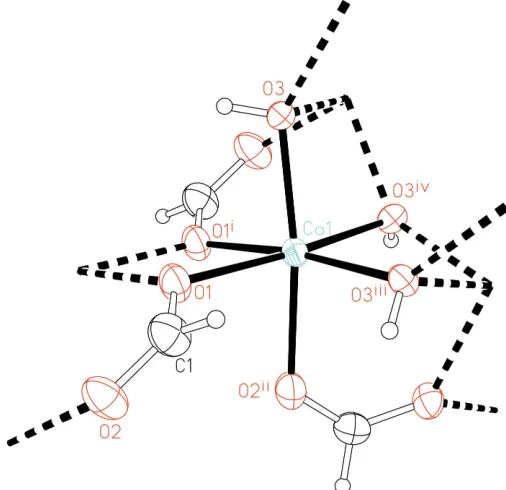
The title compound, poly[3-formato-3-hydroxo-cobalt(II)], [Co(CHO2)(OH)]n, adopts a polymeric three-dimensional network structure arising from the3-bridging mode of both the formate and the hydroxide. The Co atom lies in a fac octahedron of six O atoms, three of which belong to three different formates and the other three to different hydroxides.
Comment
The structure of cobalt(II) diformate has been reported recently; the compound was synthesized hydrothermally from cobaltous nitrate and formic acid in dimethylformamide (DMF). It crystallizes with DMF (Fuet al., 2005). With citric acid in place of formic acid, a similar synthesis affords cobalt(II) formate hydroxide, (I) (Fig. 1), the source of formic acid probably resulting from the decomposition of the citric acid. The compound adopts a three-dimensional network structure in which both the formate and hydroxide function in a 3-bridging mode; the Co
II
exists in a fac-octahedral arrangement; three of the O atoms belong to different formates and the other three to different hydroxides. The formate displays delocalized C—O bonds; one C—O bond is somewhat longer than the other. The O atom involved in the longer bond interacts with two Co atoms whereas the O atom involved in the shorter bond interacts with only one Co atom.
Experimental
A mixture of cobaltous nitrate hexahydrate (0.23 g, 0.5 mmol), citric acid (0.22 g, 1 mmol) and DMF (8 ml) was heated in a 15 ml Teflon-lined steel bomb at 400 K for 50 d. Red block-shaped crystals were obtained in about 40% yield (based on Co).
Crystal data
[Co(CHO2)(OH)] Mr= 120.96
Trigonal,R3
a= 11.698 (1) A˚
c= 11.661 (1) A˚
V= 1381.9 (2) A˚3 Z= 18
Dx= 2.616 Mg m
3
MoKradiation Cell parameters from 1480
reflections
= 2.7–28.0
= 5.37 mm1 T= 295 (2) K Block, red
0.160.110.09 mm
Data collection
Bruker APEX area-detector diffractometer
’and!scans
Absorption correction: multi-scan (SADABS; Sheldrick, 1996)
Tmin= 0.501,Tmax= 0.644
2498 measured reflections
698 independent reflections 667 reflections withI> 2(I)
Rint= 0.024 max= 27.5
h=15!7
k=15!14
l=13!14
Refinement
Refinement onF2 R[F2> 2(F2)] = 0.031 wR(F2) = 0.077 S= 1.07 698 reflections 54 parameters
All H-atom parameters refined
w= 1/[2(F
o2) + (0.0393P)2
+ 11.747P]
whereP= (Fo2+ 2Fc2)/3
(/)max= 0.001
max= 0.62 e A˚
3
min=0.50 e A˚
3
Table 1
Selected geometric parameters (A˚ ,).
Co1—O1 2.122 (2) Co1—O1 2.156 (2) Co1—O2i
2.149 (2)
Co1—O3 2.047 (2) Co1—O3ii 2.042 (2) Co1—O3iii
2.114 (2) O1—Co1—O1 79.5 (1)
O1—Co1—O2i
84.2 (1) O1—Co1—O3 92.9 (1) O1—Co1—O3ii 102.6 (1) O1—Co1—O3iii 172.5 (1) O1—Co1—O2i 83.9 (1) O1—Co1—O3 86.9 (1) O1—Co1—O3ii
171.8 (1)
O1—Co1—O3iii 93.3 (1) O2i
—Co1—O3 170.8 (1) O2i —Co1—O3ii 88.4 (1) O2i —Co1—O3iii 97.0 (1) O3—Co1—O3ii 100.8 (1) O3—Co1—O3iii 84.8 (1) O3ii —Co1—O3iii 84.9 (1)
Symmetry codes: (i) xþyþ4 3;xþ
2 3;z
1
3; (ii) yþ1;xy;z; (iii)
xyþ1 3;x
1 3;zþ
2 3.
H atoms were located in a difference Fourier map and were refined with distance restraints of O—H = 0.85 (1) A˚ and C—H = 0.95 (1) A˚; their displacement parameters were freely refined.
Data collection:SMART(Bruker, 2002); cell refinement:SAINT (Bruker, 2002); data reduction: SAINT; program(s) used to solve structure: SHELXS97(Sheldrick, 1997); program(s) used to refine structure: SHELXL97 (Sheldrick, 1997); molecular graphics: ORTEPII (Johnson, 1976); software used to prepare material for publication:SHELXL97.
The authors thank the Natural Scientific Foundation Committee of Shanxi Province (No. 20041031) and the University of Malaya for generously supporting this study.
References
Bruker (2002).SADABS,SAINTandSMART. Bruker AXS Inc., Madison, Winsonsin, USA.
Fu, Y.-L., Ji, M., Shen, X.-L. & Ng, S. W. (2005).Acta Cryst.E61, m688–m690. Johnson, C. K. (1976).ORTEPII. Report ORNL-5138. Oak Ridge National
Laboratory, Tennessee, USA.
[image:2.610.311.564.72.317.2]Sheldrick, G. M. (1997). SHELXS97 and SHELXL97. University of Go¨ttingen, Germany.
Figure 1
ORTEPII(Johnson, 1976) plot, illustrating the coordination geometry of Co and the 3-bridging modes of the —HCO2 and —OH groups.
supporting information
sup-1 Acta Cryst. (2005). E61, m2395–m2396
supporting information
Acta Cryst. (2005). E61, m2395–m2396 [https://doi.org/10.1107/S1600536805033817]
Cobalt(II) formate hydroxide
Yun-Long Fu, Jia-Lin Ren, Zhi-Wei Xu and Seik Weng Ng
poly[µ3-formato-µ3-hydroxo-cobalt(II)]
Crystal data
[Co(CHO2)(OH)]
Mr = 120.96
Trigonal, R3 Hall symbol: -R 3 a = 11.698 (1) Å c = 11.661 (1) Å V = 1381.9 (2) Å3 Z = 18
F(000) = 1062
Dx = 2.616 Mg m−3
Mo Kα radiation, λ = 0.71073 Å Cell parameters from 1480 reflections θ = 2.7–28.0°
µ = 5.37 mm−1 T = 295 K Block, red
0.16 × 0.11 × 0.09 mm
Data collection
Bruker APEX area-detector diffractometer
Radiation source: fine-focus sealed tube Graphite monochromator
φ and ω scans
Absorption correction: multi-scan (SADABS; Sheldrick, 1996) Tmin = 0.501, Tmax = 0.644
2498 measured reflections 698 independent reflections 667 reflections with I > 2σ(I) Rint = 0.024
θmax = 27.5°, θmin = 2.7°
h = −15→7
k = −15→14
l = −13→14
Refinement
Refinement on F2 Least-squares matrix: full R[F2 > 2σ(F2)] = 0.031 wR(F2) = 0.077 S = 1.07 698 reflections 54 parameters 2 restraints
Primary atom site location: structure-invariant direct methods
Secondary atom site location: difference Fourier map
Hydrogen site location: inferred from neighbouring sites
All H-atom parameters refined w = 1/[σ2(F
o2) + (0.0393P)2 + 11.747P] where P = (Fo2 + 2Fc2)/3
(Δ/σ)max = 0.001 Δρmax = 0.62 e Å−3 Δρmin = −0.50 e Å−3
Fractional atomic coordinates and isotropic or equivalent isotropic displacement parameters (Å2)
x y z Uiso*/Ueq
Co1 0.60832 (4) 0.12202 (4) 0.41991 (3) 0.0130 (2)
O1 0.5722 (2) 0.0647 (2) 0.5948 (2) 0.0185 (5)
O2 0.6642 (3) 0.0312 (2) 0.7497 (2) 0.0236 (6)
C1 0.6590 (4) 0.1012 (4) 0.6725 (3) 0.0225 (7)
H1 0.720 (4) 0.194 (1) 0.676 (5) 0.05 (2)*
H3 0.433 (4) 0.163 (4) 0.473 (3) 0.04 (2)*
Atomic displacement parameters (Å2)
U11 U22 U33 U12 U13 U23
Co1 0.0126 (2) 0.0129 (3) 0.0130 (3) 0.0061 (2) 0.0006 (2) 0.0022 (2)
O1 0.018 (1) 0.019 (1) 0.016 (1) 0.007 (1) 0.000 (1) 0.005 (1)
O2 0.023 (1) 0.020 (1) 0.025 (1) 0.008 (1) −0.009 (1) 0.002 (1)
O3 0.014 (1) 0.014 (1) 0.013 (1) 0.008 (1) 0.003 (1) 0.001 (1)
C1 0.022 (2) 0.016 (2) 0.026 (2) 0.007 (1) −0.003 (1) 0.000 (1)
Geometric parameters (Å, º)
Co1—O1 2.122 (2) Co1—Co1v 2.9942 (6)
Co1—O1i 2.156 (2) Co1—Co1iv 2.9943 (6)
Co1—O2ii 2.149 (2) O1—C1 1.264 (4)
Co1—O3 2.047 (2) O2—C1 1.240 (4)
Co1—O3iii 2.042 (2) O3—H3 0.85 (1)
Co1—O3iv 2.114 (2) C1—H1 0.95 (1)
O1—Co1—O1i 79.5 (1) O3iii—Co1—O3iv 84.9 (1)
O1—Co1—O2ii 84.2 (1) C1—O1—Co1 125.6 (2)
O1—Co1—O3 92.9 (1) C1—O1—Co1i 127.8 (2)
O1—Co1—O3iii 102.6 (1) Co1—O1—Co1i 100.5 (1)
O1—Co1—O3iv 172.5 (1) C1—O2—Co1vi 123.8 (2)
O1i—Co1—O2ii 83.9 (1) Co1vii—O3—Co1 139.0 (1)
O1i—Co1—O3 86.9 (1) Co1vii—O3—Co1v 92.2 (1)
O1i—Co1—O3iii 171.8 (1) Co1—O3—Co1v 92.0 (1)
O1i—Co1—O3iv 93.3 (1) O2—C1—O1 126.4 (3)
O2ii—Co1—O3 170.8 (1) Co1vii—O3—H3 107 (3)
O2ii—Co1—O3iii 88.4 (1) Co1—O3—H3 105 (3)
O2ii—Co1—O3iv 97.0 (1) Co1v—O3—H3 121 (4)
O3—Co1—O3iii 100.8 (1) O2—C1—H1 118 (3)
O3—Co1—O3iv 84.8 (1) O1—C1—H1 115 (3)
O3iii—Co1—O1—C1 −17.7 (3) O1—Co1—O3—Co1vii 109.8 (2)
O3—Co1—O1—C1 −119.5 (3) O1i—Co1—O3—Co1vii −171.0 (2)
O2ii—Co1—O1—C1 69.3 (3) O3iii—Co1—O3—Co1v 102.0 (1)
O1i—Co1—O1—C1 154.2 (3) O3iv—Co1—O3—Co1v 18.3 (1)
O3iii—Co1—O1—Co1i −171.9 (1) O1—Co1—O3—Co1v −154.6 (1)
O3—Co1—O1—Co1i 86.3 (1) O1i—Co1—O3—Co1v −75.3 (1)
O2ii—Co1—O1—Co1i −84.9 (1) Co1vi—O2—C1—O1 11.4 (5)
O3iii—Co1—O3—Co1vii 6.4 (2) Co1—O1—C1—O2 −138.9 (3)
O3iv—Co1—O3—Co1vii −77.4 (2) Co1i—O1—C1—O2 8.2 (6)
supporting information
sup-3 Acta Cryst. (2005). E61, m2395–m2396
Hydrogen-bond geometry (Å, º)
D—H···A D—H H···A D···A D—H···A
O3—H3···O1i 0.85 (1) 2.76 (5) 2.892 (3) 91 (4)