organic papers
o2202
Lewis and Tocher C6H14N22+2Cl2H2O doi:10.1107/S1600536805019124 Acta Cryst.(2005). E61, o2202–o2204
Acta Crystallographica Section E Structure Reports
Online
ISSN 1600-5368
A low-temperature determination of
triethylene-diaminium dichloride dihydrate
Thomas C. Lewis and Derek A. Tocher*
Department of Chemistry, University College London, 20 Gordon Street, London WC1H 0AJ, England
Correspondence e-mail: [email protected]
Key indicators
Single-crystal X-ray study T= 150 K
Mean(C–C) = 0.0017 A˚ Rfactor = 0.021 wRfactor = 0.052
Data-to-parameter ratio = 20.7
For details of how these key indicators were automatically derived from the article, see http://journals.iucr.org/e.
#2005 International Union of Crystallography Printed in Great Britain – all rights reserved
The structure determination at 150 K of triethylenediaminium dichloride dihydrate (also know as 1,4-diazaoniabicyclo[2.2.2]-octane dichloride dihydrate), C6H14N2
2+
2Cl2H2O, obtained
as part of an experimental polymorph screen on guanine, is reported here. The packing consists of a hydrogen-bonded chain structure, with one of the water molecules of crystal-lization involved in weak O—H Cl contacts.
Comment
Triethylenediamine, also known as 1,4-diazabicyclo[2.2.2]-octane, is a strong base allowing protons to be removed from other compounds to give anionic intermediates. Triethyl-enediamine has two reported anhydrous polymorphs, a room-temperature phase (Nimmo & Lucas, 1976a) and a high-temperature phase (Nimmo & Lucas, 1976b). This high-temperature structure assumes a ‘plastic’ phase, and is of interest as triethylenediamine is a one of a select group of globular molecules which undergo thermal transitions to plastic crystals because of the high degree of molecular mobility which can be achieved in the solid state (Weisset al., 1964). There are also a number of co-crystals of triethylene-diamine, including with hydroquinone (Mak et al., 1984), sulfate hemihydrate (Jayaraman et al., 2002), and bis-(hydrogen oxalate) (Vaidhyanathanet al., 2001). In addition, there are also triethylenediamine salts, including the dihydrochloride (Kennedy et al., 1987) and hydrobromide (Katrusiaket al., 1999). In this paper, we report the dihydro-chloride dihydrate salt, (I), of triethylenediamine.
In (I), atoms N1 and N2 are both protonated, with the molecule in a slightly twisted conformation, different from the symmetric cage-like structure present in the room-tempera-ture anhydrous crystal strucroom-tempera-ture of unprotonated triethyl-enediamine (Nimmo & Lucas, 1976a). The bond lengths and angles are within expected values (Allenet al., 1987), with the C–N bond lengths in the range 1.4942 (15)–1.5009 (15) A˚ , and the C—C bond lengths in the range 1.5227 (17)–1.5368 (16) A˚ .
The packing consists of a hydrogen-bonded chain structure (Fig. 2), with atom N2 hydrogen bonded to O2W, through an N—H O hydrogen bond (Table 1). Water atom O2Wacts as a hydrogen-bond donor to both Cl1 and Cl2, through O— H Cl hydrogen bonds (Table 1). The ion Cl1 is also hydrogen bonded through an N—H Cl interaction to the N1 amine group, forming the chain motif. The O1W water of crystallization forms weak hydrogen bonds to Cl2, as shown in Table 1.
Experimental
As part of an experimental polymorph screen on guanine, (I) was obtained from a saturated solution of triethylenediamine in dilute hydrochloric acid, in which approximately 0.03 g of guanine was added in an attempt to crystallize this purine base. The solution was stirred, filtered, then evaporated at room temperature (10 ml solu-tion, in 7525 mm vessels). Colourless block-shaped crystals of (I) were formed over a number of weeks. It should also be noted that large block-shaped crystals of triethylenediamine dihydrochloride were also obtained (Kennedyet al., 1987).
Crystal data
C6H14N22+2Cl
2H2O
Mr= 221.12
Orthorhombic,P212121
a= 7.1407 (8) A˚
b= 8.7188 (10) A˚
c= 16.8945 (19) A˚
V= 1051.8 (2) A˚3
Z= 4
Dx= 1.396 Mg m 3
MoKradiation Cell parameters from 7311
reflections
= 2.3–28.2 = 0.59 mm1
T= 150 (2) K Block, colourless 0.980.240.21 mm
Data collection
Bruker SMART APEX diffractometer Narrow-frame!scans
Absorption correction: multi-scan (SADABS; Sheldrick, 1996)
Tmin= 0.598,Tmax= 0.887
9177 measured reflections
2508 independent reflections 2473 reflections withI> 2(I)
Rint= 0.027
max= 28.2
h=9!9
k=11!11
l=22!22
Refinement
Refinement onF2
R[F2> 2(F2)] = 0.021
wR(F2) = 0.053
S= 1.06 2508 reflections 121 parameters
H atoms treated by a mixture of independent and constrained refinement
w= 1/[2(F
o2) + (0.0284P)2
+ 0.1405P]
whereP= (Fo2+ 2Fc2)/3
(/)max= 0.001 max= 0.27 e A˚
3 min=0.16 e A˚
3
Absolute structure: Flack (1983) Flack parameter: 0.01 (4)
Table 1
Hydrogen-bond geometry (A˚ ,).
D—H A D—H H A D A D—H A
O1W—H1W Cl2 0.84 (2) 2.50 (2) 3.2848 (12) 156 (2) O1W—H2W Cl2i
0.83 (2) 2.54 (2) 3.3537 (11) 169 (2) O2W—H3W Cl2 0.83 (1) 2.30 (1) 3.1109 (10) 167 (2) O2W—H4W Cl1 0.84 (1) 2.22 (1) 3.0585 (10) 173 (2) N1—H1 Cl1 0.91 2.16 3.0110 (11) 156 N2—H2 O2Wii 0.91 1.77 2.6634 (13) 168
Symmetry codes: (i)xþ2;yþ1
2;zþ32; (ii)xþ12;y;z12.
The triethylenediaminium H atoms were placed in geometrically idealized positions and allowed to ride on their parent atoms, whilst the water H atoms were refined, with O—H and H H distance restraints of 0.84 A˚ and 1.33 (2) A˚, respectively.
Data collection:SMART(Bruker, 2000); cell refinement:SAINT (Bruker, 2000); data reduction:SAINT(Bruker, 2000); program(s) used to solve structure: SHELXS97(Sheldrick, 1997); program(s) used to refine structure: SHELXL97 (Sheldrick, 1997); molecular graphics:ORTEP-3 for Windows(Farrugia, 1997) and MERCURY (Brunoet al., 2002); software used to prepare material for publica-tion:SHELXL97.
This research was supported by the EPSRC in funding a studentship for TCL. The authors acknowledge the Research Council’s UK Basic Technology Programme for supporting ‘Control and Prediction of the Organic Solid State’. For more information on this work, see http://www.cposs.org.uk.
References
Allen, F. H., Kennard, O., Watson, D. G., Brammer, L., Orpen, A. G. & Taylor, R. (1987).J. Chem. Soc. Perkin Trans. 2, pp. S1–19.
Bruker (2000).SMART,SAINTandSHELXTL. Bruker AXS Inc., Madison, Wisconsin, USA.
Bruno, I. J., Cole, J. C., Edgington, P. R., Kessler, M. K., Macrae, C. F., McCabe, P., Pearson, J. & Taylor, R. (2002).Acta Cryst.B58, 389–397.
Farrugia, L. J. (1997).J. Appl. Cryst.30, 565.
organic papers
Acta Cryst.(2005). E61, o2202–o2204 Lewis and Tocher C
[image:2.610.313.565.72.265.2]6H14N22+2Cl2H2O
o2203
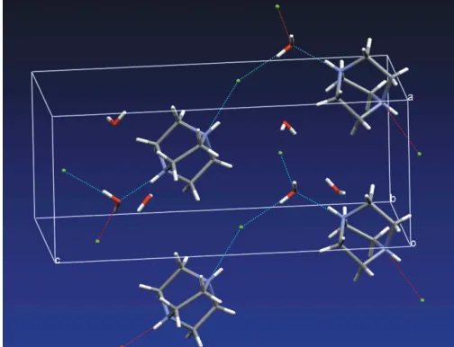
Figure 2
[image:2.610.43.297.73.175.2]The packing in (I), showing the hydrogen-bonded chain structure. The hydrogen bonds withD A > 3.2 A˚ have been omitted for clarity.
Figure 1
Flack, H. D. (1983).Acta Cryst.A39, 876–881.
Jayaraman, K., Choudhury, A. & Rao, C. N. R. (2002).Solid State Sci.4, 413– 422.
Katrusiak, A., Ratajczak-Sitarz, M. & Grech, E. (1999).J. Mol. Struct.417, 135–141.
Kennedy, S. W., Schultz, P. K., Slade, P. G. & Tiekink, E. R. T. (1987).Z. Kristallogr.180, 211–217.
Mak, T. C. W., Yip, W. H. & Book, L. (1984).J. Crystallogr. Spectrosc. Res.14, 457–465.
Nimmo, J. K. & Lucas, B. W. (1976a).Acta Cryst.B32, 348–353. Nimmo, J. K. & Lucas, B. W. (1976b).Acta Cryst.B32, 597–600. Sheldrick, G. M. (1996).SADABS. University of Go¨ttingen, Germany. Sheldrick, G. M. (1997). SHELXS97 and SHELXL97. University of
Go¨ttingen, Germany.
Vaidhyanathan, R., Natarajan, S. & Rao, C. N. R. (2001).J. Chem. Soc. Dalton Trans.pp. 699–706.
Weiss, G. S., Parkes, A. S., Nixon, E. R. & Hughes, R. E. (1964).J. Chem. Phys. 41, 3759–3767.
organic papers
o2204
Lewis and Tocher Csupporting information
sup-1
Acta Cryst. (2005). E61, o2202–o2204
supporting information
Acta Cryst. (2005). E61, o2202–o2204 [https://doi.org/10.1107/S1600536805019124]
A low-temperature determination of triethylenediaminium dichloride dihydrate
Thomas C. Lewis and Derek A. Tocher
1,4-diazaoniabicyclo[2.2.2]octane dichloride dihydrate
Crystal data
C6H14N22+·2Cl−·2H2O Mr = 221.12
Orthorhombic, P212121 Hall symbol: P 2ac 2ab a = 7.1407 (8) Å b = 8.7188 (10) Å c = 16.8945 (19) Å V = 1051.8 (2) Å3 Z = 4
F(000) = 472 Dx = 1.396 Mg m−3
Mo Kα radiation, λ = 0.71073 Å Cell parameters from 7311 reflections θ = 2.3–28.2°
µ = 0.59 mm−1 T = 150 K Block, colourless 0.98 × 0.24 × 0.21 mm
Data collection
Bruker SMART APEX diffractometer
Radiation source: fine-focus sealed tube Graphite monochromator
ω rotation with narrow frames scans Absorption correction: multi-scan
(SADABS; Sheldrick, 1996) Tmin = 0.598, Tmax = 0.887
9177 measured reflections 2508 independent reflections 2473 reflections with I > 2σ(I) Rint = 0.027
θmax = 28.2°, θmin = 2.4° h = −9→9
k = −11→11 l = −22→22
Refinement
Refinement on F2 Least-squares matrix: full R[F2 > 2σ(F2)] = 0.021 wR(F2) = 0.053 S = 1.06 2508 reflections 121 parameters 6 restraints
Primary atom site location: structure-invariant direct methods
Secondary atom site location: difference Fourier map
Hydrogen site location: inferred from neighbouring sites
H atoms treated by a mixture of independent and constrained refinement
w = 1/[σ2(Fo2) + (0.0284P)2 + 0.1405P] where P = (Fo2 + 2Fc2)/3
(Δ/σ)max = 0.001 Δρmax = 0.27 e Å−3 Δρmin = −0.16 e Å−3
Absolute structure: Flack 1983) Absolute structure parameter: 0.01 (4)
Special details
supporting information
sup-2
Acta Cryst. (2005). E61, o2202–o2204
Refinement. Refinement of F2 against ALL reflections. The weighted R-factor wR and goodness of fit S are based on F2, conventional R-factors R are based on F, with F set to zero for negative F2. The threshold expression of F2 > σ(F2) is used only for calculating R-factors(gt) etc. and is not relevant to the choice of reflections for refinement. R-factors based on F2 are statistically about twice as large as those based on F, and R- factors based on ALL data will be even larger.
Fractional atomic coordinates and isotropic or equivalent isotropic displacement parameters (Å2)
x y z Uiso*/Ueq
Cl1 0.51446 (4) −0.04577 (3) 0.554391 (16) 0.02113 (7) Cl2 1.07647 (4) 0.17252 (4) 0.656610 (17) 0.02424 (8) O1W 0.83585 (16) 0.30071 (12) 0.80854 (6) 0.0352 (2) H1W 0.924 (3) 0.283 (2) 0.7773 (11) 0.053* H2W 0.845 (3) 0.3922 (18) 0.8215 (12) 0.053* O2W 0.74444 (14) −0.03678 (11) 0.70705 (5) 0.0254 (2) H3W 0.829 (2) 0.0263 (19) 0.7005 (10) 0.038* H4W 0.674 (2) −0.034 (2) 0.6671 (9) 0.038* N1 0.16941 (14) 0.00540 (11) 0.45541 (6) 0.0185 (2)
H1 0.2499 −0.0034 0.4969 0.022*
N2 −0.05023 (13) 0.02830 (11) 0.34157 (5) 0.01770 (19)
H2 −0.1300 0.0367 0.2998 0.021*
C1 −0.01906 (17) −0.05314 (13) 0.47979 (6) 0.0202 (2)
H1A −0.0572 −0.0054 0.5291 0.024*
H1B −0.0141 −0.1632 0.4878 0.024*
C2 −0.15987 (18) −0.01430 (14) 0.41399 (7) 0.0198 (2)
H2A −0.2391 −0.1022 0.4031 0.024*
H2B −0.2389 0.0706 0.4302 0.024*
C3 0.15394 (18) 0.17045 (14) 0.43252 (7) 0.0206 (2)
H3A 0.2777 0.2146 0.4261 0.025*
H3B 0.0885 0.2273 0.4734 0.025*
C4 0.04533 (17) 0.17919 (13) 0.35432 (7) 0.0203 (2)
H4A −0.0466 0.2610 0.3567 0.024*
H4B 0.1305 0.2003 0.3109 0.024*
C5 0.24379 (18) −0.08551 (15) 0.38683 (8) 0.0253 (3)
H5A 0.2782 −0.1878 0.4041 0.030*
H5B 0.3542 −0.0361 0.3652 0.030*
C6 0.09099 (18) −0.09438 (15) 0.32414 (7) 0.0224 (2)
H6A 0.1444 −0.0789 0.2719 0.027*
H6B 0.0317 −0.1944 0.3254 0.027*
Atomic displacement parameters (Å2)
U11 U22 U33 U12 U13 U23
supporting information
sup-3
Acta Cryst. (2005). E61, o2202–o2204
C1 0.0250 (6) 0.0190 (5) 0.0166 (5) −0.0037 (5) 0.0006 (4) 0.0008 (4) C2 0.0177 (5) 0.0242 (6) 0.0175 (5) −0.0042 (5) 0.0031 (4) −0.0004 (4) C3 0.0227 (5) 0.0153 (5) 0.0237 (5) −0.0044 (5) −0.0036 (5) 0.0006 (4) C4 0.0218 (5) 0.0178 (5) 0.0212 (5) −0.0018 (4) −0.0008 (4) 0.0032 (4) C5 0.0215 (6) 0.0267 (6) 0.0277 (6) 0.0073 (5) −0.0001 (5) −0.0053 (5) C6 0.0248 (6) 0.0230 (6) 0.0195 (5) 0.0027 (5) 0.0021 (5) −0.0056 (4)
Geometric parameters (Å, º)
O1W—H1W 0.836 (15) C1—H1B 0.9700
O1W—H2W 0.829 (15) C2—H2A 0.9700
O2W—H3W 0.827 (13) C2—H2B 0.9700
O2W—H4W 0.841 (13) C3—C4 1.5339 (16)
N1—C3 1.4942 (15) C3—H3A 0.9700
N1—C1 1.4971 (15) C3—H3B 0.9700
N1—C5 1.5009 (15) C4—H4A 0.9700
N1—H1 0.9100 C4—H4B 0.9700
N2—C4 1.4976 (15) C5—C6 1.5227 (17)
N2—C6 1.4992 (16) C5—H5A 0.9700
N2—C2 1.4993 (14) C5—H5B 0.9700
N2—H2 0.9100 C6—H6A 0.9700
C1—C2 1.5368 (16) C6—H6B 0.9700
C1—H1A 0.9700
H1W—O1W—H2W 106.7 (17) H2A—C2—H2B 108.5
H3W—O2W—H4W 108.0 (15) N1—C3—C4 107.95 (9)
C3—N1—C1 109.46 (9) N1—C3—H3A 110.1
C3—N1—C5 109.58 (9) C4—C3—H3A 110.1
C1—N1—C5 110.52 (9) N1—C3—H3B 110.1
C3—N1—H1 109.1 C4—C3—H3B 110.1
C1—N1—H1 109.1 H3A—C3—H3B 108.4
C5—N1—H1 109.1 N2—C4—C3 108.10 (9)
C4—N2—C6 110.40 (9) N2—C4—H4A 110.1
C4—N2—C2 109.77 (9) C3—C4—H4A 110.1
C6—N2—C2 109.56 (9) N2—C4—H4B 110.1
C4—N2—H2 109.0 C3—C4—H4B 110.1
C6—N2—H2 109.0 H4A—C4—H4B 108.4
C2—N2—H2 109.0 N1—C5—C6 108.07 (9)
N1—C1—C2 108.30 (9) N1—C5—H5A 110.1
N1—C1—H1A 110.0 C6—C5—H5A 110.1
C2—C1—H1A 110.0 N1—C5—H5B 110.1
N1—C1—H1B 110.0 C6—C5—H5B 110.1
C2—C1—H1B 110.0 H5A—C5—H5B 108.4
H1A—C1—H1B 108.4 N2—C6—C5 108.01 (9)
N2—C2—C1 107.65 (10) N2—C6—H6A 110.1
N2—C2—H2A 110.2 C5—C6—H6A 110.1
C1—C2—H2A 110.2 N2—C6—H6B 110.1
supporting information
sup-4
Acta Cryst. (2005). E61, o2202–o2204
C1—C2—H2B 110.2 H6A—C6—H6B 108.4
Hydrogen-bond geometry (Å, º)
D—H···A D—H H···A D···A D—H···A
O1W—H1W···Cl2 0.84 (2) 2.50 (2) 3.2848 (12) 156 (2) O1W—H2W···Cl2i 0.83 (2) 2.54 (2) 3.3537 (11) 169 (2) O2W—H3W···Cl2 0.83 (1) 2.30 (1) 3.1109 (10) 167 (2) O2W—H4W···Cl1 0.84 (1) 2.22 (1) 3.0585 (10) 173 (2)
N1—H1···Cl1 0.91 2.16 3.0110 (11) 156
N2—H2···O2Wii 0.91 1.77 2.6634 (13) 168