organic papers
o1580
Warzechaet al. C16H13NO2 doi:10.1107/S1600536806010154 Acta Cryst.(2006). E62, o1580–o1581
Acta Crystallographica Section E Structure Reports Online
ISSN 1600-5368
N
-(2-Phenethyl)phthalimide
Klaus-Dieter Warzecha,* Johann Lex, Jo¨rg M. Neudo¨rfl and Axel G. Griesbeck
Institute of Organic Chemistry, University of Cologne, Greinstraße 4, D-50939 Cologne, Germany
Correspondence e-mail: [email protected]
Key indicators
Single-crystal X-ray study T= 100 K
Mean(C–C) = 0.002 A˚ Rfactor = 0.041 wRfactor = 0.095
Data-to-parameter ratio = 12.0
For details of how these key indicators were automatically derived from the article, see http://journals.iucr.org/e.
Received 8 February 2006 Accepted 20 March 2006
#2006 International Union of Crystallography All rights reserved
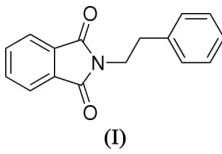
The molecule of the title compound, C16H13NO2, contains two
planar units, viz. a phthalimide system and a phenyl ring in almost parallel orientation, linked by an ethylene bridge. In the crystal structure, the molecules form centrosymmetric pairs which are held together by–interactions between the phthalimide systems. The latter are stacked in a head-to-tail fashion with an interplanar distance of 3.263 (6) A˚ .
Comment
N-(2-Phenethyl)phthalimide (2-phenethylisoindoline-1,3-dione) (I), known from early communications on the Gabriel synthesis (Ing & Manske, 1926), has received new attention as a possible agent in chemotherapy owing to its thalidomide-like effect on the production of the human tumour necrosis factor TNF- (Sasaki et al., 1995). Consequently, new synthetic approaches to this class of phthalimides have been published, such as the photoinduced electron transfer (PET) of phthali-mide anions and arylalkenes (Suauet al., 2003).
We have recently examined the photochemical properties of (I) in a laser flash photolysis study of the mechanism of PET reactions involving photo-Kolbe and pseudo-photo-Kolbe decarboxylation processes (Warzechaet al., 2006).
The molecular structure of (I) is shown in Fig. 1. The mol-ecule contains two planar subunits, viz. the phthalimide chromophore and a phenyl ring, which are linked by an ethylene bridge. The latter shows a staggered conformation
with an N1—C9—C10—C11 torsion angle equal to
[image:1.610.275.386.399.476.2] [image:1.610.206.460.631.707.2]176.69 (13). The C1—N1—C9—C10 and C9—C10—C11—
Figure 1
C12 torsion angles are close to 90 [85.46 (17) and 89.71 (17), respectively], the ethylene bridge plane being
almost orthogonal to both the phthalimide and the phenyl planes.
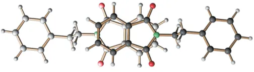
The crystal packing features pairs of molecules related by an inversion centre with parallel phthalimide planes at a distance of 3.263 (6) A˚ from each other. In these pairs, the distance between the centre of the six-membered ring of one molecule and the centre of the five-membered ring of the other molecule is as short as 3.447 (1) A˚ , which implies significant–stacking interaction (Fig. 2).
Experimental
A mixture of phthalic anhydride (5.92 g, 40 mmol) and 2-phen-ethylamine (4.84 g, 40 mmol) in an open beaker was subjected to four cycles of 1 min heating and subsequent cooling in a domestic microwave oven (800 W). Recrystallization of the resulting crude material from ethanol furnished colourless platelets of the title compound (9.04 g, 36 mmol, 90%). An almost equidimensional block suitable for X-ray diffraction, was cut from a thick platelet (m.p. 401 K).
Crystal data
C16H13NO2
Mr= 251.27
Orthorhombic,Pbca a= 28.3243 (9) A˚
b= 11.4545 (3) A˚
c= 7.6680 (2) A˚
V= 2487.81 (12) A˚3
Z= 8
Dx= 1.342 Mg m3
MoKradiation
Cell parameters from 10877 reflections
= 2.3–27.0
= 0.09 mm1
T= 100 (2) K
Block cut from a thick platelet, colourless
0.420.400.40 mm
Data collection
Nonius KappaCCD diffractometer
’/!scans
Absorption correction: none 10877 measured reflections 2700 independent reflections 1790 reflections withI> 2(I)
Rint= 0.067
max= 27.0
h=36!27
k=14!12
l=9!9
Refinement
Refinement onF2 R[F2> 2(F2)] = 0.041
wR(F2) = 0.095
S= 0.99 2700 reflections 225 parameters
All H-atom parameters refined
w= 1/[2
(Fo2) + (0.0441P)2] whereP= (Fo2+ 2Fc2)/3 (/)max= 0.019
max= 0.21 e A˚3 min=0.24 e A˚3
Extinction correction:SHELXL97
Extinction coefficient: 0.0058 (8)
All H atoms were refined freely [C—H = 0.952 (16)–1.030 (16) A˚ ]. Data collection: COLLECT (Hooft, 1999); cell refinement:
DENZO (Otwinowski & Minor, 1997); data reduction: DENZO; program(s) used to solve structure: SHELXS97 (Sheldrick, 1997); program(s) used to refine structure:SHELXL97(Sheldrick, 1997); molecular graphics: SCHAKAL99(Keller, 1999); software used to prepare material for publication: SHELXL97, PLATON (Spek, 2003) andenCIFer(Allenet al., 2004).
The generous financial support of the Deutsche
Forschungsgemeinschaft (DFG, Germany) and the Centre National de la Recherche Scientifique (CNRS, France) is gratefully acknowledged.
References
Allen, F. H., Johnson, O., Shields, G. P., Smith, B. R. & Towler, M. (2004).J. Appl. Cryst.37, 335–338.
Hooft, R. W. (1999).COLLECT. Nonius BV, Delft, The Netherlands. Ing, H. R. & Manske, R. H. F. (1926).J. Chem. Soc.pp. 2348–2351. Keller, E. (1999).SCHAKAL99. University of Freiburg, Germany. Otwinowski, Z. & Minor, W. (1997). Methods in Enzymology, Vol. 276,
Macromolecular Crystallography, Part A, edited by C. W. Carter Jr & R. M. Sweet, pp. 307–326. New York: Academic Press.
Sasaki, K., Shibata, Y., Hashimoto, Y. & Iwasaki, S. (1995).Biol. Pharm. Bull.
18, 1228–1233.
Sheldrick, G. M. (1997). SHELXL97 and SHELXS97. University of Go¨ttingen, Germany.
Spek, A. L. (2003).J. Appl. Cryst.36, 7–13.
Suau, R., Garcı´a-Segura, R., Sa´nchez-Sa´nchez, C., Pe´rez-Inestrosa, E. & Pedraza, A. M. (2003).Tetrahedron,59, 2913–2919.
[image:2.610.45.297.70.142.2]Warzecha, K.-D., Go¨rner, H. & Griesbeck, A. G. (2006).J. Phys. Chem. A,110, 3356–3363.
Figure 2
supporting information
sup-1 Acta Cryst. (2006). E62, o1580–o1581
supporting information
Acta Cryst. (2006). E62, o1580–o1581 [https://doi.org/10.1107/S1600536806010154]
N
-(2-Phenethyl)phthalimide
Klaus-Dieter Warzecha, Johann Lex, J
ö
rg M. Neud
ö
rfl and Axel G. Griesbeck
2-phenethylisoindoline-1,3-dione
Crystal data
C16H13NO2
Mr = 251.27
Orthorhombic, Pbca
Hall symbol: -P 2ac 2ab
a = 28.3243 (9) Å
b = 11.4545 (3) Å
c = 7.6680 (2) Å
V = 2487.81 (12) Å3
Z = 8
F(000) = 1056
Dx = 1.342 Mg m−3
Melting point: 401 K
Mo Kα radiation, λ = 0.71073 Å Cell parameters from 10877 reflections
θ = 2.3–27.0°
µ = 0.09 mm−1
T = 100 K
Cube cut from a thick platelet, colourless 0.42 × 0.40 × 0.40 mm
Data collection
Nonius KappaCCD diffractometer
Radiation source: fine-focus sealed tube Graphite monochromator
φ/ω Scans scans
10877 measured reflections 2700 independent reflections
1790 reflections with I > 2σ(I)
Rint = 0.067
θmax = 27.0°, θmin = 2.3°
h = −36→27
k = −14→12
l = −9→9
Refinement
Refinement on F2
Least-squares matrix: full
R[F2 > 2σ(F2)] = 0.041
wR(F2) = 0.095
S = 0.99 2700 reflections 225 parameters 0 restraints
Primary atom site location: structure-invariant direct methods
Secondary atom site location: difference Fourier map
Hydrogen site location: inferred from neighbouring sites
All H-atom parameters refined
w = 1/[σ2(F
o2) + (0.0441P)2]
where P = (Fo2 + 2Fc2)/3
(Δ/σ)max = 0.019
Δρmax = 0.21 e Å−3
Δρmin = −0.24 e Å−3
Extinction correction: SHELXL97, Fc*=kFc[1+0.001xFc2λ3/sin(2θ)]-1/4
Special details
Experimental. for X-ray diffraction, a cubical piece was cut from a thick platelet IR (neat): 1707, 1429, 1396, 1360, 1102, 1069, 991, 870, 756, 721, 709, 700 cm-1.
Geometry. All e.s.d.'s (except the e.s.d. in the dihedral angle between two l.s. planes) are estimated using the full covariance matrix. The cell e.s.d.'s are taken into account individually in the estimation of e.s.d.'s in distances, angles and torsion angles; correlations between e.s.d.'s in cell parameters are only used when they are defined by crystal symmetry. An approximate (isotropic) treatment of cell e.s.d.'s is used for estimating e.s.d.'s involving l.s. planes.
Refinement. Refinement of F2 against ALL reflections. The weighted R-factor wR and goodness of fit S are based on F2,
conventional R-factors R are based on F, with F set to zero for negative F2. The threshold expression of F2 > σ(F2) is used
only for calculating R-factors(gt) etc. and is not relevant to the choice of reflections for refinement. R-factors based on F2
are statistically about twice as large as those based on F, and R- factors based on ALL data will be even larger.
Fractional atomic coordinates and isotropic or equivalent isotropic displacement parameters (Å2)
x y z Uiso*/Ueq
supporting information
sup-3 Acta Cryst. (2006). E62, o1580–o1581
Atomic displacement parameters (Å2)
U11 U22 U33 U12 U13 U23
C1 0.0202 (9) 0.0202 (8) 0.0194 (8) −0.0004 (7) 0.0019 (6) −0.0037 (7) C2 0.0221 (9) 0.0233 (8) 0.0181 (8) 0.0005 (7) 0.0020 (6) −0.0028 (7) C3 0.0174 (8) 0.0201 (8) 0.0193 (8) −0.0012 (7) 0.0014 (6) −0.0042 (6) C4 0.0236 (10) 0.0224 (9) 0.0249 (9) −0.0026 (7) 0.0050 (7) −0.0023 (7) C5 0.0182 (9) 0.0280 (10) 0.0309 (9) −0.0036 (7) 0.0047 (7) −0.0088 (7) C6 0.0169 (9) 0.0308 (10) 0.0266 (9) 0.0043 (8) −0.0023 (7) −0.0089 (7) C7 0.0203 (9) 0.0242 (9) 0.0205 (8) 0.0040 (7) 0.0004 (6) −0.0034 (7) C8 0.0153 (8) 0.0216 (8) 0.0179 (7) 0.0004 (6) 0.0024 (6) −0.0051 (6) C9 0.0155 (9) 0.0281 (10) 0.0241 (8) 0.0009 (7) −0.0031 (6) −0.0016 (8) C10 0.0174 (9) 0.0331 (10) 0.0241 (9) 0.0000 (8) −0.0024 (7) −0.0044 (8) C11 0.0160 (8) 0.0267 (8) 0.0182 (7) 0.0009 (7) 0.0012 (6) −0.0048 (7) C12 0.0218 (9) 0.0225 (8) 0.0201 (8) 0.0030 (7) −0.0003 (6) −0.0001 (7) C13 0.0205 (9) 0.0262 (9) 0.0239 (8) −0.0040 (8) 0.0019 (7) −0.0029 (7) C14 0.0141 (9) 0.0327 (9) 0.0233 (8) 0.0010 (7) −0.0008 (6) −0.0026 (7) C15 0.0225 (9) 0.0265 (9) 0.0228 (8) 0.0037 (7) 0.0014 (7) 0.0030 (7) C16 0.0204 (9) 0.0236 (9) 0.0234 (8) −0.0019 (7) 0.0030 (6) −0.0005 (7) N1 0.0140 (7) 0.0238 (7) 0.0196 (6) 0.0002 (5) −0.0006 (5) 0.0008 (6) O1 0.0219 (7) 0.0259 (6) 0.0346 (6) −0.0017 (5) 0.0046 (5) 0.0064 (5) O2 0.0286 (7) 0.0302 (7) 0.0320 (6) 0.0016 (5) −0.0019 (5) 0.0110 (5)
Geometric parameters (Å, º)
C1—O1 1.2137 (16) C9—C10 1.522 (2) C1—N1 1.3924 (18) C9—H9A 1.015 (15) C1—C8 1.480 (2) C9—H9B 0.986 (15) C2—O2 1.2143 (17) C10—C11 1.505 (2) C2—N1 1.3928 (18) C10—H10A 0.984 (15) C2—C3 1.484 (2) C10—H10B 1.008 (17) C3—C4 1.377 (2) C11—C16 1.389 (2) C3—C8 1.391 (2) C11—C12 1.393 (2) C4—C5 1.393 (2) C12—C13 1.384 (2) C4—H4 0.962 (15) C12—H12 0.984 (15) C5—C6 1.386 (2) C13—C14 1.389 (2) C5—H5 0.999 (16) C13—H13 1.030 (16) C6—C7 1.391 (2) C14—C15 1.387 (2) C6—H6 0.952 (16) C14—H14 0.982 (16) C7—C8 1.381 (2) C15—C16 1.387 (2) C7—H7 0.984 (15) C15—H15 1.009 (17) C9—N1 1.4536 (18) C16—H16 0.988 (16)