Testosterone replacement therapy and exercise training in
males with low testosterone status and heart failure.
STOUT, Martin.
Available from Sheffield Hallam University Research Archive (SHURA) at:
http://shura.shu.ac.uk/20822/
This document is the author deposited version. You are advised to consult the publisher's version if you wish to cite from it.
Published version
STOUT, Martin. (2013). Testosterone replacement therapy and exercise training in males with low testosterone status and heart failure. Doctoral, Sheffield Hallam University (United Kingdom)..
Copyright and re-use policy
See http://shura.shu.ac.uk/information.html
Sheffield Hallam University Research Archive
Learning and IT Seances Collegiate Learning Centre Collegiate Crescent Campus
Sheffield 810 2BP
ProQuest Number: 10702931
All rights reserved
INFORMATION TO ALL USERS
The quality of this reproduction is dependent upon the quality of the copy submitted.
In the unlikely event that the author did not send a com plete manuscript and there are missing pages, these will be noted. Also, if material had to be removed,
a note will indicate the deletion.
uest
ProQuest 10702931
Published by ProQuest LLC(2017). Copyright of the Dissertation is held by the Author.
All rights reserved.
This work is protected against unauthorized copying under Title 17, United States C ode Microform Edition © ProQuest LLC.
ProQuest LLC.
789 East Eisenhower Parkway P.O. Box 1346
Testosterone replacement therapy and exercise training in males with low testosterone status and heart failure.
Martin Stout
A Thesis Submitted in Partial Fulfilment of the Requirements of Sheffield Hallam University for the Degree of Doctor of Philosophy.
August 2013.
Title page. 1
Contents. 2
Acknowledgements. 8
Statement of originality. 10
Abbreviations. 12
List of tables. 17
List of figures. 20
List of appendix. 22
Presentations and conference proceedings arising from this research. 23
Section 1: The impact of testosterone concentration in males with heart failure.
Abstract. 24
Chapter 1. General introduction. 26
Chapter 2. Literature review: Heart failure and low testosterone.
2.1: Heart failure.
2.1.1: Incidence of heart failure. 29
2.1.2: Aetiology of heart failure. 30
2.1.3: Pathophysiological considerations -heart failure. 34
2.1.4: Myocardial substrate metabolism and heart failure. 37
2.1.4.1: Normal myocardial substrate metabolism. 38
2.1.4.2: Normal myocardial fatty acid metabolism. 39
2.1.4.3: Normal myocardial ketone metabolism. 40
2.1.4.4: Electron transport chain and osidative phosphorylation.40
2.1.4.5: Myocardial substrate metabolism in heart failure. 41
2.1.5: Diastolic heart failure. 45
2.1.6: Diagnosis, signs and symptoms in heart failure. 48
2.1.7: Diagnostic considerations for diastolic heart failure. 53
2.1.8: Current treatment strategies for heart failure. 56
2.1.8.1: Ischaemic heart disease and viability. 56
2.1.8.2: Pharmacological therapies. 61
2.1.8.2.1: ACE inhibitors. 62
2.1.8.2.2: Diuretics. 63
2.1.8.2.3: p-blockers 64
2.1.8.3: Invasive procedures. 65
2.1.8.3.1: Cardiac resynchronisation. 65
2.1.8.3.2: Implantable defibrillators. 65
2.1.8.3.3: Ventricular assist devices. 66
2.1.8.3.4: Heart transplantation. 66
2.1.8.4: Rehabilitation. 66
2.1.8.5: Importance of research into new modalities. 66
2.2: Testosterone deficiency. 77
2.3: Testosterone. 77
2.3.1: Biochemistry and biosynthetic pathways. 77
2.3.2: Incidence of testosterone deficiency. 88
2.3.3: Aetiology of testosterone deficiency. 89
2.3.4: Pathophysiological considerations - testosterone deficiency. 92
2.3.5: Diagnosis, signs and symptoms - testosterone deficiency. 94
2.3.6: Treatment of testosterone deficiency. 98
2.3.7: Testosterone as a metabolic hormone. 103
2.3.8: Heart failure and testosterone deficiency. 108
2.4: Aims and objectives. 116
Chapter 3. Materials and methods. 118
3.1: Study design. 118
3.2: Patient selection and recruitment. 119
3.3: Inclusion criteria. 119
3.4: Exclusion criteria. 119
3.5: Consent procedures. 120
3.6: Withdrawal of patients. 121
3.7: Sample size. 123
3.8: Medical examination. 123
3.9: Summary of known and potential risks and benefits to patient. 124
3.10: Safety assessments. 125
3.11: Adverse events and serious adverse events. 125
3.12: Data collection and record keeping. 126
3.13: Outcome measures. 127
3.13.1: Primary outcome measure - 6 minute walk. 127
3.13.2: Secondary outcome measures. 128
3.13.2.1: Blood analysis. 128
3.13.2.2: Serum testosterone, SHBG and albumin. 129
3.13.2.3: Salivary testosterone. 132
3.13.2.4: Serum NT pro BNP. 134
3.13.2.5: Echocardiography. 135
3.13.2.6: Health related and disease specific quality of life. 146
3.13.2.8: SF36 Version 2. 147
3.13.2.9: Beck Depression Inventory. 148
3.13.2.10: Androgen deprivation in the ageing male. 148
3.14: Measurement error. 149
3.15: Analysis of results. 150
3.16: Quality control assurance and access to source documents. 150
Chapter 4. Results.
4.1: Demographic data based on total testosterone. 152
4.2: Analyses based on total testosterone.
4.2.1: Primary outcome measure - 6 minute walk. 154
4.3: Secondary outcome measures.
4.3.1: Echocardiography. 161
4.3.2: Quality of life. 169
4.3.3: NT pro BNP. 173
4.4: Analyses based on free testosterone.
4.4.1: Adjusted demographics based on free testosterone. 175
4.4.2: Primary outcome measure - 6 minute walk based on free
testosterone. 180
4.5: Secondary outcome measures.
4.5.1: Echocardiography. 181
4.5.2: Quality of life. 188
4.5.3: NT pro BNP. 190
4.6: Analyses based on bio-available testosterone.
4.6.1: Adjusted baseline demographics based on bio-available
testosterone. 192
4.6.2: Primary outcome measure - 6 minute walk based on bio-available
testosterone. 197
4.7: Secondary outcome measures.
4.7.1: Echocardiography. 198
4.7.2: Quality of life. 206
4.7.3: NT pro BNP. 208
4.8: Correlation studies. 210
4.9: Salivary testosterone. 218
4.10: Additional statistical analyses. 226
Chapter 5. Discussion. 228
Section 2: Combined testosterone therapy and exercise training in males with
heart failure. 248
Abstract. 249
Chapter 1. General introduction. 250
Chapter 2. Literature review. 252
2.1: Exercise training and heart failure. 252
2.1.1: Exercise intolerance. 252
2.2: Benefits of exercise training in heart failure.
2.2.1: Exercise capacity. 256
2.2.2: Exercise training and cardiac systolic function. 260
2.2.3: Exercise training and cardiac diastolic function. 262
2.2.4: Exercise training and muscle function. 264
2.2.5: Exercise training and endothelial function. 269
2.2.6: Exercise training and bio-chemical / inflammatory markers. 274
2.2.7: Exercise training and metabolic syndrome. 283
2.2.8: Exercise training and quality of life. 287
2.3: Testosterone supplementation in heart failure.
2.3.1: Testosterone supplementation and functional capacity. 290
2.3.2: Testosterone replacement therapy and cardiac function. 295
2.3.3: Testosterone replacement therapy and arterial function. 297
2.3.4: Testosterone replacement therapy and muscle function. 300
2.3.5: Testosterone therapy and inflammatory markers. 302
2.3.6: Testosterone replacement therapy and quality of life. 305
2.4: Aims and objectives. 306
Chapter 3. Materials and methods. 308
3.1: Trial design. 308
3.2: Randomisation. 308
3.3: Blinding. 309
3.4: Patient selection and recruitment. 310
3.5: Inclusion criteria. 310
3.6: Exclusion criteria. 311
3.7: Consent procedures. 315
3.9: Sample size. 316
3.10: Medical examination. 317
3.11: Testosterone replacement therapy. 317
3.12: Description of packaging and labelling of clinical trial medication. 318
3.13: Potential interactions with testosterone. 319
3.13.1: Warfarin. 319
3.13.2: Insulin. 320
3.13.3: Ciclosporin. 320
3.13.4: Paclitaxel. 320
3.13.5: Peanut and soya. 321
3.14: Monitoring of patient compliance. 321
3.15: Adverse and serious adverse events. 321
3.16: Safety assessments. 323
3.17: Data collection and record keeping. 323
3.18: Exercise training. 324
3.19: Outcome measures. 324
3.19.1: Incremental shuttle walk test. 325
3.19.2: Blood analysis. 326
3.19.3: Skeletal muscle contractile function. 332
3.19.4: Peak oxygen uptake and muscle oxygenation. 332
3.19.5: Echocardiography. 337
3.19.6: Arterial reactivity using flow mediated dilatation. 329
3.19.7: Quality of life. 340
3.19.7.1: CHAMPS questionnaire. 340
3.19.8: Focus groups - qualitative assessment of intervention 342
3.19.9: Focus groups - qualitative data analysis 346
3.20: Statistical analysis 347
3.21: Measurement error. 347
Chapter 4. Results. 348
4.1: Demographic data. 348
4.1.1: Measurement error. 352
4.2: Endurance performance. 354
4.3: Echocardiographic and ultrasonic parameters. 358
4.4: Quality of life. 364
4.5: Blood chemistry. 368
5.0: Correlation studies. 371
6.0: Focus group analysis. 382
Chapter 5. Discusssion. 392
Chapter 6. Feasibility and power calculation for phase II efficacy trial. 422
Chapter 7: Summary and future research. 424
Chapter 8: Overall thesis summary and conclusions. 425
8.1: Overview and contribution to knowledge. 427
8.2: Testosterone status and HF - take home message. 429
8.3: Combined ET and TS - take home message. 429
8.4: Implications for clinical practise. 430
References. 432
Acknowledgements.
I am grateful to Professors John Saxton and Kevin Channer for their continued help,
support, experience and knowledge throughout the completion and write-up of this
thesis.
In addition I would like to thank those who assisted during completion of the research.
These include Dr Emma Scott and Anouska McConnell for the administration of trial
medications and collection of blood samples. Dr Garry Tew and Dr Liam Bourke for the
assistance in supervising patient exercise sessions and in blood collection. Sue
Kesterton and Dr Helen Crank for assisting in supervising exercise sessions and in
recruiting patients onto the study. Helen Lloyd and Alan Ruddock for their technical
assistance when collecting data. These people played an important part in the smooth
running and efficiency of this research.
I am also grateful to Helen Bowler, Alexis Bedford and Lance Bum at Sheffield
Teaching Hospitals NHS Foundation Trust together with Teresa Coppinger, Carolyn
Waywell and Diane Daniel from the Heart Failure Clinic at UHSM for their expertise
and help in completing this project.
I am also indebted to Keith Pearce at the North West Heart Centre, University Hospital
of South Manchester NHS Foundation Trust for allowing time to ensure this work is
completed - often at short notice.
I thank all patients who agreed to participate in this research as without you it would not
have been completed. I also thank their families who provided them with support and
encouragement throughout the intervention period.
I would finally like to thank my wife Jess and daughter Maddie for their patience and
support during the completion of this write-up. I would also like to thank my parents for
their continued help and support.
Without you all, it would have been impossible to complete this work. Once again,
STATEMENT OF ORIGINALITY.
Section 1.
The acute and chronic use of testosterone replacement therapy in males with low
testosterone status and chronic HF has been described in studies by members of our
investigatory team (Malkin et al, 2006 and Pugh et al, 2003). More recently, researchers
in Italy have conducted similar research (Caminiti et al, 2009). Although there are
positive results regarding the use of testosterone in males with chronic HF, there is
limited information on the physiological and biochemical consequences associated with
decreased testosterone in this patient group. There are only two small correlation studies
which have demonstrated moderate relationships between testosterone concentration,
exercise capacity and right ventricular function in males with HF (Bocchi et al, 2008,
Jankowska et al, 2009). This study will be the first direct comparison of key
physiological and psychological outcomes in males with moderate to severe HF and
Tow’ or ‘normal’ levels of testosterone. For this research, the study design, regional
ethical approval, research and development approval were undertaken by myself. Also
80% of the total data collection was undertaken by myself with assistance from Mr
Keith Pearce and the HF specialist nurses at University Hospital South Manchester.
Reproducibility of the echocardiography data was undertaken by Mr Keith Pearce and
myself. Statistical analysis was performed by myself with guidance and support from
Miss Sigrid Whiteside (Medical Statistician at University Hospital South Manchester).
Section 2.
The acute and chronic use of testosterone replacement therapy in males with low
testosterone status and chronic HF has been described in studies by members of our
investigatory team (Malkin et al, 2006 and Pugh et al, 2003). More recently, researchers
in Italy have conducted similar research (Caminiti et al, 2009). However, there is no
research on the combined effects of exercise training and testosterone therapy in the
chronic HF population. This randomised controlled clinical trial aimed to study the
effects of combined exercise training and testosterone treatment on exercise capacity,
physiological function and quality of life in males with low level testosterone. During
this study, 90% of all data collection and supervision of the 12 week intervention was
undertaken by myself. I received assistance from Miss Helen Lloyd, Mr Alan Ruddock,
Professors Kevin Channer and John Saxton during the initial baseline data collection
visit. Dr Emma Scott and Miss Anouska Me Connell provided all blinded trial
medication injections. Mrs Sue Kesterton, Dr Garry Tew, Dr Liam Bourke and Dr
Helen Crank all provided cover for exercise training sessions in my absence. All
statistical analysis was performed by myself (blinded to group allocation) and Dr Helen
Doll (Medical Statistician at the University of East Anglia) under the guidance of
Professor John Saxton.
With the exception o f any statements to the contrary, all the data presented in this report are the result o f my own efforts. In addition, no parts o f this report have been copied from other sources. I understand that any evidence o f plagiarism and/or the use o f
u n a c kn o wl ed g+ U''~A — * * **• A^ta will be dealt with as a serious matter.
Signed
Abbreviations.
17pHSD 17 beta hydroxysteroid dehydrogenase
2D two dimensional
3D three dimensional
ACE angiotensin converting enzyme
ADAM Androgen deficiency in the ageing male questionnaire
ADP adenosine diphosphate
AMP adenosine monophosphate
ANCOVA analysis of covariance
ANOVA analysis of variance
AR androgen receptor
At anaerobic threshold
ATP adenosine triphosphate
AVR aortic valve replacement
BDI Beck Depression Inventory
BMI body mass index
BNP brain natriuretic pepetide
BP blood pressure
Ca2+ calcium ions
CABG coronary artery bypass graft
CAD coronary artery disease
CHAMPS Community Health Activities Model Programme for Seniors
Questionnaire
CHF chronic heart failure
Cm/s centimetres per second
co2
carbon dioxideCoA co-enzyme A
COX-1 cyclo-oxygenase-1
CRT cardiac resynchronisation therapy
c-Src proto-oncogene tyrosine protein kinase
CV cardiovascular
CVA cerebrovascular accident
DNA deoxyribonucleic acid
DSE dobutamine stress echocardiography
ED endothelial dysfunction
EDTA ethylenediaminetetraacetic acid
eNOS endothelial nitric oxide synthase
ERK-2 mitogen activated protein kinase-2
ET exercise training
f a d h2 flavin adenine dinucleotide dehydrate
Fe C 02 fraction of expired carbon dioxide
Fe 0 2 fraction of expired oxygen
F i0 2 fraction of inspired oxygen
FMD flow mediated dilatation
FT free testosterone
GLUT glucose transporter
GM-CSF granulocyte macrophage colony stimulating factor
GP general practitioner
GTP guanosine triphosphate
hCG human choriogonadotropin
HPRSD Hamilton Psychiatric Rating Scale for depression
HR heart rate
ICD implantable cardioverter defibrillator
IGF-1 insulin like growth factor -1
IHD ischaemic heart disease
IL interleukin
ISWT incremental shuttle walk test
LAEF% left atrial active emptying fraction
LA-EI left atrial expansion index
LA-PEF% left atrial passive emptying fraction
LDL low density lipoprotein
LNCaP androgen sensitive human prostate adenocarcinoma cell line
LT low testosterone
LV left ventricular
LVAD left ventricular assist device
LVEF% left ventricular ejection fraction
m metres
MCP-1 monocyte chemotactic protein-1
MET metabolic equivalent
MHz mega Hertz
MI myocardial infarction
min minutes
MLHFQ Minnesota living with heart failure questionnaire
MRI magnetic resonance imaging
MRNA messenger ribonucleic acid
NADH nicotinamide adenine dinucleotide hydroxide
NICE National Institute for Clinical Excellence
NIRS near infrared spectroscopy
nmol/L nano mo Is per Litre
NT normal testosterone
NT pro-BNP N terminal brain natriuretic peptide
NYHA New York Heart Association
PCG-la PPARa co-activator 1
PCWP pulmonary capillary wedge pressure
PDH pyruvate dehydrogenase
PDK-4 pyruvate dehydrogenase kinase isoenzyme-4
PET positive emission tomography
PPAR peroxisome proliferator activated receptor
PSA prostate specific antigen
RAAS rennin-angiotensin-aldosterone system
Raf-1 murine leukaemia viral oncogene ho mo log-1
RHH Royal Hallamshire Hospital
RV right ventricular
SAE serious adverse event
SF-36 V2 short form 36 version 2 questionnaire
SHBG sex hormone binding globulin
SHU Sheffield Hallam University
sICAM soluble intra-cellular adhesion molecule
SPECT single photon emission computed tomography
STH SheffieldTeaching Hospitals
STPD standard temperature and pressure, dry
sVCAM soluble vascular adhesion molecule
TAPSE tricuspid annular plane systolic excursion
TDI tissue Doppler imaging
TEM technical error of measurement
TNFa tumour necrosis factor alpha
TS testosterone supplementation
TT total testosterone
UHSM University Hospital South Manchester
UK United Kingdom
VE ventilation
Vmax maximum left atrial volume
Vmin minimum left atrial volume
V0 2max maximal oxygen uptake
VpreA left atrial pre-contraction volume
VSD ventricular septal defect
a alpha
B beta
List of Tables.
Table 1. Causes of heart failure. 31
Table 2. Epidemiological studies of the aetiology of heart failure as percentage. 33
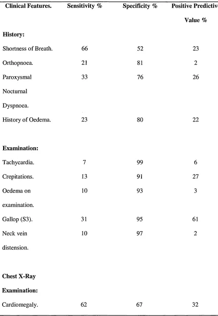
Table 3. Symptoms and signs in heart failure. 49
Table 4. Sensitivity, specificity and predictive value of signs and symptoms in heart
failure. 51
Table 5. New York Heart Association classification. 53
Table 6. Life extension and morbidity extension values in heart failure. 69
Table 7. Expanded appreciation of the aetiologies of low testosterone. 96
Table 8. Clinical formulations of testosterone. 100
Table 9. Baseline demographics of study sample based on total testosterone. 154
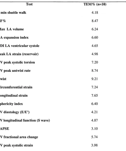
Table 10. Measurement error for key variables. 157
Table 11. 6 minute walk data based on total testosterone. 160
Table 12. LA structural and functional characteristics based on total testosterone. 162
Table 13. LV structural and functional characteristics based on total testosterone. 164
Table 14. Right heart structural and functional characteristics based on total
testosterone. 167
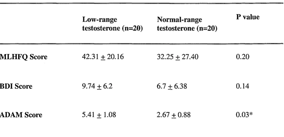
Table 15. MLHFQ, ADAM and BDI scores based on total testosterone. 173
Table 16. NT pro BNP based on total testosterone. 174
Table 17. Adjusted baseline demographics based on free testosterone. 176
Table 18. 6 minute walk data based on free testosterone. 180
Table 19. LA structural and functional characteristics based on free testosterone. 182
Table 20. LV structural and functional characteristics based on free testosterone. 184
Table 21. Right heart structural and functional characteristics based on free
testosterone. 187
Table 23. NT pro BNP based on free testosterone. 191
Table 24. Adjusted baseline demographics based on bio-available testosterone. 193
Table 25. 6 minute walk data based on bio-available testosterone. 197
Table 26. LA structural and functional characteristics based on bio-available
testosterone. 199
Table 27. LV structural and functional characteristics based on bio-available
testosterone. 202
Table 28. Right heart structural and functional characteristics based on bio-available
testosterone. 205
Table 29. MLHFQ, ADAM and BDI scores based on bio-available testosterone. 206
Table 30. NT pro BNP based on bio-available testosterone. 209
Table 31. Univariate Pearsons correlation of main outcome measures based on total
testosterone. 211
Table 32. Univariate Pearsons correlation of main outcome measures based on free
testosterone. 213
Table 33. Univariate Pearsons correlation of main outcome measures based on bio-
available testosterone. 215
Table 34. Univariate Pearsons correlation of main outcome measures based on salivary
testosterone. 222
Table 35. ANCOVA statistic comparing 6 minute shuttle walk between low and normal
total and free testosterone using age as a covariate. 226
Table 36. Pathophysiological effects of inflammatory cytokines in heart failure. 280
Table 37. Effects of testosterone supplementation on exercise capacity in
heart failure. 292
Table 38. Topic schedule for the focus groups. 345
Table 39. Baseline characteristics of the study sample for study 2. 351
Table 40. Technical error of measurement for important outcomes in study 2. 353
Table 41. Endurance and strength characteristics pre and post intervention. 355
Table 42. Echo parameters for both groups pre and post intervention period. 356
Table 43. MLHFQ, ADAM, BDI and CHAMPS questionnaire data pre and post
intervention in both groups. 367
Table 44. Blood chemistry for both groups pre and post intervention. 369
Table 45.Pearsons correlation for strength and endurance parameters based on total,
free and bio-available testosterone. 372
Table 46. Pearsons correlation for quality of life parameters based on total, free and
bio-available testosterone. 373
Table 47. Pearsons correlation for echocardiographic parameters based on total, free
and bio-available testosterone. 375
Table 48. Pearsons correlation for blood chemistry based on total, free and bio-
List of figures.
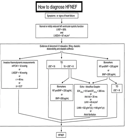
Figure 1. Flow chart for suggested diagnosis of diastolic heart failure. 54
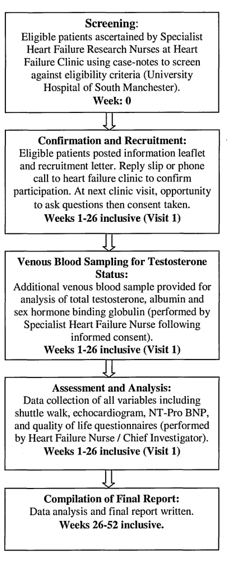
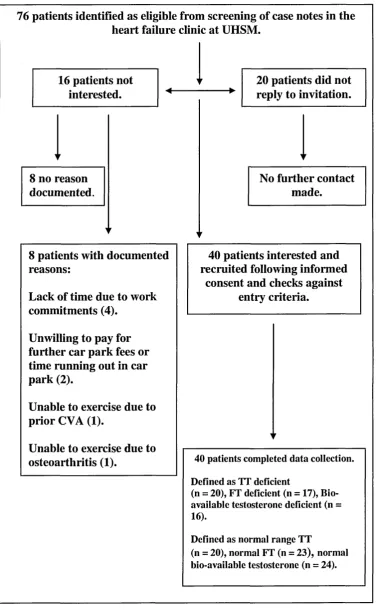
Figure 2. Patient journey for study 1. 122
Figure 3. Recruitment summary for study 1. 153
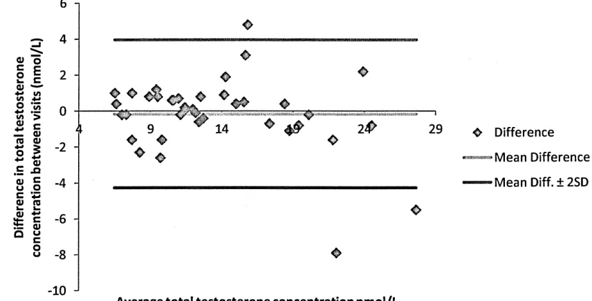
Figure 4. Bland Altman plot: agreement between two separate total testosterone
measures. 159
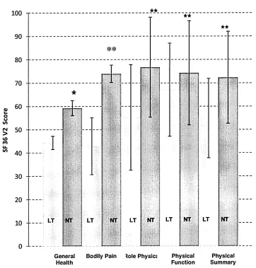
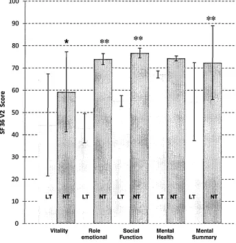
Figure 5. SF36 domains based on total testosterone. 170
Figure 6. Bland Altman plot: agreement between two separate free testosterone
measures. 179
Figure 7. SF36 domains based on free testosterone. 188
Figure 8. Blan Altman plot: agreement between two separate bio-available testosterone
measures. 196
Figure 9. Scatter plot showing relationship between total testosterone and 6 minute
walk. 217
Figure 10. Bland Altman plot: agreement between two separate salivary testosterone
measures. 218
Figure 11. Scatter plot showing the relationship between salivary and
total testosterone. 219
Figure 12. Bland Altman plot: agreement between salivary and free testosterone. 220
Figure 13. Flow chart of patient journey and important time points for study 2. 313
Figure 14. Example of lebelling of the IMP. 319
Figure 15. Recruitment summary for study 2. 349
Figure 16. FMD of femoral artery pre and post intervention for both groups. 362
Figure 17. Quality of life parameters pre and post intervention for both groups. 365
Figure 18. Bland Altman plot: agreement between two separate total testosterone
measures. 379
Figure 19. Bland Altman plot: agreement between two separate free testosterone
measures.
Figure 20. Bland Altman plot: agreement between two separate bio-available
testosterone measures.
380
List of Appendix.
Appendix 1. Ethics letter for study 1. 493
Appendix 2. Patient information leaflet for study 1. 499
Appendix 3. Case report forms for study 1. 503
Appendix 4. MLHFQ. 506
Appendix 5. Consent form example. 507
Appendix 6. SF36-V2. 508
Appendix 7. BDI. 509
Appendix 8. ADAM questionnaire. 512
Appendix 9. Ethics letter for study 2. 513
Appendix 10. Case report forms for study 2. 517
Appendix 11. CHAMPS Questionnaire. 521
Appendix 12. Patient information leaflet for study 2. 533
Appendix 13. Focus group transcription example. 540
Publications, presentations and conference proceedings arising from this research.
Innaugral Sports Medicine Meeting - Sheffield Teaching Hospitals NHS Foundation
Trust, October 2009 - invited speaker. Testosterone supplementation in heart failure.
British Association of Sport and Exercise Science: Cardiac Rehabilitation Subgroup
Meeting, Metropole Hotel, Birmingham, December 2009 - invited speaker.
Testosterone and exercise training in heart failure.
European Society of Cardiology Congress, Stockholm, Sweden, May 2010 - invited
speaker. The use of flow mediated dilatation: a clinical or research tool.
American College of Sports Medicine, Washington, U.S.A, October 2010 - abstract
presentation. Combined exercise training and testosterone supplementation in males
with low testosterone status and chronic heart failure.
European Society of Cardiology - Heart Failure Congress, Gothenburg, Sweden, May
2011 - Abstract presentation: the cardiovascular effects of testosterone and exercise
training in heart failure. European Journal of Heart Failure Supplement (2011) (10)
(suppll): S60-S104.
Stout, M., Tew, G.A., Doll, H., Zwierska, I., Woodroofe, N., Channer, K.S. and Saxton,
J.M. Testosterone therapy during exercise rehabilitation in male patients with heart
failure who have low testosterone status: A double-blind randomised controlled
Abstract.
Background: This cross-sectional study aimed to assess the effect of low testosterone
(LT) status on functional capacity, quality of life, cardiac structure and function in
males with moderate or severe stable heart failure (HF). Methods: 40 male patients with
HF (ejection fraction (EF)% <45%) with low serum total testosterone (TT) <12 nmol /
L (n =20, 71.9 ± 10.11 yrs) and normal range TT (n = 20, 66.9 ± 11.98 yrs) were
recruited from a specialist HF clinic. The primary outcome, functional capacity, was
assessed by the 6 min shuttle walk test. Health related quality of life (HRQoL) data was
assessed using a licensed version of the SF36 Version 2. Additionally, Beck Depression
Inventory (BDI), Minnesota Living with Heart Failure questionnaire (MLHFQ) and
Androgen Deficiency in the Ageing Male (ADAM) questionnaire data was collected.
Cardiac characteristics were assessed using detailed echocardiography analysis. Blood
samples were taken for assessment of NT pro BNP. Results: Normal testosterone (NT)
patients demonstrated a significantly higher 6 min walk distance (429.00 + 126.94 m;
257.65 + 65 m, p<0.01) when compared to LT. LT patients showed significantly worse
indices of quality of life using the SF36 V2 questionnaire. General health component
(31.84 + 2.85 v 59.44 + 3.24, p<0.05), overall physical component score (41.39 + 17.03
v 69.69 + 19.71, p<0.01) and overall mental component score (54.88 + 17.52 v 72.36 +
16.58, p<0.01) higher in the NT group. There were very few cardiac changes and no
differences in MLHFQ or BDI score between the groups. Conclusion: This is the first
study to compare important prognostic outcomes in matched patients with HF and low
testosterone. Additionally, this is the first paper to report significantly adverse HRQoL
in male patients with low testosterone and HF when compared to normal range
testosterone counterparts. LT in HF is associated with reduced functional capacity,
together with attenuated general, physical and mental components of quality of life.
Further research is warranted to assess the impact of testosterone supplementation on
these important prognostic outcomes in male patients with HF and low testosterone
Section 1: The impact of testosterone concentration in males with heart failure.
Chapter 1: General introduction.
Heart failure (HF) is a multi-organ disease, involving the musculoskeletal, respiratory
and endocrine systems. It is a common, debilitating condition and a major public health
and financial burden in the Western world. The incidence and prevalence of congestive
HF has increased together with the resulting morbidity and mortality. Worldwide it is
estimated that 15 million individuals suffer from this condition, with 400,000 new cases
each year in the United States of America and a total of 4.5 million Americans effected
(Erikson, 1995). In the United Kingdom (UK), it is estimated that 900,000 people have
HF (Al-Mohammed et al, 2010). Surveys in North West London show prevalence rates
of 0.6 per 1000 patients aged less than 65 years and 28 per 1000 patients aged 65 years
and over (Parameshwar et al, 1992).
As males age, there is a gradual decline in circulating bio-available testosterone
(Betocchi, 2005). There is a general consensus that testosterone levels decline about 1%
per year from as early as age 30 years. Noticeable declines are common after the age of
50 years but there is great individual variability (Morales and Lunenfield, 2002).
HF also appears to be associated with decreased levels of plasma testosterone,
supported by the fact that about 25% of men of all ages with HF have biochemical
evidence of testosterone deficiency (Malkin et al, 2009). Low levels of testosterone
have been correlated with disease progression in HF and may also be responsible for
some of the features of HF, such as reduced skeletal muscle mass and function,
cachexia, fatigue and depressed mood (Malkin et al, 2006). Furthermore, myocardial
cachexia, a syndrome with poor prognosis, is characterised by low levels of testosterone
(Aukrust et al, 2009).
In addition to this it is well known that HF as a metabolic syndrome can adversely alter
numerous endocrine, metabolic and inflammatory parameters (Jackson et al, 2000,
Noutsias et al, 1999). The alterations can include changes in levels and sensitivity to
insulin, growth hormone and importantly testosterone (Von Haehling et al, 2007).
Reduced insulin sensitivity and the development of diabetes could be a major
complicating factor of HF. It has been described that more than 40% of patients with
HF have manifest disorders of glucose metabolism which appear independent of fat
distribution and obesity (Aukrust et al, 2009).
Interestingly, testosterone replacement therapy at physiological doses has been shown to
improve indices of physical function, cardiac function and also quality of life in HF
patients (Malkin et al, 2006, Caminiti et al, 2009). These studies have shown modest
improvements in cardiac output, maximal oxygen uptake, muscular strength and
improved disease and health specific quality of life markers. Additionally, testosterone
supplementation has been shown to improve arterial function via reduction in systemic
vascular resistance (Pugh et al, 2003). Animal models have shown positive associations
between testosterone supplementation and reduced levels of biochemical inflammation
(Zhang et al, 2007) and also improvements in cardiac contractile performance (Scheuer
et al, 1997). These studies are important because they suggest that when testosterone
level is returned to the normal range there are significant improvements in important
Recent correlation studies have suggested that in HF, testosterone level is independently
related to peakv ° 2, oxygen pulse and also right ventricular function (Jankowska et al,
2009 and Bocchi et al, 2008). However, there is no research that directly compares
parameters of physical fitness, cardiac function, quality of life and biochemical
inflammation in male HF patients with low or normal testosterone levels.
Contradictory results have been published regarding the successful administration of
testosterone therapy to males with HF (Caminiti et al, 2009, Malkin et al, 2006) and no
large scale trials have aimed to determine the efficacy of supplementation. Based on
previous observations, the efficacy of testosterone supplementation in a male HF
population with low testosterone status is uncertain. This study aims for the first time to
determine the effects of testosterone concentration on exercise capacity, quality of life
and cardiac function in males with low testosterone status in comparison with normal
testosterone in moderate or worse HF.
Chapter 2: Literature review - Heart failure and low testosterone.
2.1: Heart failure.
2.1.1 Incidence of HF.
There has been a dramatic decline in the incidence of mortality and morbidity from
many cardiovascular diseases over recent decades (Sytkowski et al, 1990). Conversely,
the incidence and prevalence of congestive HF, together with the resultant morbidity
and mortality have shown an increase throughout this time. Worldwide it is estimated
that 15 million individuals suffer from this condition, with 400,000 new cases each year
in the United States of America and a total of 4.5 million Americans effected (Eriksson,
1995). In the United Kingdom (UK), surveys in North West London show prevalence
rates of 0.6 per 1000 patients aged less than 65 years and 28 per 1000 patients aged 65
years and over (Parameshwar et al, 1992). This statistic is reinforced by the
Framingham Study which described a doubling in the incidence of HF for every decade
of life reaching 3% in those aged 85-94 years (Ho et al, 1993). Large surveys have also
been conducted in the UK during the 1990’s aiming to study left ventricular (LV)
dysfunction using echocardiography in large populations of Glasgow and the West
Midlands. In Glasgow the prevalence of significantly impaired LV systolic dysfunction
in participants 25-74 years was 2.9% and in the West Midlands 1.8% in patients aged 45
years and over (Lip et al, 1997 and Me Donagh et al, 1997). Economists have studied
the costs of HF to the National Health Service in the UK. It has been found that in the
year 2000 HF cost the Health Service 905 million pounds (1.91% of total UK Health
Service expenditure). The most predominant factor for this cost was hospital admission
(69%) followed by prescriptions for important treatment (18%). Additionally, the cost
for secondary hospital admission and nursing home care for patients with HF was 750
2.1.2: Aetiology of HF.
The aetiology of HF differs according to the population being studied. For example, in
Western society, the most common causes of HF are coronary artery disease (CAD) and
hypertension. In the developing countries, valvular heart disease and nutritional heart
disease are most common (Lip et al, 2000). There are also a host of other conditions
which can cause HF and these are listed in the summary table 1 below.
Table 1. Causes of HF adapted from Lip et al, 2000.
Causes of Heart Failure.
Coronary artery disease:
• Myocardial Infarction.
• Ischemia.
Hypertension.
Cardiomyopathy:
• Dilated (congestive).
• Hypertrophic / obstructive.
• Restrictive (e.g. amyloidosis, sarcoidosis, haemochromatosis).
• Obliterative.
Valvular and Congenital Heart Disease:
• Mitral valve disease.
• Aortic valve disease.
• Atrial septal defect / ventricular septal defect.
Arrhythmias:
• Tachycardia.
• Bradycardia (complete heart block, sick sinus syndrome).
• Loss of atrial transport (e.g. atrial fibrillation).
Alcohol and Drugs:
• Alcohol.
• Cardiac depressants (e.g. p blockers, calcium channel blockers).
‘High output9 cardiac failure:
• Anaemia, thyrotoxicosis, Paget’s Disease, arteriovenous fistulae.
• Constrictive pericarditis.
• Pericardial effusion.
Primary Right Heart Failure:
• Pulmonary hypertension (e.g. pulmonary embolism, cor pulmonale).
• Tricuspid incompetence.
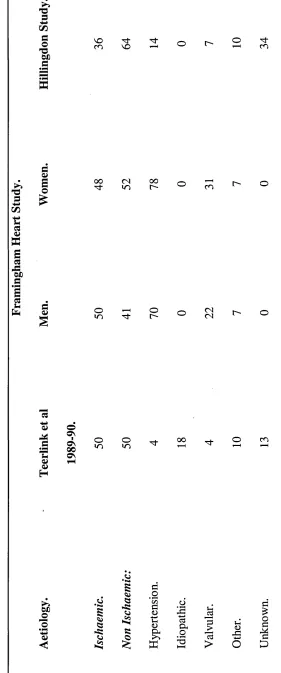
Table 2 summarises the percentages of common aetiologies of HF in the
western world.
e 2. Ep id emi ol ogi cal s tu di es of the ae tio lo gy of HF as pe rc en ta ge (a da pt ed fro m Lip et al, 2 00 0) . 3 CO 15a © 8 cz fa0£ 3 03 U fa -4-> C/3 3 © 01) 3 3 © a © £ 3 -4J© fa3 1272 © © H 0£ o ©
VOm vo cn
oo CNin 00l> CO
o
in o CNCN
t'-o
©VI OS
00
OS
o oin oo cn
53 J>a s 53 -SCO Jo I 3 .2 ’m3 ©
4—< l- l
[image:36.613.108.389.52.725.2]2.1.3: Pathophysiological considerations - Heart failure.
In systolic HF, there is impairment in left ventricular ejection fraction (LV EF%)
resulting in a decline in cardiac output. In response to this there is activation of various
neuro-hormonal compensatory mechanisms which attempt to improve the mechanical
efficiency of the heart. The phenomenon of which is addressed below in more detail.
Stimulation of the renin-angiotensin-aldosterone system (RAAS) via neuro-hormonal
activation and altered autonomic control increases levels of renin, plasma angiotensin II
and aldosterone. Angiotensin II has been found to be a potent vasoconstrictor of the
renal efferent arterioles and systemic circulation where it stimulates release of nor
adrenaline from sympathetic nerve terminals, inhibits vagal tone and promotes release
of aldosterone (Jackson et al, 2000). This activation results in retention of sodium and
water together with increased release of potassium. Furthermore, angiotensin II has
been shown to cause anorexia and weight loss in experimental models using animals
(Brink et al, 1996). Additional research has also suggested an increase in the levels of
endothelin (another potent vasoconstrictor peptide) in HF, constituting adverse
prognosis (Tsutamoto et al, 1995). Blood plasma levels of Endothelin - 1 have been
shown to be a reliable correlate to pulmonary artery capillary wedge pressure (PCWP),
hospital admission and death (McMurray et al, 1992). As an early compensatory
mechanism to provide inotropic support to the failing cardiac muscle, low and high
pressure baroreceptors further stimulate the sympathetic nervous system to increase
heart rate (HR) and hence cardiac output. Adverse consequences of prolonged elevation
of adrenergic drive to the cardiac muscle involve a direct toxic effect of nor-epinephrine
on cardiac myocytes (Mann et al, 2002), facilitation of the development of ventricular
arrhythmias (Schwartz et al, 1994), alterations in p-adrenoceptor function (Bristow et al,
p- adrenoceptor function increases sympathetic activity and also down-regulates
autonomic modulation of the sinus node leading to the notion that reduced HR
variability is an important prognostic marker in patients with HF (Jackson et al, 2000).
Atrial and Brain natriuretic peptides are released by cardiac muscle in HF primarily due
to volume overload and the corresponding atrial and ventricular stretch. These peptides
have an antagonistic effect to those of angiotensin II on vascular tone, aldosterone
secretion and renal-tubule sodium reabsorption (Jackson et al, 2000). As a result, levels
of these circulating mediators in HF have become an important diagnostic marker and
are also the subject of research interest in developing new medications that inhibit the
enzyme that metabolises atrial natriuretic peptide (neutral endopeptidase) (Jackson et al,
2000). In untreated congestive cardiac failure, high levels of atrial and brain natriuretic
peptide correlate closely with adverse prognosis and ultimately mortality.
HF also results in increased levels of circulating cytokines which include tumour
necrosis factor-alpha (TNF-a), interleukins-1( IL-1), 2 (IL-2), 6 ( IL-6), 8 (IL-8) and
their receptors (Berry and Clarke, 2000). In more detail, research has shown that TNF-a
and IL-6 levels are elevated in relation to deteriorating functional classification and in
this regard should also be recognised to be associated with some of the classic
hallmarks of HF including worsening LV dysfunction, pulmonary oedema and cardiac
remodelling (Millar et al, 1982). In relation to this, in patients with severe HF, elevated
levels of soluble TNF receptor are a stronger predictor of early mortality when
compared to plasma nor adrenaline, atrial natriuretic peptide or (New York Heart
Association) NYHA classification (Berry and Clarke, 2000). Physiologically in HF,
there is an increased production of soluble adhesion molecules via the endothelium;
inflammatory response (Oppenheimer et al, 1991). This physiological process relates to
the earlier notion that endothelial injury is associated with an adverse prognosis in HF.
Other researchers have discovered that during hypoxia (an occurrence related to the
reduced cardiac output in HF) IL-6 and the soluble adhesion molecule - 1 (sICAM-1)
are secreted by injured endothelial cells (Sluiter et al, 1993) and vascular xanthine
dehydrogenase enzyme is converted to xanthine oxidase. This catalyses the production
of uric acid and its reactive bi-product superoxide (Yan et al, 1997). It has been
hypothesised that injured vascular endothelium in HF patients may be a source of free
radical production, triggering leukocyte activation and increased cytokine production
(Leyva et al, 1998).
Researchers and clinicians have observed widespread endothelial cell activation
followed closely by endothelial dysfunction (ED) in HF (Brutsaert, 2003). This ED has
been thought to be partly responsible for the early fatigue and exercise intolerance in
HF. This may be attributed to inappropriate, endothelial mediated, vasoconstrictor
responses with reduced vasodilatory capacity contributing to elevated peripheral
vascular resistance (Drexler et al, 1992). In relation to this, reduced gene expression of
endothelial nitric oxide synthase (eNOS) and cyclo-oxygenase-1 (COX-1) has also been
shown to be a factor in the early onset of ED in cardiac failure (Smith et al, 1996).
Experimental research in patients with cardiac dysfunction has provided direct evidence
of ED in both conductive and resistance coronary arteries (Treasure et al, 1990). In
more detail, these patients displayed impaired dilatory responses to acetylcholine and
bradykinin within the coronary circulation. The coronary ED has been attributed to
reduced synthesis of coronary endothelial nitric oxide. The concept has emerged that
coronary vascular ED could trigger coronary vasoconstriction, smooth muscle
coronary thrombosis. It is evident that these processes would accelerate the
development CAD, resulting in decreases in myocardial perfusion and indirectly
contributing to cardiac failure.
Skeletal muscle physiology and function is abnormal in patients with HF. Muscle bulk
is decreased and there are documented reductions in muscular strength and endurance
(Massie et al, 1987). Early studies have found that during leg exercise, oxygen
extraction to the exercising muscle and lactate efflux were increased, together with
diminished total oxygen utilisation (Donald et al, 1961). This was attributed to represent
the metabolic consequences of reduced blood flow to exercising muscle. To support this
notion, research has suggested that skeletal muscle is histologically abnormal with a
tendency towards anaerobic (type II) muscle fibres (Mancini et al, 1989). This research
has also identified abnormal mitochondrial structure with reduced cristae volume and
reduction in the key enzymes involved in the Kreb’s cycle and the oxidative chain. This
can be supported by other research which has shown more rapid declines of
phosphocreatine and rises in inorganic phosphate in HF patients when compared with
healthy controls. The slope of the relationship between phosphocreatine and inorganic
phosphate versus muscular power output (an index of mitochondrial oxidative
metabolism) was found to be steeper in HF patients suggesting impaired oxidative
capacity (Wilson et al, 1985).
2.1.4: Myocardial substrate metabolism and heart failure.
In order to understand the abnormalities related to myocardial substrate metabolism in
HF, it is important to review myocardial metabolism in a normal heart. In non-
heart is derived from oxidative phosphorylation in the mitochondria with minor
contribution from glycolysis and guanosine triphosphate (GTP) formation via the citric
acid cycle (Stanley et al, 2005). The initial step in the energetic pathway is the cellular
uptake of and utilisation of available substrate, their breakdown by P-oxidation and
glycolysis, resulting in formation of acetyl coenzyme A (CoA) which is then fed into
the Krebs cycle producing nicotinamide adenine dinucleotide hydroxide (NADH) and
carbon dioxide (CO2) (Neubauer, 2007). Under normal perfusion approximately 60-
90% of CoA results from p-oxidation of free-fatty acids and 10-40% from pyruvate
oxidation via glycolysis and lactate oxidation (Stanley et al, 2005).
2.1.4.1: Normal myocardial carbohydrate metabolism.
Glycolytic substrate is utilised from exogenous glucose and glycogen stores. Glucose
transport into cardiac myocytes is facilitated by trans-membrane gradients and
sarcolemma glucose transporters (GLUT-4 and GLUT-1) (Stanley et al, 2005). Insulin
stimulation, increased cardiac demand or myocardial ischaemia result in a shift in
glucose transporters from intra-cellular vesicles to the sarcolemma therefore increasing
glucose transport and uptake into the cell (Stanley et al, 1997). In addition, GLUT-4 re
location into the sarcolemma may be facilitated by activation of adenosine
monophosphate (AMP) -activated protein kinase, particularly during ischaemia or in
response to exercise (Russell et al, 1999 and Russell et al, 2004). Intra-cellular glycogen
stores may be used as an additional source of glucose 6 phosphate for uptake into the
glycolytic pathway. Previous studies have suggested that glycogen concentration may
be increased by an elevated supply of exogenous substrate together with, or solely by,
increased levels of insulin (Kruszynska et al 1991). Glycogenolysis can therefore be
activated by adrenergic stimulation, decrease in tissue adenosine triphosphate (ATP)
concentration and increase in inorganic phosphate - occurring typically with stressors
such as exercise or ischaemia (Stanley et al, 1997).
Pyruvate, formed from glycolysis, may be converted to lactate, decarboxylated to CoA
or carboxylated to oxaloacetate or malate (Stanley et al, 2005).The decarboxylation of
pyruvate is considered to be the first irreversible step in carbohydrate oxidation,
catalysed by the enzyme pyruvate dehydrogenase (PDH) (Randle, 1986). Importantly,
higher levels of circulating lipids, together with increased accumulation of long chain
fatty acid moieties (e.g. associated with diabetes or fasting) have been shown to increase
phosporylation inhibition of PDH and decrease oxidation of pyruvate from the
glycolytic pathway and lactate oxidation (Huang et al, 2002). Furthermore, glucose and
pyruvate oxidation, together with related PDH activity, are attenuated by increased rates
of fatty acid oxidation; for instance when there are elevated levels of plasma free fatty
acids. Conversely to this, pyruvate oxidation is increased when fatty acid oxidation is
attenuated (Kruszynska et al, 1991).
2.1.4.2: Normal myocardial fatty acid metabolism.
The concentration of plasma non-esterified fatty acid levels mainly determines the rate
of fatty acid uptake by the myocardium (Stanley et al, 2005). As such, metabolic
stressors such as ischaemia, diabetes or starvation which increase plasma free fatty acid
concentration, result in increased rate of myocardial free fatty acid uptake (Stanley et al,
2005). Regulation of plasma free fatty acid concentration is via their net release from
triglycerides in adipocytes, facilitated by the action of hormone-sensitive lipase and
synthesis by glycerolphosphate acyltransferase. Hormone-sensitive lipase is activated
by catecholamines and inhibited by insulin. Accordingly, when insulin concentration is
hence myocardial free fatty acid uptake and oxidation are high (Lopaschuk et al, 1994).
Uptake of free fatty acids into the cardiomyocyte is promoted by passive diffusion or
protein mediated transport across the sarcolemma (Stanley et al, 2005). Once within the
sarcolemma, non-esterified fatty acids bind to fatty acid binding protein to be activated
by esterification to fatty acyl-CoA via fatty acyl-CoA synthase (Stanley et al, 2005).
Acyl-CoA transportation into the mitochondria is facilitated by a carnitine-dependent
transport system, with carnitine palmitoyltransferase-I serving as the key regulator for
the rate of this uptake (Lopaschuk et al, 1994). Fatty acid P-oxidation occurs within the
mitochondria. This process repeatedly cleaves two carbon acetyl-CoA units in order to
generate NADH and flavin adenine dinucleotide dihydroxide (FADH2). Bing and co
workers in 1954 suggested four reactions, involving different, but specific enzymes, to
facilitate fatty acid p-oxidation dependent on long, medium or short-chain fatty
intermediates. Initially, catalysed by acyl-CoA dehydrogenase, followed by 2-enoyl-
CoA hydratase and 3-hydroxyacyl-CoA dehydrogenase. Finally, 3-ketoacyl-CoA
thiolase regenerates acyl-CoA for further P-oxidation and releases acetyl-CoA for the
citric acid cycle.
2.1.4.3: Normal myocardial ketone body metabolism.
Normal arterial plasma concentration of ketone bodies is low. Fatty acid ketone body
formation in the liver therefore, plays a minor role as a substrate for the myocardium
(Stanley et al, 2005). It should be highlighted however, that during starvation or poorly
controlled diabetes, plasma ketone bodies become elevated due to low insulin and high
fatty acid levels and as such become a major substrate for myocardial metabolism (Hall
et al, 1996).
2.1.4.4: Electron transport chain and oxidative phosphorylation in HF.
Research in both human and animal models of HF have shown that there is a decreased
concentration of tissue ATP, increased concentration of adenosine diphosphate (ADP)
and reduction in phosphorylation potential (Montgomery et al, 1992 and Shen et al,
1999). As a result, there is significant impairment of the kinetic mechanisms of ATP
utilisation for myocardial cell contraction and relaxation via myosin ATPase and
sarcoplasmic reticulum Ca2+- ATPase (Stanley et al, 2005). It has previously been noted
that HF attenuates the ability of the creatine kinase system to transfer mitochondrial
ATP to the myofibril. In addition to this, impairment of the electron transport chain may
be detrimental to the mitochondrial and cytosolic redox state, adversely affecting the
concentration of ATP, ADP and phosphate and therefore, reducing the rate of influx
through key metabolic enzymes such as PDH or phosphofructokinase (Ye et al, 2001).
Abnormalities at the level of the mitochondria in HF are abundant. Previous studies in
both humans and animals have suggested there is a greater incidence of mitochondrial
membrane disruption and matrix depletion (Sabbah et al, 1992), a lower capacity for
respiration with a variety of substrates (Sharov et al, 2000), electron transport chain
defects together with decreased capacity for oxidative phosphorylation (Casademont
and Miro, 2002). Although there are different theories as to the exact processes involved
in electron transport chain complex dysfunction in HF, it is widely evident that there is
a major disruption in oxidative metabolism at this level. To date, there is no consensus
regarding the exact site of the lesions of electron transport chain dysfunction, whether
the effects of this dysfunction are isolated to a group of myocytes or all cardiac
myocytes as a whole or finally, whether dysfunction is localised to subsarcolemmal or
intrafibrillar components of the mitochondria (Stanley et al, 2005).
Results are conflicting regarding myocardial substrate metabolism in heart failure,
particularly in human models. There is however, a consensus that HF reduces the
capacity to transduce foodstuffs into ATP (Stanley et al, 2005). Initially, in the early
stages of HF, fatty oxidation rate is maintained. However, Paolisso et al (1994)
observed that in established HF with NYHA Class II and III, there was increased
extraction and uptake of plasma free fatty acid and decreased glucose uptake.
Furthermore, it has also been established that HF increases myocardial lipid oxidation
by as much as 50% when compared to healthy aged matched controls (Paolisso et al,
1994). The same study also showed that in HF patients there is a significant decrease in
myocardial carbohydrate oxidation when compared to controls. Increased plasma nor
adrenaline concentrations corresponding to increased levels of free fatty acid where also
noted in the HF population. This has been attributed to enhanced p-adrenergic
stimulation and also higher levels of plasma insulin, both factors which may facilitate
glucose uptake and oxidation by the myocardium (Stanley et al, 2005). Using other
methods, researchers have been able to develop radiolabelled fatty acid / deoxyglucose
analogues and with positive emission tomography (PET) have discovered increased
uptake of fatty acid analogue and decreased uptake of deoxyglucose analogue in
patients with NYHA class III HF (Taylor et al, 2001). In contradiction to this, Yazaki et
al (1999) have suggested that in HF patients with idiopathic dilated cardiomyopathy, the
process is reversed with increased myocardial glucose uptake and decreased fatty acid
oxidation when compared to healthy controls. Davila-Roman et al (2002) have re
affirmed the findings of Yazaki and colleagues by using l8F-deoxyglucose 6 phosphate
infusion during PET to estimate glucose uptake. This study determined that there was
increased myocardial glucose metabolism and decreased fatty acid utilisation in their
population of HF patients.
Studies of patients with end-stage HF have consistently suggested that there is down
regulation of myocardial fatty acid oxidative enzymes - a feature relating to the
conversion of substrate metabolism from fatty acid oxidation towards glucose oxidation
(Stanley et al, 2005). In more detail, explanted hearts at transplant, have demonstrated
reduced messenger ribonucleic acid (mRNA) for the fatty acid oxidation enzymes long-
chain acyl-CoA dehydrogenase and medium-chain acyl-CoA dehydrogenase together
with significantly reduced protein levels of medium-chain acyl-CoA dehydrogenase
without down-regulation of mRNA for the glycolytic enzyme glyceraldehydes-3-
phosphate dehydrogenase (Sack et al, 1996). Pacing induced HF in a canine model, has
also showed similar reductions in mRNA levels of key enzymes involved in the fatty
acid oxidation pathway, but in addition, GLUT-1, GLUT-4, glyceraldehydes-3-
phosphate dehydrogenase, PDH and pyruvate dehydrogenase kinase isoenzyme-4
(PDK-4), providing evidence that the failing heart attenuates the expression of all
metabolic enzymes rather than selectively suppressing fatty acid oxidation enzyme and
potentiating the carbohydrate pathway (Razeghi et al, 2001).
In HF, impaired oxidative phosphorylation is detrimental to cardiac function due to an
inadequate supply of ATP to cardiac myocytes. Mitochondria in HF have been found to
have abnormal structure and contribute to a substantial reduction in oxygen
consumption and derangement of energy production in a failing myocardium. (Ide et al,
2001). In more detail, the action of electron-transport chain complexes and ATP
synthase capacity are attenuated together with the regulation of oxidative
phosphorylation by phosphate ADP, AMP and creatine (Lewandowski, 2002 and
In advanced, end-stage HF, myocardial ATP levels decrease by up to 40% (Beer et al,
2002). However, it should be noted that average ATP levels remain sufficient for
myosin-ATPase usage and, as such, do not contribute to pump failure (Beer et al, 2002).
Phosphocreatine and total creatine levels have been shown to decline at an earlier stage
and by greater values up to 70%, probably due to down-regulation of creatine-transport
function (Ten-Hove et al, 2005). In relation to this, mitochondrial creatine kinase
activity has been shown to reduce to 20% of normal activity and myofibrillar creatine
kinase activity can decrease by as much as 50%. These processes clearly result in a
drastic decline in ATP transfer, energy flux within the mitochondrial structure and a
combined up to 70% reduction in energy delivery to myofibrils in a HF model (Liao et
al, 1996). Increased catecholamine secretion in HF results in high workload states that
promote artificial elevation in free ADP concentration to values twice that of normal
human myocardium (Neubauer, 2007). Increased free ADP accumulation in the
perimyofibrillar compartments together with compartments adjacent to the sarcoplasmic
reticulum and sarcolemmal ion pumps serves to limit the inotropic contractile reserve of
the myocardium, resulting clinically with dyspnoea during high workload states e.g.
exertion (Neubauer, 2007).
31P-MR spectroscopy has been widely utilised to assess myocardial energetics in HF
and is a robust indicator of the energetic state of the myocardium by comparing the ratio
of phosphocreatine to ATP. As such, the creatine kinase reaction equilibrium favours
ATP synthesis rather than phosphocreatine synthesis by a factor of 100 (Neubauer,
2007). Resultantly, if demand for ATP outweighs ATP synthesis, then phosphocreatine
levels decline initially with ATP only decreasing when phosphocreatine levels are
substantially reduced. In HF however, total creatine also decreases serving to reduce the
phosphocreatine / ATP ratio (Hardy et al, 1991). This ratio is particularly important in
HF because it has been consistently shown to Correlate well with NYHA score (Hardy
et al, 1991), indexes of systolic and diastolic LV impairment (Neubauer et al, 1995,
Lamb et al, 1991) and ultimately has been suggested as the strongest predictor of
mortality when compared to clinical and functional characteristics (Neubauer et al,
1997).
The nuclear receptors of the peroxisome proliferator-activated receptor (PPAR) family
(isoforms: PPARa, PPARp and PPARy) play an important role in cardiac lipid
metabolism (Neubauer, 2007). PPARa has been suggested to be the key determinant to
cardiac lipid metabolism by controlling the expression of enzymes directly involved in
fatty acid oxidation. In human models, PPARa expression decreases in conjunction with
the attenuation of fatty acid oxidation and, as such, is thought to be the principle
mechanism in the switch from fatty acid substrate utilisation to glycolytic metabolism
(Sack et al, 1996). PPARy coactivator-1 (PCG-la) is a nuclear receptor co-activator and
plays a crucial role in mitochondrial metabolic function (Huss et al, 2004). PCG-la is
able to activate genes responsible for fatty acid uptake and oxidation and also for
oxidative phosphorylation. Primarily, these genes include PPARa and PPARp. PCG-la
inhibition due to increased catecholamine levels in HF results in down-regulation of
mitochondrial gene expression and therefore directly contributes to impaired oxidative
phosphorylation (Garnier et al, 2003). Furthermore, development of HF is accelerated
by inhibition or deficiency of PCG -la which suggests that this nuclear receptor co
activator may play an important cardio-protective role (Aranzy et al, 2006).
2.1.5: Diastolic heart failure.
Previously, diastolic HF was considered a more benign condition than the systolic
More recently, perspectives have altered and the prevalence of diastolic HF has
increased to 54% of all cases of HF (Owan et al, 2006). Importantly, recent evidence
also suggests that diastolic HF carries a similar prognosis to that of systolic HF (Cleland
et al, 2003). Pre-disposing conditions to diastolic HF include advancing age, female
gender, diabetes, obesity, arterial hypertension and left ventricular hypertrophy (Fischer
et al, 2003). As a result of these pre-disposing conditions and the evolution of
Westernised society, diastolic HF is clearly becoming more dominant.
Historically, diastolic impairment was referred to as heart failure with a normal ejection
fraction. In this sub-group of patients, evidence for diastolic impairment was pr