Nicotinamide Cofactor Specificity
Thesis by
Jackson Kenai Blender Cahn
In Partial Fulfillment of the Requirements for the Degree of
Doctor of Philosophy
CALIFORNIA INSTITUTE OF TECHNOLOGY Pasadena, California
2016
© 2016
Jackson Kenai Blender Cahn
ACKNOWLEDGEMENTS
First, I’d like to thank my advisors Frances Arnold and Steve Mayo for their support and guidance over the last four years. In their labs, I’ve had a great deal of freedom to pursue this work down the various twists and turns it has taken, but Frances and Steve have channeled my natural urge to dabble down the most productive avenues. A particular thank-you to Frances for her guidance in taking my ideas and data and turning them into the narratives that follow.
Sabine Brinkmann-Chen brought me into the Arnold Lab, introduced me to cofactor-switching, and taught me nearly everything I know about experimental biochemistry. She has been my main collaborator on everything in this thesis, and none of this would have been possible without her constant assistance and valuable suggestions. I have also been so lucky to have had a number of excellent students who have assisted me. Ruchi Jahagirdar, Lisa Mears, Armin Baumschlager, Nelson Chou, and Caroline Werlang: thank you. Without your observations, your pipetting prowess, and your knowledge of HTML I would still be far from graduating.
There are too many members of the Arnold and Mayo Labs for me to adequately thank all of them for their assistance. Devin Trudeau and Martin Engqvist deserve particular gratitude for their experimental wisdom; Bernardo Sosa Padilla Araujo, Emzo de los Santos, and Alex Nisthal gave me valuable python pointers; Sheel Dodani, Andy Buller, and Seth Leiblich have been incredibly valuable for helping me work through ideas. Dozens of others, in my labs and beyond, have supported me with their friendship over these years, celebrating my successes and commiserating my setbacks. Thank you to all of you. You have made my time here a joy.
Beyond the lab, I owe gratitude to Pavle Nikolovski and Jens Kaiser of the Molecular observatory for their able assistance in all things X-ray, and to Neil Fromer and Heidi Rusina of the Resnick Sustainability Institute, not only for their assistance in obtaining a graduate fellowship but also for their guidance in presenting my research to a broader audience.
ABSTRACT
Oxidoreductases, the enzymes that catalyze the transfer of electrons between molecules, represent the largest group of enzymes in metabolism, and the vast majority of these enzymes use the functionally-equivalent cofactors nicotinamide adenine dinucleotide (NAD) or nicotinamide adenine dinucleotide phosphate (NADP) for the storage and transport of the electrons. Understanding the interactions of these proteins with their cofactors is therefore crucial to the engineering of biological pathways and systems that involve these enzymes. In particular, because cells tightly regulate the levels of oxidized and reduced NAD and NADP, it is often valuable to engineer the specificity of enzymes to better integrate them into particular metabolic contexts.
The first section of this thesis focuses on this specificity, taking as a model system the ketol-acid reductoisomerase (KARI) enzyme family. Prior to the work described here, all known members of the KARI enzyme family displayed a strict specificity for NADP over NAD. However, the use of these enzymes in a constructed pathway for the production of medium-chain alcohols created a clear need for NAD-specific KARIs to improve yields. Chapter 1 briefly summarizes the prior state of the art in nicotinamide cofactor specificity engineering before describing how a previous switching of the KARI from E. coli was extended to create a simple recipe for the specificity reversal of any KARI enzyme. Chapter 2 then uses the insights into cofactor specificity in KARIs afforded by the engineering in Chapter 1 to search databases of KARI sequences and predict naturally NAD-specific KARIs, resulting in the discovery of extremophilic NAD-utilizing KARIs with properties that outstrip those of the best engineered enzymes. Chapter 3 extends this prediction approach into another enzyme family, xylose reductases, and discusses its strengths and limitations across diverse enzyme folds and families.
structural classes, replicating in a class I KARI the structural duplication that produced the class II KARI fold, and demonstrating a remarkable retention of enzymatic activity. Chapter 6 explores a curious sensitivity to mutations observed around the adenine moiety of several KARIs, and extends this observation to a range of other NADP- and NAD-dependent enzymes, with implications both for engineering these proteins and for understanding protein evolution more generally.
PUBLISHED CONTENT
(* indicates equal contributions by authors)
Brinkmann-Chen S.*, Flock T.*, Cahn J. K. B., Snow C. D., Brustad E. M. McIntosh J. A., Meinhold P., Zhang L., Arnold F. H. (2013). General approach to reversing ketol-acid reductoisomerase cofactor dependence from NADPH to NADH, Proceedings of the National Academy of Sciences 110(27), 10946-10951. doi:0.1073/pnas.1306073110
J.K.B.C. participated in the design and performance of research, analysis of data, and writing of the paper.
Brinkmann-Chen S.*, Cahn J. K. B.*, and Arnold F. H. (2014). Uncovering rare NADH-preferring ketol-acid reductoisomerases, Metabolic Engineering 26, 17-22. doi:10.1016/j.ymben.2014.08.003
J.K.B.C. participated in the design and performance of research, analysis of data, and writing of the paper.
Cahn J. K. B.*, Brinkmann-Chen S.*, Spatzal T., Wiig J. A., Buller A. R., Einsle O., Hu Y., Ribbe M. W., Arnold F. H. (2015). Cofactor specificity motifs and the induced fit mechanism in class I ketol-acid reductoisomerases, Biochemical Journal 468(3), 475-484. doi:10.1042/BJ20150183
J.K.B.C participated in the conception of the project, solved and analyzed the crystal structures, prepared the data, and participated in the writing of the manuscript.
Cahn J. K. B.*, Brinkmann-Chen S.*, Buller A. R., Arnold F. H. (2016). Artificial domain duplication replicates evolutionary history of ketol-acid reductoisomerases, Protein Science Epub ahead of print. Doi:10.1002/pro.2852
Cahn J. K. B.*, Baumschlager A.*, Brinkmann-Chen S., Arnold F. H. (2016). Artificial domain duplication replicates evolutionary history of ketol-acid reductoisomerases, Protein Engineering, Design and Selection 29(1), 31-38. doi:10.1093/protein/gzv057
TABLE OF CONTENTS
Acknowledgements ... iii
Abstract ... iv
Published Content ... vi
Table of Contents ... viii
List of Illustrations and/or Tables ... xii
Section I ... 1
Chapter 1: Reversing cofactor preference in the ketol-acid reductoisomerase enzyme family ... 2
Abstract ... 2
Introduction ... 2
Materials and Methods ... 10
Cloning and library construction ... 10
Kinetic assays and high-throughput screening ... 10
KARI sequence alignment ... 10
Crystallization and data collection ... 11
Structure determination and refinement ... 11
Results and Discussion ... 11
Transfer of a 12-residue cofactor switch solution to seven- and six-residue β2αB-loops ... 14
Recovering catalytic activity with cofactor-switched enzymes ... 16
Cofactor switch guide for the KARI enzyme family ... 18
Application of the cofactor switch guide ... 18
Molecular determinants of cofactor specificity in KARIs ... 22
Conclusions: General cofactor binding principles for KARIs ... 23
References ... 26
Chapter 2: Uncovering rare NAD-preferring ketol-acid reductoisomerases ... 30
Abstract ... 30
Introduction ... 30
Materials and Methods ... 32
Cloning, variant construction, expression, and kinetic assays ... 32
Sequence alignment ... 33
Results and Discussion ... 33
Chapter 3: Discovery of an NAD-preferring xylose reductase ... 45
Abstract ... 45
Introduction ... 45
Materials and Methods ... 46
Results and Discussion ... 47
References ... 55
Section II ... 56
Chapter 4: Cofactor specificity motifs and the induced fit mechanism in class I ketol-acid reductoisomerases ... 56
Abstract ... 56
Introduction ... 56
Materials and Methods ... 60
Cloning, expression, and purification of KARIs ... 60
Crystallization and data collection of AaKARI, UaKARI, and IaKARI 60 Crystallization and data collection of AvKARI ... 61
Structure solution ... 61
Structure analysis ... 61
IaKARI thermostability determination ... 62
Results and Discussion ... 62
Structural diversity of the specificity loop: Structure of AaKARI’s six-residue specificity loop ... 66
Structural diversity of the specificity loop: Structure of the naturally NAD-preferring UaKARI ... 69
Structural diversity of the specificity loop: The unusual β2αB-loop of the bispecific IaKARI ... 70
Conformational changes in class I KARIs: Induced fit in IaKARI ... 72
Conformational changes in class I KARIs: Induced fit in other class I KARIs ... 77
Conclusions ... 80
References ... 81
Chapter 5: Artificial domain duplication replicates the evolutionary history of ketol-acid reductoisomerases ... 85
Abstract ... 85
Introduction ... 85
Materials and Methods ... 88
Cloning and expression ... 88
Protein purification and characterization ... 88
Crystallization and X-ray data collection ... 89
Structure solution ... 89
Results ... 90
Design of constructs ... 90
Characterization of the 2IaKARIs ... 93
X-ray crystal structure ... 95
Discussion ... 102
References ... 104
Chapter 6: Mutations in adenine-binding pockets enhance catalytic properties of NAD(P)-dependent enzymes ... 109
Abstract ... 109
Introduction ... 109
Materials and Methods ... 112
Cloning and library construction ... 112
Heterologous gene expression for high-throughput screening and protein purification ... 112
Enzyme assays and high-throughput screening ... 113
Protein purification and enzyme kinetics ... 114
Protein crystallization and structure determination ... 114
In vivo growth assays ... 115
Results ... 115
Improvement of catalytic properties through mutations around adenine N6 ... 115
Structural alterations in EcFucO ... 122
Discussion ... 126
References ... 129
Section III ... 134
Chapter 7: A general tool for nicotinamide cofactor specificity engineering ... 135
Abstract ... 135
Introduction ... 135
Approach ... 139
Structure analysis ... 139
Library design ... 142
Activity recovery ... 142
Materials and Methods ... 143
CSR-SALAD development ... 143
Structure analysis ... 143
Library screening ... 144
Enzyme expression, purification, and kinetic measurements ... 146
Results ... 146
Comparison to previus studies ... 146
Experimental validation ... 147
Discussion ... 152
References ... 154
Chapter 8: Cofactor Specificity Reversal – Structural Analysis and Library Design (CSR-SALAD) users manual ... 158
Introduction ... 158
How to use CSR-SALAD ... 158
How to use CSR-SALAD results in the lab ... 164
How CSR-SALAD works ... 166
Structural analysis ... 166
Residue classification ... 171
Library design ... 176
Activity recovery ... 180
References ... 182
Number Page
Figure 1-1: NAD and NADP ... 5
Figure 1-2: KARI sequence and structural diversity ... 13
Figure 1-3: KARI cofactor switch guide ... 20
Figure 1-4: Catalytic efficiency ratios ... 21
Figure 1-5: SeKARI and SeKARIDDV ... 25
Table 1-1: Previous NADP-to-NAD switches ... 6
Table 1-2: Previous NAD-to-NADP switches ... 8
Table 1-3: Kinetics of engineered KARIs ... 17
Table 1-4: Crystal parameters for SeKARIs ... 24
Figure 2-1: KARI specificity loop diversity ... 38
Figure 2-2: Canonical NADP-binding motif ... 38
Figure 2-3: Putative NAD-utilizing KARIs ... 39
Table 2-1: Kinetics of NAD-utilizing KARIs ... 40
Table 2-2: KARI nearest neighbors ... 41
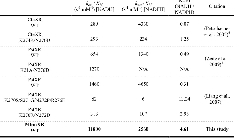
Figure 3-1: Xylose reductase cofactor specificity ... 50
Table 3-1: Relative cofactor consumption ... 51
Table 3-2: MbmXR kinetics ... 51
Table 3-3: MbmXR compared to engineered XRs ... 51
Figure 4-1: KARI reaction and substrates ... 59
Figure 4-2: Class I KARI topology ... 59
Figure 4-3: Annotated KARI sequence alignment ... 64
Figure 4-4: Specificity loop alignment ... 68
Figure 4-5: β2αB-loops of KARIs ... 68
Figure 4-6: Dehydroxylation of IpoHA ... 71
Figure 4-7: IaKARI Cα-Cα distances ... 74
Figure 4-8: Bending of the α1-helix ... 75
Figure 4-10: Rearrangemetns in IaKARI ... 76
Figure 4-11: Rearrangements in AvKARI/SeKARI ... 79
Figure 4-12: Rearrangements in EcKARI ... 79
Table 4-1: KARI class and specificity loop length ... 60
Table 4-2: Crystal parameters for KARIs ... 63
Table 4-3: List of KARI crystal structures ... 64
Figure 5-1: Cartoon of KARI classes ... 87
Figure 5-2: 2Ia_KARI schematics ... 92
Figure 5-3: EcKARI symmetry deviations ... 92
Figure 5-4: Gel filtration analysis ... 94
Figure 5-5: Dilution of 2Ia_KARI-DD ... 95
Figure 5-6: Structures of class I and II KARIs ... 98
Figure 5-7: Backbone deviation localization ... 100
Figure 5-8: Backbone deviation plot ... 101
Figure 5-9: Two-state α5+1-helix ... 102
Table 5-1: Kinetics of 2Ia_KARIs ... 93
Table 5-2: Cofactor specificity of 2Ia_KARIs ... 94
Table 5-3: Crystal parameters for 2Ia_KARI-DD ... 99
Figure 6-1: Adenine N6 and surrounding residues ... 111
Figure 6-2: DmADH pH activity profiles ... 121
Figure 6-3: Growth rates ... 122
Figure 6-4: Structures of EcFucOs ... 125
Table 6-1: Expression and screening of enzymes ... 113
Table 6-2: Enzymes tested ... 119
Table 6-3: Kinetics of enzymes tested ... 120
Table 6-4: Thermostability of enzymes tested ... 121
Table 6-5: Crystal parameters for EcFucOM185C ... 124
Figure 7-1: CSR-SALAD framework ... 138
Figure 7-2: Structural classifications ... 141
Table 7-1: Expression, lysis and assay of enzymes ... 145
Table 7-2: Mutant recapitulation summary ... 147
Table 7-3: Enzymes tested ... 149
Table 7-4: Kinetics of enzymes tested ... 150
Figure 8-1: Atom labels for cofactors ... 163
Figure 8-2: Components of x’y’z’-space ... 175
Table 8-1: Mixed base-pair notations ... 163
Table 8-2: Structural classification details ... 169
Table 8-3: Key atoms for pseudocenter calculations ... 171
Table 8-4: Atom pairs foc v calculation ... 175
Table 8-5: Codons for NADP-to-NAD switching ... 176
Table 8-6: Codons for NAD-to-NADP switching ... 178
Table S1-1: Analysis of KARI MSA ... 187
Table S1-2: KARI sequence identities ... 188
Supplementary Material S1-1: Citations for Tables 1-1 and 1-2 ... 183
Table S2-1: Table 2-1 with errors ... 190
Supplementary Material S2-1: Python script ... 189
Figure S4-1: PaKARIapo modeling error ... 192
Table S4-1: AvKARI sequence identities ... 191
Table S4-2: Amino acid numberings ... 191
Figure S5-1: 2Ia_KARI-DD crystal packing ... 193
Figure S5-2: 2Ia_KARI sequence alignment ... 194
Table S7-1: NADP-to-NAD mutant recapitulation ... 195
Section I
Chapter 1: Reversing cofactor preference in the
ketol-acid reductoisomerase enzyme family
Chapter 2: Uncovering rare NAD-preferring ketol-acid
reductoisomerases
C h a p t e r 1
REVERSING COFACTOR PREFERENCE IN THE KETOL-ACID REDUCTOISOMERASE ENZYME FAMILY
Material from this chapter appears in Brinkmann-Chen S., Flock T., Cahn J. K. B., Snow C. D., Brustad E. M. McIntosh J. A., Meinhold P., Zhang L., Arnold F. H. (2013). General approach to reversing ketol-acid reductoisomerase cofactor dependence from NADPH to NADH,
Proceedings of the National Academy of Sciences 110(27), 10946-10951, and is reprinted by permission from the National Academy of Sciences.
Abstract
To date, efforts to switch the cofactor specificity of oxidoreductases from nicotinamide adenine dinucleotide phosphate (NADP) to nicotinamide adenine dinucleotide (NAD) have been made on a case-by-case basis with varying degrees of success. Here we present a straightforward recipe for altering the cofactor specificity of one family of NADP-dependent oxidoreductases, the ketol-acid reductoisomerases (KARIs). Combining previous results for an engineered NAD-dependent variant of Escherichia coli KARI with available KARI crystal structures and a comprehensive KARI-sequence alignment, we identified key cofactor specificity determinants and used this information to construct five KARIs with reversed cofactor preference. Additional directed evolution generated two enzymes having NAD-dependent catalytic efficiencies that are greater than the wild-type enzymes with NADP. High-resolution structures of a wild-type/variant pair reveal the molecular basis of the cofactor switch.
Introduction
hydroxyl of the adenosine ribose, generally more than 15 Å from the chemically active carbon of the nicotinamide. This phosphate has no effect on the reduction potential of the molecule, yet the vast majority of enzymes display a strict specificity for one cofactor or the other. This allows the cell to partition metabolic reactions into two groups for separate regulation, but can pose an engineering hurdle.
For purposes of in vitro catalysis NADPH (the reduced form of NADP) is 16 times more expensive than NADH and has a shorter shelf life.1,2 Even more importantly, for the
purposes of in vivo biocatalysis, NADP-dependent enzymes cannot be used under the anaerobic conditions favored for industrial biotechnology because in many organisms NADPH is only produced during aerobic metabolism.3,4 As bioengineering and metabolic
engineering have developed as fields, cofactor specificity has proven a common engineering hurdle. Because of this, NAD(P)-binding enzymes have been the subject of many studies attempting to understand the molecular determinants of cofactor specificity and to engineer specificity reversal (Tables 1-1 and 1-2). However, despite the use of numerous approaches including combinatorial active-site saturation,5 computational
design,6 and homology guided design,7 no single method has developed which has been consistently successful in the reversal of cofactor specificity, and many of the published results have been only marginally successful.
Two factors contribute to the challenge of cofactor switching. First, the ubiquity and proposed ancient origin of NAD(P) – potentially dating back to the ‘RNA world’8 – have lead to the evolution of cofactor binding in diverse folds and orientations.9,10 Even
within single enzyme families, the binding of the cofactor molecule can use different geometries between homologues,11,12 and this structural diversity has limited the
development of general methods. Furthermore, a survey of the studies in Tables 1 and 1-2 shows that nearly all of these proteins have required multiple simultaneous mutations to achieve reversal of specificity;3 as a result many traditional protein engineering techniques, which operate by walking up a fitness landscape one beneficial mutation at a time, cannot be used because of the enormously non-additive nature of the specificity fitness landscape.
converting (S)-2-acetolactate (S2AL) or 2-aceto-2-hydroxybutyrate to (R )-2,3-dihydroxy-isovalerate (RDHIV) and (R)-2,3-dihydroxy-3-methylvalerate, respectively,13 essential intermediates in the biosynthesis of branched-chain amino acids (BCAAs).14,15 The demand
for these essential amino acids, used in the preparation of animal feed, human dietary supplements, and pharmaceuticals, is currently estimated to exceed 1,500 tons per year.16 In addition, the BCAA pathway has been engineered to produce fine chemicals and biofuels, including 1-butanol and isobutanol.17,18 Under the anaerobic conditions preferred for
large-scale fermentations, biosynthesis of BCAAs and other products that use this pathway is limited by the pathway’s cofactor imbalance and reduced cellular production of NADPH.3,19 One approach to overcoming the cofactor imbalance is to engineer KARI to
use NADH generated in glycolysis, thereby enabling anaerobic production of BCAA pathway products.3,19
The three prior reports of cofactor-switched KARIs from two different organisms show few commonalities in terms of approach or even residues targeted for engineering.3,19,20 A general recipe for switching KARI cofactor specificity would allow
metabolic engineers to take advantage of the natural sequence diversity of the KARI family and the concomitant diversity in properties such as expression level, pH tolerance, or thermal stability. By combining a systematic analysis of all Swiss-Prot-annotated21 KARIs,
information from previous work in the Arnold Lab on switching the cofactor specificity of the Escherichia coli KARI,3 and available KARI structures, we have identified a subset of
Figure 1-1. The nicotinamide cofactors, nicotinamide adenine dinucleotide (here shown in its reduced (hydride) form (NADH)), and nicotnimamide adenine dinucleotide phosphate (NADP). The differences between the molecules are highlighted in magenta, illustrating the spatial and covalent distance between the 2’ recognition element (the phosphate of NADP) and the chemically relevant element (the hydride).
N NH2 O O HO OH O P O OH O P O OH O O HO OH N N N N NH2
Nicotinamide adenine dinucleotide hydride (NADH)
H H N NH2 O O HO OH O P O OH O P O OH O O HO O N N N N NH2
Nicotinamide adenine dinucleotide phosphate (NADP)
P
O OH
Table 1-1. Previous reports of NADP-to-NAD cofactor specificity reversal. Structures followed by (h) are homology models, while those followed by another PDB accession code use the cofactor from that protein and (m) denotes a structure of a mutant protein. For citation information, see Supplementary Material S1-1.
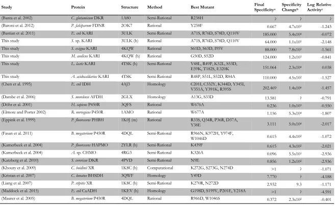
Study Protein Structure Method Best Mutant Final Specificitya Specificity Changeb Log Relative Activityc
(Banta et al. 2002) C. glutamicum DKR 1A80 Semi-Rational R238H ? ? ?
(Baroni et al. 2012) P. falciparum FDNR 2OK7 Rational Y258F 0.667 4.7x101 -1.243
(Bastian et al. 2011) E. coli KARI 3ULK Semi-Rational A71S, R76D, S78D, Q110V 185.000 5.4x10⁴ -0.072
This study S. sp. KARI 3ULK (h) Rational A71S, R76D, S78D, Q110V 64.000 1.1x10⁵ -2.148
This study S. exigua KARI 4KQW Rational S61D, S63D, I95V 88.000 7.8x10³ -1.561
This study M. aeolicus KARI 4KQW (h) Rational G50D, S52D 124.000 1.2x10³ -0.841
This study L. lactis KARI 4TSK (h) Semi-Rational V48L, R49P, K52L, S53D,
E59K, T182S, E320K 151.064 2.3x10⁴ 0.038 This study A. acidocaldarius KARI 4TSK Semi-Rational R48P, S51L, S52D, R84A 110.000 4.5x10² -1.527 (Chen et al. 1995) E. coli IDH 4AJ3 Homology C201I, C332Y, K344D, Y345I,
V351A, Y391K, R395S 202.469 1.4x10⁶ -1.457
(Dambe et al. 2006) S. morelense AFDH 2GLX Homology A13G, S33D 13.581 ? -0.791
(Döhr et al. 2001) H. sapiens P450R 3QFS Rational W676A 0.236 1.0x10³ -0.550
(Elmore and Porter 2002) R. norvegicus P450R 1AMO Rational W677A 1.156 5.3x10⁴ -1.807
(Eppink et al. 1999) P. fluorescens PHBH 1K0J (m) Rational R33S, Q34R, P36R, D37A,
Y38E 3.111 5.0x10⁴ -2.017
(Fasan et al. 2011) B. megaterium P450R 4DQL Semi-Rational R966N, K972H, Y974F,
W1046D 0.615 4.4x10² -1.072
(Kamerbeek et al. 2004) P. fluorescens HAPMO 2YLR (h) Semi-Rational K439F 0.615 4.3x10² -2.021
(Kamerbeek et al. 2004) A. sp. CHMO 4RG3 Semi-Rational K326A 0.096 5.5x101 -2.936
(Katzberg et al. 2010) S. cerevisae DKR 4PVD Semi-Rational N9E 0.856 1.2x10² -2.936
(Khoury et al. 2009) C. boidinii XR 1K8C (h) Computational K272G, S273G, N274D >1 ? -1.071
(Kristan et al. 2007) C. lunatus BHSDH 3QWF Homology Y49D 7.770 ? -4.188
(Liang et al. 2007) P. stipitis XR 1K8C (h) Semi-Rational K270R, N272D 2.932 9.3 -1.171
(Maddock et al. 2015) E. coli CaADH 1KEV (h) Homology G198D, S199V, P201E, Y218A >1 ? -4.591
Table 1-1. continued
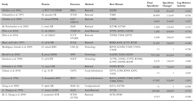
Study Protein Structure Method Best Mutant Final Specificitya Specificity Changeb Log Relative Activityc
(Medina et al. 2001) A. PCC7119 FDNR 2BSA Rational S223D 0.120 8.1x10³ -5.352
(Nakanishi et al. 1997) M. musculus CR 1CYD Rational T38D 30.909 1.3x10³ -0.510
(Paladini et al. 2009) P. sativum FDNR 4AF7
(1QGA) Rational Y308S 0.015 3.3x10² -1.627
(B. Petschacher et al. 2005) C. tenuis XR 1K8C Rational K274R, N276D 1.249 1.9x101 -1.170
(Pick et al. 2014) E. coli AdhZ3 1YQD (h) Semi-Rational S199N, S200N, N201D 1.245 6.9x101 -0.754 (Pick et al. 2014) E. coli AdhZ2 1UUF
(1YQD) Rational T205D, T206I, S207N 2.188 3.8x101 0.000
(Rane and Calvo 1997) E. coli KARI 3ULK Rational R68D, K69L, K75V, R76D 31.435 5.8x10⁴ -0.548 (Rodriguez-Arnedo et al. 2005) H. volcanii IDH 1AI2 (h) Homology R291S, K343D, Y344I, V350A,
Y390P >1 ? -0.768
(Rosell et al. 2003) R. perezi ADH8 1P0F Homology G223D, T224I, H225N 7571.333 8.1x10⁴ 0.551 (Scrutton et al. 1990) E. coli GTR 1GET Homology A179G, A183G, V197E, R198M,
K199F, H200D, R204P 8.133 1.8x10⁴ -1.482
(Shiraishi et al. 1998) N. crassa CbR Rational S920D, R932S 65.000 7.2x10⁴ -2.626
(Takase et al. 2014) S. sp. A1-R 3AFN Loop Substitution H37N, G38S, R39H, K40V,
A41D >1 ? -3.241
(Yaoi et al. 1996) T. thermophilus IDH 2D1C Loop Substitution R231A, K283D, Y284I, N287G,
V288I, I290A 67.692 5.2x10⁴ -1.051
(Zeng et al. 2009) P. stipitis XR 1K8C (h) Computational K21A, N272D >1 ? -0.098
(L. Zhang et al. 1999) V. harveyi ALDH 1EZ0 Semi-Rational T175E 129.545 4.8x10³ -0.763
(R. Z. Zhang et al. 2009) C. parapsilosis SCR 3CTM
(1CYD) Rational S67D, P69D 0.313 4.6 -0.180
Catalytic efficiency, CE, is given as kcat/KMwhen available, or vmax/KM otherwise. aFinal specificity is defined as 𝑪𝑬
𝒎𝒖𝒕
𝑵𝑨𝑫 𝑪𝑬
𝒎𝒖𝒕
𝑵𝑨𝑫𝑷
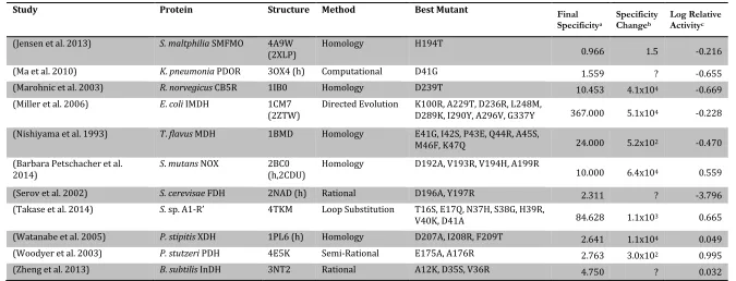
Table 1-2. Previous reports of NAD-to-NADP cofactor specificity reversal. Structures followed by (h) are homology models, while those followed by another PDB accession code use the cofactor from that protein and (m) denotes a structure of a mutant protein. For citation information, see Supplementary Material S1-1.
Study Protein Structure Method Best Mutant Final
Specificitya
Specificity Changeb
Log Relative Activityc
(Ashida et al. 2004) S. sp AlaDH 2VHW (h) Rational D198A 13.846 3.4x10⁴ -‐0.963 (Bernard et al. 1995) L. delbruckii LDH 1J49 Rational D175A 1.043 4.4x101 -‐0.962 (Bocanegra et al. 1993) E.coli DHLDH 4JQ9
(1GEU) Homology E205V, M206R, F207K, D208H, P212R >1 ? 0.668 (Bubner et al. 2008) P. fluorescens M2DH 1M2W Rational E68K, D69A 18.667 4.5x10³ 0.766 (Capone et al. 2011) C. symbiosum GDH 1BGV
(4XGI) Rational F238S, P262S 0.323 5.8x101 -‐3.323 (Chen et al. 1996) T. thermophilus IMDH 2ZTW Loop Substitution N/A 1000.000 8.7x10⁴ 0.187 (Clermont et al. 1993) B. stearothermophilus
GAPDH 3CMC Rational D32A, L187A, P188S 1.556 ? -‐1.699
(Cui et al. 2015) G. oxydans Gox2181 3AWD
(2WDZ) Computational Q20R, D43S 1.457 ? 0.073
(Ehrensberger et al. 2006) G. oxydans XDH 1ZEM Rational D38S, M39R >1 ? 0.699 (Ehsani et al. 2009) S. cerevisae BDH 2D8A (h) Homology E221S, I222R, A223S >1 ? 0.059 (Feeney et al. 1990) B. stearothermophilus
LDH 1LDN Rational D53S 0.152 3.4 -‐1.313
(Friesen et al. 1996) P. mevalonii HMG-‐
CoAR 4I4B Rational D146A, L148K 0.135 7.6x10⁴ -‐3.683
(Galkin et al. 1997) T. intermedius LuDH 1LEH
(h,1BW9) Homology D203A, I204R, D210R 74.286 ? -‐1.636 (Gul-‐Karaguler et al. 2001) C. methylica FDH 2FSS
(2NAD) Rational D195S 0.024 6.1x10³ -‐1.479
(Hoelsch et al. 2013) M. vaccae FDH 2GSD (h) Rational C145S, A198G, D221Q, C225V 13.254 ? ? (Holmberg et al. 1999) B. stearothermophilus
LDH 1LDN Homology I51K, D52S 2.200 4.9x101 -‐1.582
Table 1-2. continued
Study Protein Structure Method Best Mutant Final
Specificitya
Specificity Changeb
Log Relative Activityc
(Jensen et al. 2013) S. maltphilia SMFMO 4A9W
(2XLP) Homology H194T 0.966 1.5 -‐0.216
(Ma et al. 2010) K. pneumonia PDOR 3OX4 (h) Computational D41G 1.559 ? -‐0.655 (Marohnic et al. 2003) R. norvegicus CB5R 1IB0 Homology D239T 10.453 4.1x10⁴ -‐0.669 (Miller et al. 2006) E. coli IMDH 1CM7
(2ZTW) Directed Evolution K100R, A229T, D236R, L248M, D289K, I290Y, A296V, G337Y 367.000 5.1x10⁴ -‐0.228 (Nishiyama et al. 1993) T. flavus MDH 1BMD Homology E41G, I42S, P43E, Q44R, A45S,
M46F, K47Q 24.000 5.2x10² -‐0.470 (Barbara Petschacher et al.
2014) S. mutans NOX 2BC0 (h,2CDU) Homology D192A, V193R, V194H, A199R 10.000 6.4x10⁴ 0.559 (Serov et al. 2002) S. cerevisae FDH 2NAD (h) Rational D196A, Y197R 2.311 ? -‐3.796 (Takase et al. 2014) S. sp. A1-‐R’ 4TKM Loop Substitution T16S, E17Q, N37H, S38G, H39R,
V40K, D41A 84.628 1.1x10³ 0.665
(Watanabe et al. 2005) P. stipitis XDH 1PL6 (h) Homology D207A, I208R, F209T 2.641 1.1x10⁴ 0.049 (Woodyer et al. 2003) P. stutzeri PDH 4E5K Semi-‐Rational E175A, A176R 2.763 3.0x10² 0.995 (Zheng et al. 2013) B. subtilis InDH 3NT2 Rational A12K, D35S, V36R 4.750 ? 0.032
Catalytic efficiency, CE, is given as kcat/KMwhen available, or vmax/KM otherwise. aFinal specificity is defined as 𝑪𝑬
𝒎𝒖𝒕
𝑵𝑨𝑫 𝑪𝑬
𝒎𝒖𝒕
𝑵𝑨𝑫𝑷
Materials and Methods
Cloning and library construction
The genes encoding S. exigua SeKARI, L. lactis LlKARI, and Shewanella sp. ShKARI were obtained from DNA2.0. The genes encoding M. aeolicus MaKARI and A. acidocaldarius AaKARI were obtained as gBlocks from Integrated DNA Technologies. For each gene, the gBlocks were assembled via PCR, using T7 promoter and terminator primers and Phusion polymerase following the manufacturer’s instructions (Thermo Scientific). Site-saturation mutagenesis libraries were made by splicing by overlap extension PCR, as described.3,22 Error-prone PCR was performed according to a published protocol,23 using commercial T7 promoter and terminator primers. All KARIs and libraries
were cloned into pET22(b)+, using NdeI and XhoI in frame with the C-terminal his-tag for expression in E. coli. Heterologous protein expression, high-throughput expression, and purification were conducted as described.3
Kinetic assays and high-throughput screening
For the high-throughput assays, E. coli cells were lysed with 100 mM potassium phosphate at pH 7, 750 mg/L lysozyme, and 10 mg/L DNaseI. KARI activities were then assayed by monitoring NAD(P)H consumption in the presence of S2AL at 340 nm in a plate reader. The assay buffer contained 100 mM potassium phosphate at pH 7, 1 mM DTT, 200 µM NAD(P)H, 12.5 mM S2AL for LlKARI and 2.5 mM for the other KARIs, and 10 mM MgCl2. The LlKARI error-prone PCR library was screened at 5 mM S2AL.
The EcIlvC6E6 library was screened at 1 mM S2AL. The SeKARIDD library was screened at
100 µM NADH and 2.5 mM S2AL.
KARI sequence alignment
Manually annotated and reviewed sequence data for ketol-acid reductoisomerases (E.C. 1.1.1.86) were retrieved from the UniProt Database.24 Clustal Omega25,26 was used to
Crystallization and data collection
N-hydroxy-N-isopropyloxamate was prepared as described.29 High-throughput screening of crystallization conditions for SeKARI and SeKARIDDV was conducted at the
Beckman Molecular Observatory at the California Institute of Technology. For SeKARI with NADPH, the best condition was an unbuffered 0.2 M di-ammonium tartrate solution containing 20% (wt/vol) polyethylene glycol (PEG) 3350 as precipitant. For SeKARIDDV with NADH and N-hydroxy-N-isopropyloxamate as inhibitor, the best condition was an unbuffered 0.1 M potassium thiocyanate solution with 30% (wt/vol) PEG monomethyl ether 2000 as precipitant. The crystals were soaked in Fomblin oil for cryoprotection before flash-freezing in liquid nitrogen. Diffraction data were collected using a Dectris Pilatus 6M detector on beamline 12–2 at the Stanford Synchrotron Radiation Laboratory at 100 K. Diffraction datasets were integrated with XDS30 and scaled using SCALA.31
Structure determination and refinement
For SeKARI, the structure of Pseudomonas aeruginosa KARI (PDB code 1NP332)
was used as for molecular replacement. A multiblock refinement was applied dividing the model in six subparts according to secondary structure elements (residues 1–202, 203–228, 229–252, 253–278, 279–308, and 309–337) to allow automated standard refinement with Phenix (CCP4 suite). Refinement was conducted by iterating automatic refinement with Refmac5 (CCP4 suite) and manual refinement using Coot.33 We used the refined wild-type
structure as a model for molecular replacement to obtain the structure for SeKARIDDV. After placement of the inhibitor, several iterations of automated refinement with Refmac5 and manual refinement in Coot were performed. The structures were submitted to the protein database as PDB 4KQW (SeKARI) and PDB 4KQX (SeKARIDDV).
Results and Discussion
In previous work, Bastian et al. described E. coli KARI variant EcIlvC6E6 with four
mutations (A71S, R76D, S78D, and Q110V) that resulted in a 54,000-fold reversal in cofactor specificity for NAD over NADP.3 This variant was also highly active when using
NAD (85% of wild-type activity using NADP). Structural analysis of wild-type EcIlvC with and without bound cofactor (PDB codes 3ULK and 1YRL34,35) showed that three of
2-strand and the αB-helix of the Rossmann fold,36 herein referred to as the β2αB-loop. R76
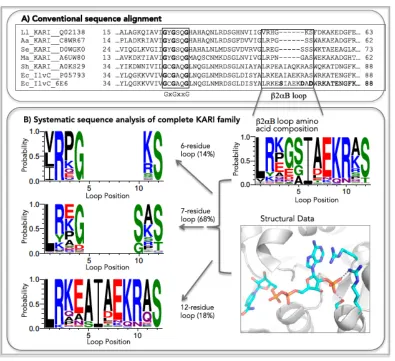
and S78 are in direct contact with the 2′-phosphate of NADP. The existence of β2αB-loops of varying lengths obscured sequence patterns of NADP specificity in KARI multiple sequence alignments.13,19,20,37 To find the commonalities among KARI β2αB-loops, we
systematically analyzed the loop regions of the entire enzyme class. We generated a multiple sequence alignment of all 643 Swiss-Prot-annotated21 KARI sequences (Table S1-1) and used structural data to identify and refine the β2αB-loop region in the alignment. An excerpt (Figure 1-2a) of the alignment of a few representative KARIs shows the well-known, conserved GxGxxG motif38 and, 18 amino acids downstream, the β2αB-loop,
which is diverse in both length and amino acid sequence.
On the basis of the sequence alignment alone, the diversity of this region might seem to argue against a conserved function for loop residues (Figure 1-2a). However, analysis of the 643 KARI sequences shows three different loop lengths: six (14%), seven (68%), and 12 residues (18%) (Fig. 1-2b). On subalignment of KARIs according to loop length, common conservation patterns emerge: a positively charged residue (73% arginine and 9% lysine) usually appears at the N-terminal end of the β2αB loop, and small, polar residues such as serine (85%) or threonine (11%) predominate at the C-terminal end. The last two residues in the six-residue loops are conserved (lysine/arginine and serine), and the last three residues follow the pattern SxS in seven-residue and RxS in 12-residue loops.
Comparison of the β2αB-loop segment of all available KARI structures (seven structures from four different organisms32,34,37,39-41) provides an explanation for the trends
found in the sequence alignment. Three of the seven structures have NADP cocrystallized, and in all those cases, the small polar C-terminal residues interact with the NADP 2′ -phosphate. The conserved N-terminal arginine is homologous to R68 in EcIlvC6E6, which has recently been shown to form a cation–pi interaction with the NADP adenine ring34 and
Figure 1-2. Excerpt of a multiple sequence alignment (a) of L. lactis KARI (LlKARI, UniProt ID Q02138), A. acidocaldarius KARI (AaKARI, C8WR67), S. exigua KARI (SeKARI, D0WGK0), M. aeolicus KARI (MaKARI, A6UW80), Shewanella sp. KARI (ShKARI, A0KS29), E. coli KARI (EcIlvC, P05793), and EcIlvC6E6. The GxGxxG motif is
[image:28.612.111.506.80.441.2]Transfer of a 12-residue cofactor switch solution to seven- and six-residue β2αB-loops
We examined the transferability of mutations previously reported for EcIlvC6E6, which has a 12-residue loop, to KARIs with shorter loops using SeKARI from S. exigua
(seven residues) and LlKARI from L. lactis (six residues) as models. On inspection of the sequence alignment in Figure 1-2a, we identified residues S61 and S63 of SeKARI as corresponding to residues R76 and S78 in EcIlvC (R76D and S78D in EcIlvC6E6). EcIlvC mutation A71S had no match in the SeKARI amino acid sequence because of the latter enzyme’s shorter β2αB-loop. Grafting both aspartates from EcIlvC6E6 into SeKARI resulted
in variant SeKARIDD (S61D and S63D) and a 7,800-fold reversal of cofactor specificity from NADPH to NADH. SeKARIDD (Table 1-3) had an eightfold decreased k
cat with
NADPH (from 0.8 to 0.1 s−1), whereas the kcat with NADH increased from 0.4 to 1.0 s−1.
The mutations increased the KM for NADPH 880-fold, but only 2.5-fold for NADH.
Overall, the catalytic efficiency for NADH remained the same, whereas catalytic efficiency for NADPH was reduced 7,300-fold to 0.11 mM−1s−1. That switching SeKARI cofactor
specificity could be achieved with only two mutations suggests that KARIs with seven-residue and 12-seven-residue β2αB-loops share similar cofactor specificity determinants involving interactions with small, polar residues at the end of the β2αB-loop.
LlKARI is a representative of the 14% of the 643 KARI sequences with a six-residue β2αB-loop. Whereas in 12- and seven-residue β2αB-loop KARIs the antepenultimate and ultimate residues are highly conserved, in six-loop KARIs the ultimate conserved serine is usually preceded by a positively charged residue at the penultimate position. We hypothesized that residues K52 and S53 of LlKARI were equivalent in function to residues R76 and S78 in EcIlvC. LlKARIDD variant with mutations K52D and
To develop a recipe for switching cofactor specificity in KARIs having a six-residue β2αB-loop, we generated single-site-saturation mutagenesis libraries at each of the six loop residues in LlKARI (V48, R49, H50, G51, K52, and S53), expecting that the
β2αB-loop also is key to specificity in this KARI family. On screening for activity with both cofactors, we found no single mutation that resulted in an NAD-preferring variant (rate of NADH consumption > rate of NADPH consumption at saturating substrate conditions). Mutations were identified at R49 (proline) and V48 (leucine) that increased activity with both cofactors. Saturation mutagenesis at H50, G51, K52, and S53 did not yield variants with improved NADH activity. Convinced that NADP specificity in KARIs with a six-residue β2αB loop is conveyed in a similar manner as in KARIs with seven- and 12-residue loops, we built a dual-site library by saturation mutagenesis at Lys52 and Ser53
while also incorporating the V48L and R49P mutations. Screening this library to ∼80%
coverage, we identified two variants, LlKARILPLD (mutations V48L, R49P, K52L, and
S53D) and LlKARILPED (mutations V48L, R49P, K52E, and S53D), with 46-fold and 54-fold specificity for NADH over NADPH (Table 1-3), representing specificity shifts of 6,600 (LlKARILPLD) and 7,700 (LlKARILPED).
These cofactor-switched KARIs with six-residue loops contained mutations to four of the six loop residues, suggesting that KARIs with six-residue loops are slightly more difficult templates for engineering the cofactor switch. Although mutation of the conserved arginine at the beginning of the β2αB-loop was not required to switch cofactor specificity in seven- and 12-loop KARIs, variant LlKARILRLD with proline reverted to arginine showed that the arginine contributes to NADP cofactor preference in six-residue loop KARIs. Reversion of P49 to arginine not only reduced the cofactor KM values (twofold for NADH
and threefold for NADPH, Table 1-3) but also decreased the kcat on NADH (fourfold),
thereby reducing the 46-fold preference for NADH over NADPH to only sixfold. Reversion of L48 to valine (LlKARIVPLD) lowered the catalytic efficiency on NADH
threefold (because of a reduction in kcat), whereas the NADPH KM was reduced twofold.
2′-phosphate than in the seven- and 12-residue loop KARIs, possibly because of its closer packing and proximity to the cofactor. The six-amino acid loop KARI structure with cofactor that we describe in Chapter 4 will help explain these differences.
Recovering catalytic activity of cofactor-switched enzymes
Shifting cofactor preference often decreased the overall activity (i.e., catalytic efficiency using NADH) relative to the wild-type enzyme using NADPH in our KARIs, which is also true for other cofactor-switched enzymes.20,42-44 To demonstrate that the activity of the cofactor-switched variants can be improved to match or even surpass the wild-type enzyme, we randomly mutated SeKARIDD, LlKARILPLD, and EcIlvC6E6 and screened for higher total activity in cell lysate. For SeKARI, we identified variant SeKARIDDV with mutation I95V.
Interestingly, this mutation corresponds to Q110V, which was previously found in EcIlvC6E6, and is speculated to confer general activation by optimizing cofactor orientation
for catalysis. With LlKARI, we isolated variant LlKARI2G6, which contained three new mutations: E59K, T182S, and E320K. These mutations effectively restored the enzyme to
wild-type levels of activity; the catalytic efficiency of LlKARI2G6 with NADH was ∼10%
higher than wild-type LlKARI with NADPH (71 vs. 65 mM−1s−1). The EcIlvC6E6 random
mutant library yielded variant EcIlvCP2D1-A1, also with three additional mutations (D146G, G185R, and K433E) and an approximately twofold greater catalytic efficiency on NAD than the wild-type on NADP (Table 1-3). The random mutations in EcIlvCP2D1-A1 and
Table 1-3. Biochemical properties of EcIlvc (12 residues), ShKARI (12 residues), SeKARI (seven residues), MaKARI (seven residues), LlKARI (six residues), AaKARI (six residues), and variants. T50 values are given for some enzymes.
KM for cofactors [µM] kcat for cofactors [s-1] kcat/KM [mM-1s-1] NADH/NADPH
of kcat/KM
Enzyme Mutations T50 [°]a NADH NADPH NADH NADPH NADH NADPH
EcIlvC -- 44.0 ± 0.1 1,075 ± 370 41 ± 3 0.3 ± 0.0 3.6 ± 0.4 0.3 ± 0.1 88 ± 11 0.003 ± 0.001
EcIlvC6E6 A71S, R76D, S78D, Q110V 43.7 ± 0.2 30 ± 6 650 ± 80 2.3 ± 0.2 0.20 ± 0.02 74 ± 15 0.40 ± 0.05 185 ± 50
EcIlvCP2D1-A1 A71S, R76D, S78D, Q110V, D146G, G185R, K433E 41.3 ± 0.2 26 ± 1 > 1,400 4.3 ± 0.3 0.54 ± 0.20 165 ± 22 < 0.4 > 412 ± 162
ShKARI -- 415 ± 44 1.0 ± 0.1 1.1 ± 0.1 4.5 ± 0.1 2.6 ± 0.3 4,500 ± 450 0.0006 ± 0.0001
ShKARIDD R76D, S78D 90 ± 24 > 1,000 1.30 ± 0.01 0.10 ± 0.02 14 ± 4 0.12 ± 0.02 > 119 ± 46
ShKARI6E6 A71S, R76D, S78D, Q110V 75 ± 10 600 ± 130 2.40 ± 0.01 0.30 ± 0.05 32 ± 4 0.5 ± 0.1 73 ± 23
SeKARI -- 51.3 ± 0.2 45 ± 10 1.0 ± 0.1 0.41 ± 0.02 0.8 ± 0.1 9 ± 2 800 ± 100 0.01 ± 0.00
SeKARIDD S61D, S63D 44 ± 1 113 ± 4 880 ± 523 0.97 ± 0.01 0.10 ± 0.01 9 ± 1 0.11 ± 0.07 78 ± 49
SeKARIDDV S61D, S63D, I95V 47 ± 15 > 1000 1.01 ± 0.01 0.25 ± 0.04 22 ± 7 0.25 ± 0.04 87 ± 32
bMaKARI -- 59 ± 4 17.3 ± 0.2 0.3 ± 0.0 0.7 ± 0.1 4.6 ± 0.3 43 ± 6 0.12 ± 0.02
bMaKARIDD G50D, S52D 26 ± 2 80 ± 9 0.15 ± 0.01 0.004 ± 0.001 6.2 ± 0.6 0.05 ± 0.01 124 ± 24
LlKARI -- 50 ± 1 285 ± 30 13 ± 1 0.10 ± 0.01 0.8 ± 0.1 0.43 ± 0.06 65 ± 11 0.007 ± 0.002
LlKARILPLD V48L, R49P, K52L, S53D 46.5 ± 0.3 108 ± 9 1,000 ± 100 0.40 ± 0.01 0.08 ± 0.01 3.7 ± 0.3 0.08 ± 0.01 46 ± 8
LlKARILRLD V48L, K52L, S53D 60 ± 8 306 ± 70 0.09 ± 0.00 0.09 ± 0.00 1.7 ± 0.2 0.30 ± 0.07 6 ± 1
LlKARIVPLD R49P, K52L, S53D 105 ± 7 447 ± 91 0.13 ± 0.01 0.07 ± 0.00 1.2 ± 0.1 0.15 ± 0.03 8 ± 2
LlKARILPED V48L, R49P, K52E, S53D 128 ± 9 1,180 ± 280 0.35 ± 0.12 0.06 ± 0.01 2.7 ± 0.9 0.05 ± 0.01 54 ± 26
LlKARI2G6 V48L, R49P, K52L, S53D, E59K, T182S, E320K 15 ± 4 749 ± 95 1.01 ± 0.03 0.35 ± 0.08 70 ± 19 0.47 ± 0.12 153 ± 57
bAaKARI -- 28 ± 2 18.0 ± 0.2 0.26 ± 0.01 0.66 ± 0.01 9 ± 1 37 ±1 0.24 ± 0.02
bAaKARIPLD R48P, S51L, S52D 43 ± 6 > 1,000 0.03 ± 0.00 0.013 ± 0.0004 0.7 ± 0.1 < 0.013 > 54 ± 20 bAaKARIPLDA R48P, S51L, S52D, R84A 27 ± 3 > 1,000 0.03 ± 0.00 0.01 ± 0.00 1.1 ± 0.2 < 0.01 > 110 ± 20 bAaKARILS Loop switch (LS) of LlKARILPLD to AaKARI:
R48P, P49H, S51L, S52D
46 ± 6 > 1,000 0.03 ± 0.00 0.009 ± 0.006 0.6 ± 0.1 < 0.01 > 55 ± 8
Mutations are given relative to each wild-type sequence. Each value represents the average of three independent measurements. Mutations located within the β2αB-loop are highlighted in bold.
aHalf-denaturation temperature (T
50) determination: 30 µL aliquots of purified enzyme were transferred to PCR tubes. Each tube was assigned a specific incubation temperature on the
block of an Eppendorf master cycler PCR machine. The measurements were conducted in duplicates. The tubes were incubated in their slots for 15 min, and the reactions were quenched on ice. Residual activity was determined with the activity assay. T50 is defined as the termperature at which 50% of the initial activity is retained after 15 min incubation. aMaKARI, AaKARI, and their variants show cooperative behavior, and their kinetics follow the Hill equation with a Hill coefficient of 2.0 instead of the Michaelis-Menten equation:
Cofactor switch guide for the KARI enzyme family
We propose the following guide for switching KARI cofactor specificity, which does not require a priori knowledge of the KARI structure (Figure 1-3). The first step is the identification of the β2αB-loop and its length via sequence alignment against the KARIs reported in this work or a multiple sequence alignment of KARIs. If the target KARI has a 12- or seven-residue loop, replacement of the last and third-to-last residue of the loop with aspartates is likely to achieve a switch in cofactor specificity. In the case of the six-residue loop, a modified approach is required. The last polar loop residue should be mutated to aspartate, and the conserved charged residue near the N-terminus of the loop should be mutated to proline. Simultaneously, the penultimate loop residue should be targeted for site-saturation mutagenesis. This approach led to reversed cofactor preference in both test cases. Last, to achieve wild-type-like activity for NAD, additional mutations that fine-tune cofactor orientation, as exemplified by Q110V or I95V, may be introduced, as will be discussed further in Chapter 6. Additional enhancement of activity can be achieved by random mutagenesis and screening.
Application of the cofactor switch guide
We tested the proposed protocol on three additional KARIs representing the β2α B-loop lengths (twelve, seven, and six residues) and composition. With the addition of these three KARIs, we covered the different phylogenetic subbranches of the KARI enzyme family. Representative of 12-residue β2αB-loop KARIs was ShKARI from Shewanella sp. Methanococcus aeolicus MaKARI45 exemplifies the seven-residue type, and AaKARI from
Alicyclobacillus acidocaldarius has a six-residue loop (Table 1-3). We also used this last enzyme to test the transferability of the Q110/I95 position by making and screening a site-saturation library at position R84.
By introducing a customized set of two to four mutations based on the guide in Figure 1-3, we obtained variants with the desired cofactor specificity for KARI family members sharing as little as 20% sequence identity (Table S1-2). Low catalytic efficiency in cofactor-switched variants can be remedied by directed evolution, as demonstrated for EcIlvCP2D1-A1 and LlKARI2G6. Mutations (alanine or valine) at positions corresponding to
Molecular determinants of cofactor specificity in KARIs
We solved the crystal structures of SeKARI wild-type enzyme (1.39 Å, PDB 4KQW) and variant SeKARIDDV (1.8 Å, PDB 4KQX) with their respective cofactors. The
crystallographic parameters are summarized in Table 1-4. These structures confirm that only the β2αB-loop is involved in interactions with the respective 2′-moiety, and thus is responsible for specificity (Figure 1-5). In the wild-type structure, three residues form direct interactions with the 2′-phosphate of NADP: R58, S61, and S63. The structures support the suggested dual role of R58: the positively charged guanidinium moiety is 3.5 Å from the adenine moiety of the cofactor in both structures, forming cation–pi stacking interactions,46 as reported for E. coli KARI.34 At the same time, this side chain could form a salt bridge to
the negatively charged 2′-phosphate of NADP (possibly also involving the oxygen of the phosphoester bond) and a hydrogen bond to the 2′-hydroxyl of NAD.
The residues that were mutated to alter cofactor preference, S61 and S63, are in a position to hydrogen bond directly to at least a single oxygen atom of the phosphate. The high-resolution structures revealed water molecules surrounding the 2′-phosphate group, enabling additional, indirect interactions with the side chains of R58, S61, and S63 (Figure 1-5). The side chain of S62 stabilizes this network of water molecules, as does the R58 backbone. In the mutant structure, the β2αB-loop is moved slightly closer toward the cofactor. Mutations S61D and S63D would interrupt the serine hydrogen bonding interactions and also result in electrostatic repulsion to the 2′-phosphate of NADP. The carboxyl groups of the two aspartates compensate for the missing 2′-phosphate by filling the pocket and substituting its negative charge. In addition, the side-chain carboxylate moiety of S61D is at an ideal distance for hydrogen bonding to the 2′-hydroxyl group of NAD. As in the wild-type structure, water molecules link the 2′-hydroxyl moiety of the ribose sugar with R58 and D63 by hydrogen bonds. All other interactions of SeKARIDDV with the cofactor, for
instance, involving the GxGxxG motif, remain the same. The remaining loop residues L57, E59, and G60 are not involved in binding the cofactor.
moiety to shift about 1 Å toward the side chain of residue 95. This compensates exactly the distance the β2αB-loop is reoriented inward in the mutant structure at position R58 and preserves the cation–pi stacking of the adenine moiety and the side chain of R58, which is in the same rotamer conformation in the wild-type and in the cofactor-switched mutant. We propose that this movement compensates for the slightly different conformations of NADP and NAD and readjusts the catalytically active nicotinamide moiety of NAD to take on a more favorable position for electron transfer. A similar activating mechanism is speculated for EcIlvC6E6’s Q110V mutation.3
Conclusions: General cofactor binding principles for KARIs
Table 1-4. Crystallographic parameters of wild-type SeKARI and Variant SeKARIDDV.
SeKARI SeKARIDDV
Protein Data bank code 4KQW 4KQX
Space group P 1211 P 212121
Monomers per asymmetric unit 2 2
Water molecules per monomer, n 443 140
Cocrystallized cofactor NADPH NADH
Cocrystallized compounds L-(+)-tartarate N-hydroxy-N-isopropyloxamate
Unit-cell parameters (Å) a = 52.205 a = 49.985 b = 118.934 b = 105.346
c = 62.158 c = 122.357
α = 90.00 α = 90.00
β = 101.17 β = 90.00
γ = 90.00 γ = 90.00
Resolution range (Å) 38.81-1.39 79.83-1.80
Number of unique reflections 130,511 78,635
Rwork 0.1595 0.2014
Rfree 0.1866 0.2520
Refinement parameters RMSD from ideal
Bond (Å) 0.0293 0.0210
Angle (°) 2.7444 2.1084
Chirality 0.1858 0.1499
Ramachandran parameters
Favored 95.85% 94.33%
Allowed 4.15% 5.51%
Outliers 0.00% 0.15%
Figure 1-5. Crystal structures of SeKARI wild-type enzyme with cocrystallized NADP (left, cyan) and variant SeKARIDDV with cocrystallized NAD (right, green). The β2αB-loop is
highlighted, and side chains involved in defining cofactor-specificity are shown as sticks. Introduced mutations (S61D, S63D, and I95V) are shown with red labels. In SeKARIDDV,
References
1. Flock, T. (2012) Investigation of Cofactor Specificity-Determining Amino Acid Residues of Ketol-Acid Reductoisomerases: Structure-Guided Evolution to Swith the Nicotinamide Cofactor Preference. M.S., California Institute of Technology
2. Woodyer, R., van der Donk, W. A., and Zhao, H. M. (2003) Relaxing the nicotinamide cofactor specificity of phosphite dehydrogenase by rational design. Biochemistry42, 11604-11614
3. Bastian, S., Liu, X., Meyerowitz, J. T., Snow, C. D., Chen, M. M. Y., and Arnold, F. H. (2011) Engineered ketol-acid reductoisomerase and alcohol dehydrogenase enable anaerobic 2-methylpropan-1-ol production at theoretical yield in Escherichia coli. Metabolic Engineering13, 345-352
4. Verho, R., Londesborough, J., Penttila, M., and Richard, P. (2003) Engineering redox cofactor regeneration for improved pentose fermentation in Saccharomyces cerevisiae. Appied and Environmental Microbiology69, 5892-5897
5. Liang, L., Zhang, J. Q., and Lin, Z. L. (2007) Altering coenzyme specificity of Pichia stipitis xylose reductase by the semi-rational approach CASTing. Microbial Cell Factories6
6. Khoury, G. A., Fazelinia, H., Chin, J. W., Pantazes, R. J., Cirino, P. C., and Maranas, C. D. (2009) Computational design of Candida boidinii xylose reductase for altered cofactor specificity. Protein Science18, 2125-2138
7. Rosell, A., Valencia, E., Ochoa, W. F., Fita, I., Pares, X., and Farres, J. (2003) Complete reversal of coenzyme specificity by concerted mutation of three consecutive residues in alcohol dehydrogenase. Journal of Biological Chemistry 278, 40573-40580
8. Denessiouk, K. A., Rantanen, V. V., and Johnson, M. S. (2001) Adenine recognition: a motif present in ATP-, CoA-, NAD-, NADP-, and FAD-dependent proteins. Proteins44, 282-291
9. Carugo, O., and Argos, P. (1997) NADP-dependent enzymes. 1: Conserved stereochemistry of cofactor binding. Proteins: Structure, Function, and Genetics28, 10-28
10. Bellamacina, C. R. (1996) The nicotinamide dinucleotide binding motif: A comparison of nucleotide binding proteins. FASEB Journal 10, 1257-1269
11. Cahn, J. K. B., Brinkmann-Chen, S., Spatzal, T., Wiig, J. A., Buller, A. R., Einsle, O., Hu, Y., Ribbe, M. W., and Arnold, F. H. (2015) Cofactor specificity motifs and the induced fit mechanism in class I ketol-acid reductoisomerases. Biochemical Journal468, 475-484
13. Dumas, R., Biou, V., Halgand, F., Douce, R., and Duggleby, R. G. (2001) Enzymology, structure, and dynamics of acetohydroxy acid isomeroreductase. Accounts of Chemical Research34, 399-408
14. Arfin, S. M., and Umbarger, H. E. (1969) Purification and properties of acetohydroxy acid isomeroreductase of Salmonella typhimurium. Journal of Biological Chemistry244, 1118-1127
15. Umbarger, H. E., and Davis, B. (1960). The Bacteria3, 167
16. Park, J. H., and Lee, S. Y. (2010) Fermentative production of branched chain amino acids: A focus on metabolic engineering. Applied Microbiology and Biotechnology 85, 491-506
17. Atsumi, S., Cann, A. F., Connor, M. R., Shen, C. R., Smith, K. M., Brynildsen, M. P., Chou, K. J. Y., Hanai, T., and Liao, J. C. (2008) Metabolic engineering of Escherichia coli for 1-butanol production. Metabolic Engineering10, 305-311 18. Atsumi, S., Hanai, T., and Liao, J. C. (2008) Non-fermentative pathways for
synthesis of branched-chain higher alcohols as biofuels. Nature451, 86-89
19. Hasegawa, S., Uematsu, K., Natsuma, Y., Suda, M., Hiraga, K., Jojima, T., Inui, M., and Yukawa, H. (2012) Improvement of the redox balance increases L-valine production by Corynebacterium glutamicum under oxygen deprivation conditions. Applied and Environmental Microbiology78, 865-875
20. Rane, M. J., and Calvo, K. C. (1997) Reversal of the nucleotide specificity of ketol acid reductoisomerase by site-directed mutagenesis identifies the NADPH binding site. Archives of Biochemistry and Biophysics338, 83-89
21. Boeckmann, B., Bairoch, A., Apweiler, R., Blatter, M. C., Estreicher, A., Gasteiger, E., Martin, M. J., Michoud, K., O'Donovan, C., Phan, I., Pilbout, S., and Schneider, M. (2003) The SWISS-PROT protein knowledgebase and its supplement TrEMBL in 2003. Nucleic Acids Research31, 365-370
22. Kunkel, T. A., Roberts, J. D., and Zakour, R. A. (1987) Rapid and efficient site-specific mutagenesis without phenotypic selection. Methods in Enzymology 154, 367-382
23. Bloom, J. D., Labthavikul, S. T., Otey, C. R., and Arnold, F. H. (2006) Protein stability promotes evolvability. Proceedings of the National Academy of Sciences 103, 5869-5874
24. Un Consortium (2012) Reorganizing the protein space at the Universal Protein Resource (UniProt). Nucleic Acids Research40, D71-D75
25. Goujon, M., McWilliam, H., Li, W. Z., Valentin, F., Squizzato, S., Paern, J., and Lopez, R. (2010) A new bioinformatics analysis tools framework at EMBL-EBI. Nucleic Acids Research38, W695-W699
27. Schneider, T. D., and Stephens, R. M. (1990) Sequence logos – A new way to display consensus sequences. Nucleic Acids Research18, 6097-6100
28. Crooks, G. E., Hon, G., Chandonia, J. M., and Brenner, S. E. (2004) WebLogo: A sequence logo generator. Genome Research14, 1188-1190
29. Aulabaugh, A., and Schloss, J. V. (1990) Oxalyl hydroxamates as reaction-intermediate analogs for ketol-acid reductoisomerase. Biochemistry29, 2824-2830 30. Kabsch, W. (2010) XDS. Acta Crystallographica Section D - Biological
Crystallography66, 125-132
31. Evans, P. (2006) Scaling and assessment of data quality. Acta Crystallographica Section D - Biological Crystallography62, 72-82
32. Ahn, H. J., Eom, S. J., Yoon, H. J., Lee, B. I., Cho, H. J., and Suh, S. W. (2003) Crystal structure of class I acetohydroxy acid isomeroreductase from Pseudomonas aeruginosa. Journal of Molecular Biology328, 505-515
33. Emsley, P., and Cowtan, K. (2004) Coot: Model-building tools for molecular graphics. Acta Crystallographica Section D - Biological Crystallography 60, 2126-2132
34. Wong, S.-H., Lonhienne, T. G. A., Winzor, D. J., Schenk, G., and Guddat, L. W. (2012) Bacterial and plant ketol-acid reductoisomerases have different mechanisms of induced fit during the catalytic cycle. Journal of Molecular Biology424, 168-179 35. Tyagi, R., Duquerroy, S., Navaza, J., Guddat, L. W., and Duggleby, R. G. (2005)
The crystal structure of a bacterial class II ketol-acid reductolsomerase: Domain conservation and evolution. Protein Science14, 3089-3100
36. Rossmann, M. G., Moras, D., and Olsen, K. W. (1974) Chemical and biological evolution of nucleotide-binding protein. Nature250, 194-199
37. Dumas, R., Curien, G., Derose, R. T., and Douce, R. (1993) Branched-chain-amino-acid biosynthesis in plants – Molecular cloning and characterization of the gene encoding acetohydroxy acid isomeroreductase (ketol-acid reductoisomerase) from Arabidopsis thaliana (thale cress). Biochemical Journal294, 821-828
38. Wierenga, R. K., De Maeyer, M. C. H., and Hol, W. G. J. (1985) Interaction of pyrophosphate moieties with α-helixes in dinucleotide-binding proteins. Biochemistry24, 1346-1357
39. Biou, V., Dumas, R., Cohen-Addad, C., Douce, R., Job, D., and Pebay-Peyroula, E. (1997) The crystal structure of plant acetohydroxy acid isomeroreductase complexed with NADPH, two magnesium ions and a herbicidal transition state analog determined at 1.65 angstrom resolution. EMBO Journal16, 3405-3415 40. Leung, E. W. W., and Guddat, L. W. (2009) Conformational changes in a plant
ketol-acid reductoisomerase upon Mg2+ and NADPH binding as revealed by two
crystal structures. Journal of Molecular Biology389, 167-182
42. Kristan, K., Stojan, J., Adamski, J., and Rizner, T. L. (2007) Rational design of novel mutants of fungal 17 β-hydroxy steroid dehydrogenase. Journal of Biotechnology129, 123-130
43. Petschacher, B., Leitgeb, S., Kavanagh, K. L., Wilson, D. K., and Nidetzky, B. (2005) The coenzyme specificity of Candida tenuis xylose reductase (AKR2B5) explored by site-directed mutagenesis and X-ray crystallography. Biochemical Journal