Directed Evolution of Cytochrome P450 for
Small Alkane Hydroxylation
Thesis by
Mike Ming Yu Chen
In Partial Fulfillment of the Requirements
for the Degree of
Doctor of Philosophy
California Institute of Technology
Pasadena, California
2011
© 2011 Mike Ming Yu Chen
ACKNOWLEDGEMENTS
It has been a privilege to be a part of the Arnold lab at Caltech for the last six years. I want to thank my advisor, France Arnold, for providing a great work environment and allowing me the freedom to learn so many new techniques and test new ideas. I am grateful to the members of my committee, Mark Davis, John Bercaw, and Jay Labinger, for advice throughout this process. I am also grateful to the National Science Foundation for the graduate research fellowship that supported my graduate work.
Since the very first days of being a member of the Arnold lab, I have been fortunate to be around a group of wonderful researchers and helpful labmates who are far too numerous for me to name. I particularly owe many thanks to Matt Peters, Peter Meinhold, Michelle Meyer, and Marco Landwehr for teaching me the basics of molecular biology and protein engineering. I am also grateful to the many collaborators that I had the fortune to work with, Rudi Fasan, Daniel Koch, Andrew Sawayama, Chris Snow, Christina Vizcarra, Jorge Rodriguez, Jared Lewis, and Pedro Coelho, amongst many others.
ABSTRACT
Methane is an ideal alternative to petroleum refining as a chemical feedstock source since it is highly abundant an inexpensive. However, the lack of selective methane oxidation catalysts has limited such utilization. Starting from cytochrome P450 CYP102A1 (BM3) from Bacillus megaterium, which prefers C12-C20 fatty acids as its substrates, I investigated several protein engineering approaches to shift the enzyme’s substrate specificity toward small gaseous alkanes, with the ultimate goal of methane. By continuing previous directed evolution efforts in our group, a variant with wild-type-like affinity and catalytic efficiency for propane, P450PMO, was isolated. To alleviate the loss of protein thermostability (~ 10 oC) as a result of this approach, mutations were targeted to the BM3 active site with site saturation mutagenesis, targeted mutagenesis with a reduced set of amino acids, and computationally guided library designs. From these enzyme libraries, variants were identified that replicated much of the P450PMO activities with a minimal number of mutations while maintaining wild-type thermostability.
THESIS SUMMARY
Selective hydroxylation of small alkanes is a long-standing problem for which few practical catalysts are available. The lack of catalysts that can efficiently convert gaseous alkanes into transportable liquid commodities has been a barrier to broader utilization of these resources. In particular, methane, the principal component of natural gas, is an ideal alternative to petroleum as a chemical feedstock source since it is highly abundant and inexpensive. Currently, methane is converted to methanol via an energy intensive, endothermic, and costly process that first converts methane into synthesis gas, followed by methanol synthesis from this intermediate. This process is economically feasible only on a large scale, which in combination with general transportation limitations of a gas commodity prevent methane recovery from many sources.
The selective oxidation of small alkanes is difficult because of the inertness of the alkane C-H bond, which requires highly reactive radical or ionic species to cleave. However, as the desired partial oxidation products, alcohol and aldehyde, have weaker C-H bonds compared to the alkane, they are susceptible to further oxidation to CO2. While this transformation has been achieved only by a limited set of transition-metal-based catalyst systems, a variety of alkane hydroxylases found in alkanotrophic microorganisms support selective alkane oxidation at ambient conditions using oxygen as the oxidant. Chapter 1 of this thesis provides an introduction to enzymatic alkane oxidation by monooxygenases, highlighting the structure and mechanisms of three major enzyme classes, methane monooxygenases (MMOs), non-heme (di-iron) monooxygenases, and cytochrome P450s (P450s).
step in hydrocarbon metabolism. Unfortunately, since the majority of these hydroxylases function as a part of a larger enzyme complex and are membrane associated, their potential for industrial applications is limited. For these reasons, we have been engineering well-expressed, soluble, bacterial P450s, in particular CYP102A1 (BM 3) isolated from Bacillus megaterium, for small alkane hydroxylation.
Chapter 2 describes the continuation of laboratory evolution efforts aimed at converting BM3 into a small alkane hydroxylase, starting from variant 35E11. As a result of the previous ten rounds of mutagenesis and screening, variant 35E11 displayed a significantly lower thermostability (T50 = – 11.6 oC) compared to the wild-type enzyme. To reverse this loss in thermostability, which is known to reduce the ability of a protein to acquire beneficial mutations that are destabilizing, known stabilizing mutations from a P450 peroxygenase were grafted onto variant 35E11 singly and in combination. The resulting thermostablized variant was subjected to a domain-based protein-engineering strategy (developed by Rudi Fasan), in which the three domains of BM3 were mutated individually using both random and site-saturation mutagenesis. Beneficial mutations identified through high-throughput screening for dimethyl ether demethylation were verified to improve propane and ethane hydroxylation in the context of the holoenzymes. Using this strategy, re-specialization of BM3 for propane hydroxylation was achieved with variant P450PMO, a proficient P450 propane monooxygenase. This variant displays substrate affinity and coupling of cofactor consumption rivaling those of the natural P450s with their preferred substrates. In addition, we were able to demonstrate in vivo propane hydroxylation using these BM3 variants in resting E. coli cells reaching activities surpassing those reported for natural alkane hydroxylases acting on their preferred substrates.
acids and two structure-based computational library design approaches, we identified variants supporting both propane and ethane hydroxylation. Although, none of the obtained variants reached the level of specialization that was previously obtained with P450PMO, the range of obtained propane TON and coupling of cofactor consumption corresponds to those values of generalist intermediates of P450PMO lineage obtained after 10 – 12 rounds of mutagenesis and screening. These results suggest semi-rational library design can be an effective strategy to move away from a specialist enzyme toward generalist enzymes, but functional specialization still requires optimization through several rounds of random mutagenesis and screening.
terminal hydroxylation. These results demonstrated the usefulness of this in vivo selection system, which could be generally applied to directed evolution of enzymes for small alkane hydroxylation.
To apply selection pressure for the main goal of this research, selective hydroxylation of ethane and methane, we developed a high-throughput screen to directly assay for P450 alkane hydroxylation, described in Chapter 5. With the use of a pressurizable 96-well reactor, the P450 alkane hydroxylation reaction was conducted in high throughput and the alcohol product was quantified spectroscopically by a coupled enzyme assay. Applying this screen to BM3 variants generated in our laboratory, we identified variant E31 as the best candidate for further engineering, since it displayed both the highest activity in the screen and wild-type-like thermostability. Subsequent rounds of site-saturation and random mutagenesis resulted in improved variants demonstrating the efficacy of the screen. However, none of the identified BM3 variants were able to produce ethanol or methanol in whole-cell alkane bioconversions using growth-arrested E. coli cells. In contrast, CYP153A6, a natural terminal alkane hydroxylase, was able to produce ethanol in whole-cell alkane bioconversions. The inability of BM3 variants to produce ethanol in vivo reflects their poor affinity for ethane and indicates they still lag behind a natural P450 alkane hydroxylase in terminal hydroxylation of small alkanes.
reactions. Using iodosylbenzene, 3-chloroperoxybenzoic acid, and hydrogen peroxide as oxidants, we investigated the ability of the compound I of five P450s (BM3, P450PMO, P450cam, CYP153A6, and CYP153A6-BMO1) to hydroxylate alkanes ranging from methane to octane. From these terminal oxidant-supported P450 reactions, we found the compound I of CYP153A6, and CYP153A6 BMO-1 to be able to break the methane C-H bond using PhIO as the oxidant. This demonstrates both the feasibility of P450 methane oxidation and the use of terminal oxidant-supported P450 reactions as an assay to investigate the compatibility of P450 active sites for small alkane oxidation. By chemically generating the active radical, we eliminated the requirement for substrate binding to initiate P450 catalysis, which enabled us to determine the innate substrate range of each P450 active site.
TABLE OF CONTENTS
Acknowledgements iii
Abstract iv
Thesis Summary v
Table of Contents xii
Figures and Tables xiii
Abbreviations xvi
Chapters
Chapter 1 Introduction: enzymatic alkane oxidation by monooxygenases 1 Chapter 2 Engineered alkane-hydroxylating cytochrome P450 BM3
exhibiting native-like catalytic properties 48 Chapter 3 Active site engineering of P450 BM3 for
small alkane hydroxylation 65
Chapter 4 In vivo evolution of butane oxidation by AlkB and
CYP153A6terminal alkane hydroxylases 95
Chapter 5 Directed evolution of P450 BM3 for ethane hydroxylation 118 Chapter 6 P450 alkane hydroxylation using terminal oxidants 141 Chapter 7 Panel of cytochrome P450 BM3variants to produce
drug metabolites and diversify lead compounds 160
Chapter 8 Materials and methods 181
Appendix
Appendix A Sequence and activities of cytochrome P450 BM3 variants 216 Appendix B Corbit and CRAM algorithm and evaluation of mutations 223 Appendix C Candidate high-throughput screens for small alkane hydroxylation 229 Appendix D Chapter 6 supplemental material 233 Appendix E Variant selection for production of drug metabolites
FIGURES AND TABLES
Figure 1.1 The crystal structure of pMMO 7
Figure 1.2 The crystal structure and mechanism of sMMO 10 Figure 1.3 The crystal structure and mechanism of P450s 20
Figure 2.1 Outline of the domain engineering strategy 52
Table 2.1 Thermostablized variants of 35E11 53
Table 2.2 In vitro propane oxidation activities of representative BM3 variants 55 Figure 2.2 Mapping of the activity-enhancing reductase domain mutations 57 Figure 2.3 Whole-cell biotransformation of propane 58 Table 2.3 In vivo propane oxidation activities of P450 BM3 variants 59 Figure 2.4 Propanol profile during P450 biotransformation of propane 60
Figure 3.1 Structure of the BM3 active site highlighting mutagenesis targets 70 Table 3.1 Active site mutagenesis library designs and properties 71 Figure 3.2 DME activity profiles of active site mutagenesis libraries 75 Figure 3.3 Histogram of propane and ethane hydroxylating variants identified
from active site mutagenesis libraries and correlation of alkane
hydroxylation activity with DME demethylation activity 78 Figure 3.4 Amino acid distribution of propane hydroxylating variants from the
CRAM library 81
Figure 3.5 Structural alignment of BM3 with BM3-A328V 88
Figure 4.1 Growth of P. putida GPo12(pGEc47B) with primary and secondary
linear alcohols 100
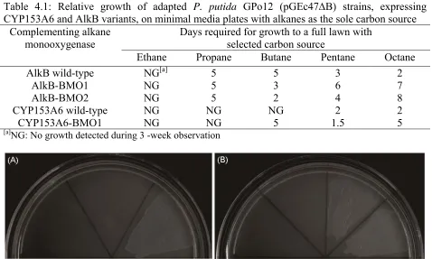
Table 4.1 Growth on alkanes of adapted P. putida GPo12 (pGEc47B) strains
expressing CYP153A6 and AlkB variants 104
Figure 4.4 Whole-cell bioconversions of resting E. coli BL21(DE3) cells
expressing CYP153A6 and AlkB variants 107
Figure 4.5 Mapping of beneficial mutation of CYP153A6 and AlkB
homology models 112
Figure 5.1 High-throughput alkane hydroxylation assay 123 Figure 5.2 Comparison of ethanol quantification by GC-FID
with enzymatic colorimetric assay 124
Table 5.1 High-throughput ethane screening results for selected variants 126 Figure 5.3 Ethane hydroxylation validation with a monoclonal 96-well plate 128 Table 5.2 Ethane TON of select variants as cell-free extract and purified enzyme 129 Figure 5.4 Whole-cell propane bioconversion of select P450 variants 134
Figure 6.1 Reaction scheme for terminal oxidant-supported
P450 alkane hydroxylation 145
Table 6.1 Alkane hydroxylation by P450s utilizing terminal oxidants 148
Figure 6.2 Alkane induced spin-shift of A6 153
Table 6.2 A6 kinetic parameters for alkane hydroxylation 155
Table 7.1 Verapamil metabolites generated by human P450s and BM3 variants 167 Table 7.2 Astemizole metabolites generated by human P450s and BM3 variants 169 Table 7.3 LY294002 metabolites generated by human P450s and BM3 variants 171 Table 7.4 Substrate hydrophobicity preference of BM3 variants 173 Table 7.5 Production of astemizole metabolites by 9-10A F87L variants 174
Table 8.1 CRAM and Corbitlibrary designs 192
Figure A.1 Nucleotide sequence of full-length, wild-type cytochrome P450 BM 3 218
Figure A.2 Amino acid sequence of full-length, wild-type cytochrome P450 BM 3 219
Table A.1 Sequence and activities of BM 3 variants identified from
active site mutagenesis libraries 220
Table B.1 Frequency table for the most stable 20,000 sequences
as determined by Corbit 225
Table B.2 Repulsive van der Waal energy as determined by ROSETTA 227 Figure C.1 Colorimetric screen for chloromethane dehalogenation 230 Figure C.2 High-throughput methanol oxidation screen 232 Figure D.1 GC/MS-SIM chromatogram of 12C and 13C methanol
calibration standards 234
Figure D.2 GC/MS-SIM chromatogram of PhIO-supported 12C-methane reactions 235 Figure D.3 GC/MS-SIM chromatogram of PhIO-supported A6 methane
reactions with 12C-and 13C-methane 235
Figure D.4 GC/MS-SIM chromatogram of terminal oxidant-supported
A6 methane reactions with 16O-and 18O-water 236 Figure D.5 UV/Vis spectra of purified FdrA6 and FdxA6 237 Figure D.6 Co-factor consumption in the presence and absence of octane at
varying concentrations of FdrA6 and FdxA6 238 Figure D.7 Michaelis-Menten plots of initial rate for A6 hydroxylation of
hexane, octane, ethane, iodomethane, and d3-iodomethane 239 Figure D.8 UV/Vis difference spectra of alkane induced spin-shift of A6 240 Table E.1 Identity of engineered P450 BM3 variant panel: enzyme family,
name, sequence, number of mutations from closest wildtype parent 242 Table E.2 Amino acid sequence of blocks 1 – 8 of the cytochrome P450 chimeras 246 Table E.3 Complete list of active enzymes and their metabolite distributions
with verapamil 247
Table E.4 Complete list of active enzymes and their metabolite distributions
with astemizole 248
Table E.5 Complete list of active enzymes and their metabolite distributions
ABBREVIATIONS
MMO Methane monooxygenase M. c. Bath Methyloccus capsulatus Bath M. t. OB3b Methylosinus trichorium OB3b E. coli Escherichia coli
BM3 Cytochrome P450 BM3 (CYP102A1)
A6 CYP153A6
CAM CYP101
PMO P450PMO
ET Electron transfer
PCET Proton coupled electron transfer KIE Kinetic isotope effect
SRS Substrate recognition site TON Turnover number
EPPCR Error-prone polymerase chain reaction
SOEPCR Splicing by overlap extension polymerase chain reaction SSM Site-saturation mutagenesis
CAST(ing) Combinatorial active site saturation test
NADH Nicotinamide adenine dinucleotide, reduced form NAD+ Nicotinamide adenine dinucleotide, oxidized form
NADPH Nicotinamide adenine dinucleotide phosphate, reduced form NADP+ Nicotinamide adenine dinucleotide phosphate, oxidized form FAD Flavin adenine dinucleotide
FMN Flavin mononucleotide DME Dimethyl ether
Chapter 1
Introduction: Enzymatic Alkane Oxidation by Monooxygenases
A. Introduction
Petroleum and natural gas are the primary energy resources currently utilized to meet the world’s energy needs (1). In addition to its use as a fuel source, the conversion of crude oil to olefins and aromatics through refining has also allowed petroleum to act as a major feedstock for the chemical industry. This ability to generate chemical precursors—through processes such as cracking, dehydrogenation, and reforming—differentiates petroleum from natural gas, which has been limited to usage as a fuel. However, as the world’s known reserves of crude oil are shrinking (2), the need to find alternative sources for chemical feedstocks, such as natural gas, is becoming more pressing. This search for alternative feedstocks is also motivated by the environmental impact of petroleum refining. As the reactions to produce olefins and aromatics from petroleum are endothermic, CO2 is released during both the generation of these chemical precursors and in the subsequent partial oxidation steps to produce the desired oxygenated compounds (e.g., aldehydes, alcohols, carboxylic acids).
all these favorable factors, methane is still underutilized as a feedstock owing to a lack of economical and sustainable strategies for its selective oxidation (5).
The selective oxidation of methane to oxygenated products represents a significant challenge, as the methane C-H bond is extremely inert (105 kcal/mol) (6). Therefore, highly reactive radical or ionic species are required to cleave the methane C-H bond. However, as the desired partial oxidation products, methanol and formaldehyde, have weaker C-H bonds compared to methane, they are susceptible to further oxidation to CO2. To overcome these challenges, research toward partial methane oxidation and improved methane utilization has taken several different approaches: (1) the one-step oxidation of methane to methanol or formaldehyde, (2) oxidative and non-oxidative coupling of methane, (3) Fischer-Tropsch synthesis of hydrocarbons from synthesis gas (syngas), generated from steam reformation of methane. Currently, industrial conversion of methane to methanol falls into the latter category, utilizing an energy intensive, endothermic, and costly process to first convert methane into syngas, followed by methanol synthesis from this intermediate (1, 7). While there is a variety of mixed metal-oxide heterogeneous catalysts capable of the desired methane partial oxidation (8) and coupling reactions (9 – 10), these catalysts currently lack the reactivity and selectively necessary for commercialization (5).
the reactivity for the methane C-H bond is substantially greater than that of a product C-H bond, such as H-CH2OH or H-CH2SO4H (15). The mechanism of the Shilov systems occurs in three steps: (1) electrophilic activation of the R-H bond by Pt(II) to form a Pt(II)-alkyl intermediate, (2) oxidation of the Pt(II)-alkyl complex by [PtCl6]2- to give a Pt(IV)-alkyl species, (3) nucleophilic SN2 attack of water at Pt-C bond results in the formation of the alcohol product and regenerates the Pt(II) catalyst.
Advancement of the original system has been made by Periana et al., which has replaced the oxidant [PtCl6]2- with sulfuric acid (15). Using an Hg2+complex in sulfuric acid, a one-pass yield of 40% conversion of methane to methyl hydrogensulfate was obtained at > 90% selectivity (14). An improved system utilizing Pt(II) chelated by 2,2’-bipyrimidine, which is more thermodynamically robust, resulted in a one-pass yield of greater than 73% (15). While these yields are the highest reported for direct partial oxidation of methane, several key disadvantages have prevented commercialization: low turnover frequency (16), costly methanol recovery from concentrated sulfuric acid, and catalyst poisoning by water and oxidation products (5).
scaffolds. The synthesis and characterization of multiple di-iron FeIV=O complexes modeled after the Q intermediate of MMOs have been reported (19 – 22). To date, these complexes have been shown to activate C-H bonds as strong as 100 kcal/mol, however, the obtained reaction rates were much lower than those observed with metalloenzymes (20, 22).
B. Alkane Oxidizing Enzymes
B.1. Methane monooxygenases (MMOs)
While a catalyst that supports efficient conversion of methane to methanol has so far eluded transition metal chemistry, Nature found a solution to utilize methane as an energy source long ago. Methanotrophic bacteria found in a variety of environments including methane vents in the deep sea, gastrointestinal tracts of cows, and landfills are unique in their ability to utilize methane as their sole carbon and energy source (23). Methanotrophs, comprising 13 different genera within the and protobacteria (24), are defined by their expression of a methane monooxygenase (MMO) that directly converts methane to methanol. The methanol product is further oxidized to formaldehyde by a methanol dehydrogenase and is used both for biomass synthesis (23) and as a source of ATP through further oxidation reactions (23).
with ~ 4 M copper present, pMMO is expressed along with the developments of extensive, intracytoplasmic membranes (27 – 28).
B.2. pMMO
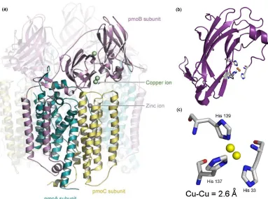
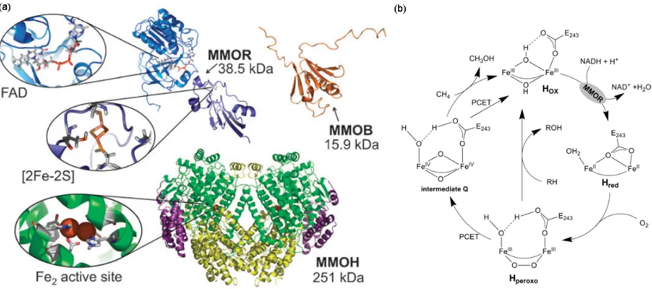
Figure 1.1: The pMMO (M. c. Bath) structure (pdb: 1YEW); (a) the full structure with one protomer highlighted, reproduced from ref 29; (b) the soluble domain pmoB; (c) the first coordination sphere of the dicopper metal center
pMMO has been shown to oxidize only alkanes and alkenes up to five carbons in length (38 – 39). Interestingly, for these multi-carbon substrates, sub-terminal oxidation at the C-2 position is preferred (40). Studies using chiral alkanes have given evidence to suggest the pMMO mechanism for oxygen insertion occurs in a concerted fashion rather than involving radical or cationic intermediates, as with sMMO or cytochrome P450s (41 – 42). In addition, an absence of a carbon kinetic isotope effect in the oxidation of propane also suggests little or no structural rearrangement occurs at the carbon center during the rate-limiting step (43). Unfortunately, attempts to determine the pMMO mechanism have been sparser compared to similar efforts with sMMO, and much of the mechanism is still not well understood.
B.3. sMMO
Due to both its unique ability to oxidize methane as well as its high substrate promiscuity, i.e., the ability to hydroxylate more than 50 different compounds including aromatics (17, 35), sMMO has been a favored target for research (39). sMMO has been purified from M. t. OB3b (44), M. c. Bath (45), and several other strains of methanotrophs (46). It belongs to the family of bacterial multi-component monooxygenases (BMMs) (EC.1.14.13.25), which includes toluene monooxygenase, phenol hydroxylase, and alkene monooxygenase (47), that enable their hosts to utilize a variety of hydrocarbons as their sole carbon and energy source (47 – 48). Using a common carboxylate-bridged di-iron center in their hydroxylase, BMMs are able to activate oxygen for formal insertion into the substrate C-H bond, which initiates the metabolism of these hydrocarbons.
shuttles electrons from the NADH cofactor to the MMOH active site, and a regulatory protein (MMOB), which is required for methane oxidation (17). The MMOH subunit consist of three polypeptides arranged as an 222 dimer, Figure 1.2 (a). The di-iron active site is embedded in a four-helix bundle and coordinated by four carboxylates and two imidazoles from two E(D/H)XXH binding motifs.
Figure 1.2: The sMMO structure and mechanism. (a) The structure of MMOH (pdb: 1MTY), MMOB (pdb: 1CKV), and MMOR (pdb: 1JQ4) with the cofactors highlighted, reproduced from ref 17. (b) The sMMO catalytic cycle, see text for details, (PCET: proton coupled electron transfer)
MMOH is only active in the presence of a protein cofactor, MMOB, which when complexed with MMOH changes its structure and reactivity. For example, MMOH from M. t. OB3b oxidizes alkanes and nitrobenzene to form secondary alcohols and m-nitrophenol products in the absence of MMOB (62). Upon MMOB addition, the product ratios shift such that mostly primary alcohols and p-nitrophenol are formed. In addition, MMOB must be present for efficient generation of MMOH intermediates in the reaction cycle, which suggests that binding of MMOB initiates the electron transfer (ET) and O2 binding steps (63 – 64). The presence of MMOB has been generally reported to enhance ET between MMOH and MMOR (63), but when chemically reduced MMOR was added to premixed solutions of MMOH and MMOB the same ET between MMOH and MMOR was inhibited (65). These apparently conflicting results have led investigators to suggest that slow structural changes associated with MMOB and MMOR binding to MMOH may result in hysteresis in MMOH activity (62). A current hypothesis is that the interaction of one hydroxylase component of MMOH with MMOR or MMOB could be dependent on the presence of MMOR or MMOB bound to the other component of MMOH (66). As a consequence of this dependence, the oxidative phase of the catalytic cycle may only occur at one of the two active sites at a time. This hypothesis has been experimentally verified by observing a ~ 50% maximal conversion of the initial di-iron (II) protein during reactions of MMOH with oxygen (53).
(68), and -halogenated primary alcohols (69) has revealed the presence of multiple hydrophobic substrate binding pockets that trace a contiguous pathway from the protein surface to the di-iron center. The entry of the substrate appears to pass through several such cavities in its path from aqueous solution to the enzyme’s active site (69). Finally, as many as eleven binding sites have been identified with Xe, which has similar polarity, water solubility, and van der Waals radius as methane. The binding of these surrogate substrates of methane did not induce significant side-chain displacement in the enzyme; therefore it appears that methane and other sMMO substrates are bound in pre-formed hydrophobic pockets.
hydrogen atom transfer mechanism, whereas those of Q are extensively non-classical and involve hydrogen atom tunneling.
This difference could be particularly important for methane oxidation, as methane is kinetically stable with a large barrier height for its oxidation. For the reaction with the Q intermediate, tunneling across this barrier could lead to progression along the reaction coordinate, whereas the reaction with the Hperoxo intermediate may not proceed due to absence of tunneling. While this explanation could resolve why sMMO homologs cannot activate methane while possessing nearly the same di-iron active site, unfortunately, KIE studies for the sMMO methane reaction which would determine if tunnel effects were present have yielded varied results. Under single-turnover conditions, KIE values of 23 to 50 have been reported (50, 70), which indicates proton tunneling in the transition state. However, under steady-state conditions, a KIE of only 1.7 was observed, when comparing Vmax (or kcat) values (70 – 71), which suggests an absence of tunneling.
B.4. Using methanotrophs/MMOs for methanol synthesis
While methanotrophs and MMOs have been focus of extensive research over the past decades, successful attempts to use either the organisms or enzymes for methanol synthesis have been sparse. The inability to express either pMMO or sMMO in a heterologous host severely limits their utilization in industrially relevant organisms as well as the ability to use standard molecular biology methods to engineer desired protein properties. In addition, the multi-component nature of MMOs is also a hindrance to evolving more active or more stable variants. One successful strategy for methanol biosynthesis using methanotrophs is to inhibit the downstream enzyme in methanol metabolism, methanol dehydrogenase (MDH). Using NaCl as a MDH inhibitor, 7.7 mM of methanol were accumulated in M. t. OB3b cultures after 20 hours (73). Optimization of the growth conditions as well as the addition of ethylene diamine tetra-acetic acid to further inhibit MDH resulted in 13.2 mM methanol accumulation after 12 h batch fermentations with an overall activity of 0.036 U/mg cell mass (1 U = 1 mol methanol/min). While this strategy is successful in producing methanol, significant yield improvements and reduction of the product loss to the natural methanol metabolism of the methanotroph host are hard to envision.
available crystal structures of MMOH have been solved in the absence of MMOB, which modulates the MMOH tertiary structure directly affecting both substrate access and the first coordination sphere of the diiron center. It is therefore questionable if the observed active site configurations reflect that of the active configuration during methane oxidation.
B.5. AlkB and non-heme di-iron alkane monooxygenases
Expanding the search for potential methane biocatalysts beyond MMOs, two other class of enzymes, non-heme di-iron alkane monooxygenases and cytochrome P450s, are also able to activate oxygen and perform O-atom insertion into inert alkane C-H bonds. The family of non-heme di-iron alkane hydroxylases has been identified in bacteria and fungi utilizing C5 – C16 n -alkanes as their sole carbon source (75). Exemplified by the most studied alkane hydroxylase isolated from Pseudomonas putida GPo1, the non-heme di-iron alkane hydroxylase is a three-component system consisting of (1) a soluble NADH-rubredoxin reductase (AlkT) (76), (2) a soluble rubredoxin (AlkG) (77), and (3) the integral membrane oxygenase (AlkB) (78 – 79). Although AlkB can be functionally expressed in Escherichia coli as lipoprotein vesicles, purification and maintenance of activity in the purified state is difficult, which has limited its mechanistic and structural analysis (80).
quantitatively oxidized back to its resting state by enzymatic turnover in the presence of substrate and oxygen (82). Further evidence for the similarities between the AlkB and sMMO mechanisms has been provided through studies with the use of norcarane as a chemical probe (83). From these studies, the AlkB reaction has been shown to be consistent with an oxygen-rebound mechanism via a substrate-centered radical, analogous to the proposed P450 and sMMO mechanisms, exhibiting limited rearranged products (83).
The ability to functionally express AlkB heterologously in E. coli certainly makes it a potentially better industrial biocatalyst compared to MMOs and also more amenable to enzyme engineering. However, the integral membrane nature of AlkB limits the enzyme’s expression to the available membrane surface area. In addition, the lack of a crystal structure and knowledge of both the second coordination sphere of the diiron center and the component interactions are significant hindrances to directed evolution efforts to shift the AlkB substrate range from C5-C16 alkanes to methane.
B.6. Cytochome P450s
Cytochrome P450s, which utilize a thiolate-ligated heme (iron protoporphyrin IX) prosthetic group in their active sites (84), represent an entirely different solution to diiron centers for catalytic oxygen insertion into C-H bonds. Unlike MMOs and non-heme diiron alkane hydroxylases, which are only found in methanotrophs and alkanotrophs, the superfamily of cytochrome P450s is one of the most prevalent enzyme families found across all three domains of life. To date, over 10,000 P450 enzymes have been identified (data source: http://drnelson.utmem.edu/CytochromeP450.html). P450s are involved in the metabolism of
functional groups to facilitate further metabolism or excretion. This defense mechanism is particularly prominent in plants, which require P450s to break down herbicides due to their immobile nature (85 – 86). This is exemplified by the presence of over 400 P450 genes in rice (87). In their other role, P450s are responsible for synthesis of a variety of steroid hormones and the conversion of polyunsaturated fatty acids to biologically active molecules implicated in development and homeostasis.
The defining reaction P450s is the reductive activation of molecular oxygen as it is one of the few oxygenases possessing the requisite “FeIV=O.+” state for alkane C-H bond activation. In this reaction, one oxygen atom is inserted into the substrate while the other is reduced to water. The overall equation for the reaction is RH + NAD(P)H + O2 + H+ ROH + NAD(P)+ + H2O, where RH is the substrate. In addition to this canonical reactivity, due to the existence of multiple oxidants in the P450 catalytic cycle, P450s can also catalyze epoxidation, dealkylation, sulfoxidations, desaturation, carbon-carbon bond scission, and carbon-carbon bond formation among other known reactivities (88 – 89).
(93), CYP174A1 (94), and CYP231A2 (95) as well as BM3 for its unique self-sufficiency and high catalytic rates (96 – 100).
B.7. P450 structure
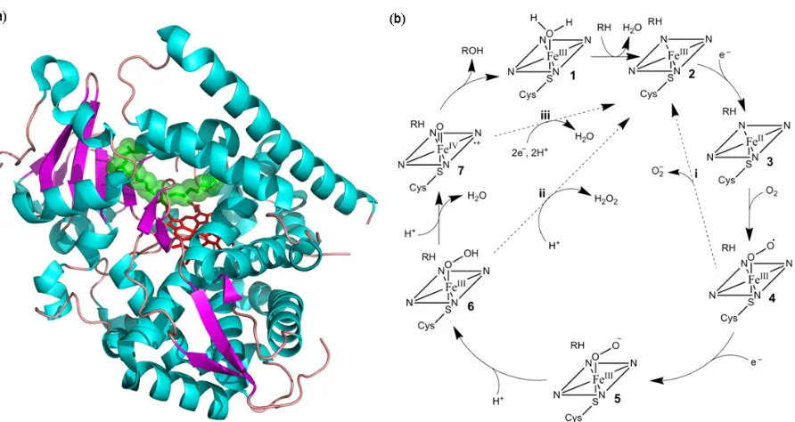
The overall P450 fold (Figure 1.3 (a)) is retained across the enzyme superfamily even though members can share less than 20% sequence identity (101). The core four-helix bundle composed of three parallel helices (D, L, and I) and the antiparallel E helix are conserved in all P450s (102). The prosthetic heme group is ligated to the absolutely conserved cysteine located on a loop containing a highly conserved FxxFx(H/R)xCxG binding motif. This thiolate ligation gives rise to the 450 nm Soret absorbance maximum for the ferrous-CO complex for which P450s were named (103). The other common feature among P450s is a kink at the center of the I helix, which contains the amino acid sequence (A/G)Gx(E/D)T that has been implicated in oxygen binding and protonation (104 – 105).
B.8. P450 catalytic mechanism
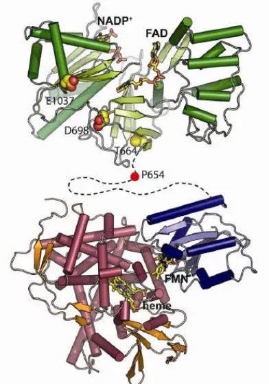
Figure 1.3: Cytochrome P450 structure and catalytic cycle; (a) the structure of the hydroxylase domain of CYP102A1 (BM3) with the heme shown in red and the substrate N-palmitoyl glycinesubstrate shown in green (pdb: 1JPZ); (b) the P450 catalytic cycle (see text for details)
The electrons for the reduction step of the P450 catalytic cycle are provided by either (a) cytochrome P450 reductase (CPR), a soluble flavoprotein with FAD and FMN prosthetic groups, or (b) an iron-sulfur protein that shuttles electrons from a flavoprotein with a single FMN prosthetic group, or (c) a P450 reductase-like domain fused to the P450 heme domain. In each case, the electron donor uncouples the two electrons provided by NAD(P)H and transfers them singly to the P450 enzyme. Since the final reducing agent for the catalytic cycle is NAD(P)H, which has a midpoint potential of -320 mV (115), the resting state of the heme iron with a midpoint potential of ca. -300 mV (116 – 117) is reduced slowly in the absence of substrate. The substrate binding event triggers a change in the spin state of the heme iron from LS to HS, which induces a positive shift of 100 to 300 mV in the heme reduction potential allowing for rapid electron transfer (118). This mechanism clearly acts as a safeguard against the unproductive consumption of NAD(P)H and the formation of superoxide and peroxides. This substrate-induced initiation of electron transfer represents a specific P450 regulatory mechanism and is a clear departure from the initiation of the MMO catalytic cycle through binding of MMOB.
position to amino acids capable of hydrogen-bonding interactions. The function of this proton delivery network is also dependent on substrate binding. For non-natural, poorly fitting substrates, their binding is insufficient to expel excess water from the active site, and protonation of the hydroperoxide anion (6) can occur at the proximal position, resulting in peroxide release. In fact, the uncoupling of proton and electron transfer does not even require a poorly fitting substrate; simply blocking the site of hydroxylation with fluoro-groups is sufficient to result in normal cofactor consumption with only water or peroxide production (121).
B.9. P450 substrate binding and substrate specificity
As mentioned previously, P450 substrate binding occurs by an “induced fit” model as proposed by Koshland (122) in which the enzyme accommodates different substrates in its active site by virtue of having a high level of flexibility to undergo appropriate conformational changes. Comparison of the X-ray structures of cytochrome P450s crystallized in substrate-free and substrate-bound forms (109) shows large structural rearrangements induced by substrate binding, which suggests that the SRSs are quite flexible and can provide a variety of substrates access to the heme. The absence of charged and hydrogen-bonding groups in the typical P450 substrate, as well as in the active sites of most P450 enzymes, requires such binding mechanisms as an alternative means to stabilize the substrate-enzyme complex. In many cases, different substrate analogues bind tightly to P450 enzymes simply due to their poor solubility in water rather than the presence of specific interactions with active site residues (123).
by the C-H bond strength, and (c) steric constraints imposed by the active site geometry. While the compatibility of a substrate within a P450 active site and steric constraints of binding modes are case specific, lipophilicity has been shown to be directly correlating to KM or Kd for sets of similarity structured compounds (124 – 125). The general preference of P450 oxidation occurs with the following order of C-H bonds: tertiary>secondary>primary, which was determined using several small molecular probes that minimized the effect of the P450 active site structure in controlling the site of oxidation (126). This preference is reinforced by DFT calculations for the activation barriers for hydrogen abstraction, which predict a similar reactivity preference: benzylic or allylic>tertiary >secondary>primary (127 – 128).
B.10. P450 reactions using terminal oxidants
In addition to the normal P450 “turnover” conditions utilizing oxygen and NAD(P)H, the P450 catalytic cycle can also be accessed through the branching/shunt pathways using a variety of terminal oxidants including hydrogen peroxide, alkyl peroxides, acyl peroxides, and iodosobenzene. Early studies with alkylperoxides provided evidence for the formation of a ferric alkylperoxo complex (FeIII-OOR) (129) as well as a compound II-like ferryl (FeIV=O) species, which is one oxidation equivalent higher than the resting ferric state (130 – 131) but little evidence for the formation of a CMP I-like ferryl porphyrin radical. However, recent works have confirmed the formation of both a compound-II – like and a compound-I – like species as transient intermediates (132 – 133).
to the intrinsically destructive nature of peroxides as well as the lack of acid-base catalytic residues in the P450 structure, with the exception of P450s which naturally utilize peroxides as their oxidant (134). In natural peroxidases, such as chloroperoxidase, the formation of the ferryl-oxo intermediate involves proton transfer from the proximal to the distal oxygen atom of the bound hydrogen peroxide, which is aided by a conserved His-Arg or His-Asp amino acid pair (135). P450s have highly hydrophobic active sites that lack these acid-base catalytic residues in close proximity to the oxygen binding pockets.
The peroxide-driven P450 reactions proceed through the formation of CMP 0, which after protonation and heterolytic O-O cleavage generates CMP I. In contrast, P450 reactions driven with iodosobenzene (PhIO) produce only CMP I as an oxidant without any potential involvement of peroxo-iron species, since PhIO is a single oxygen donor (136). The initial finding of solvent oxygen incorporation through experiments with 18O-labeled water in reactions supported by PhIO led researchers to question if the oxidation proceeded via a ferryl intermediate (137 – 138). However, subsequent work has shown that the oxygen of PhIO readily exchanges with the medium through a porphyrin-oxidant complex, [(Porp)FeIII-OIPh]+ (139). Nevertheless, whether PhIO-meditated reaction is a faithful mimic of the P450 reaction remains contentious due to differences observed in regio- and chemoselectivities (136) and kinetic isotope effects (140) between reactions supported by PhIO and NAD(P)H/O2 (141).
B.11. H-abstraction and mechanistic comparisons with MMOs
considering the energy requirements for breaking the inert alkane C-H bonds. In the consensus radical rebound mechanism for the O-atom insertion step, the ferryl oxygen initially abstracts a hydrogen from the substrate, leaving a carbon radical, which in turn recombines with the oxo radical coordinated to the iron atom (145). This mechanism is supported by observed large intramolecular isotope effects as well as the partial loss of stereo-chemistry at the carbon center for reactions with chemical probes (146 – 148).
orbitals), which results in four distinct reaction pathways with intermediates: 3TS, 3TS, 5TS and 5TS.
Regardless of the spin-state of the reaction pathway, the transition state for the H-abstraction step presents the largest barrier in the reactions involving CMP I, or Q (58, 127). For methane, this barrier height is 26.7 kcal/mol for P450 CMP I, which is significantly higher than the ca.19 kcal/mol barrier for known P450 substrate camphor. The barrier heights for other small gaseous alkanes, ethane (21.6 – 21.8 kcal/mol) and propane (terminal: 21.6 kcal/mol, subterminal: 19 kcal/mol) are also much lower than methane (127, 149 – 151). As a comparison, this transition state barrier for H-abstraction from a methane C-H bond has been calculated to be as low as 13.8 kcal/mol (60) and as high as 23.2 kcal/mol (152) for sMMO. While these calculations show the barrier heights for P450 hydroxylation of known substrates and sMMO hydroxylation of methane are comparable, it is difficult to draw meaningful conclusions for the potential of P450s to oxidize methane.
C. Cytochrome P450 Enzyme Engineering
The limiting factors toward broader utilization of wild-type P450s as biocatalysts include poor expression, being membrane-bound, and lacking known redox partner proteins. For these reasons, protein engineering efforts have been focused on expanding the substrate range of well-expressed bacterial P450s with known redox partners to accept desired target compounds. Much of this work has been focused on P450cam and BM3 (163). P450cam is a type I P450 requiring a putidaredoxin containing a [2Fe-2S] cluster and an FAD-containing putidaredoxin reductase for the highly stereoselective hydroxylation of its physiological substrate (1R)-camphor to (1R)-5-exo-hydroxycamphor. BM3, as mentioned previously, is a type II P450 with its FAD and FMN containing reductase fused to its heme domain as a single polypeptide chain (92). BM3’s native function is believed to be the detoxification of polyunsaturated fatty acids (164 – 165). Its unique domain architecture has been credited for its high hydroxylation and epoxidation rates (~ 17,000 min-1 (166)) on long-chain fatty acid substrates (92, 166). In addition, these two proteins were also the first P450s of which crystal structures in the presence (167 – 168) and absence of substrates (169 – 173) were solved, which has also aided protein engineering efforts.
C.1. P450cam
substrates has proved to be relatively easy, i.e., requiring just a few mutations, the resulting variants generally exhibited poor coupling between cofactor consumption and product formation, typically ranging from 5% to 32%.
Far fewer examples exist for P450cam variants with nearly wild-type coupling efficiency for non-native substrates, as these variants require multiple rounds of mutagenesis. For example, simply introducing a Y96F mutation will increase P450cam’s ability to epoxidize styrene 25-fold, with 32% coupling compared to only 7% coupling for the wild-type (186). Placing an additional V247L mutation in the active site increases the coupling efficiency to 60% (175). The best example for the ability of P450cam to be engineered to accept new substrates with high coupling efficiency was provided by Xu et al. (187) with the engineering of P450cam to hydroxylate short-chain alkanes. Over the course of multiple studies (176, 187 – 188), the authors gradually decreased the active site volume by incrementally introducing bulky, hydrophobic residues. A P450cam quadruple mutant, F87W/Y96F/T101L/V247L, was able to oxidize butane at 750 min-1 with 95% coupling compared to 4% for wild type (176). To achieve 85% coupling of product formation with NADH consumption for propane oxidation, five additional mutations, L244M/L294M/T185M/L1358P/G248A, were required (187). This variant with nine active site mutations was also able to oxidize ethane at 78.2 min-1 with 10.5 % coupling (187).
product formation, they fall short of the optimal free enzyme activity obtained at higher ratios of electron transfer components, i.e., 1: > 4 : > 12 (121).
C.2. CYP102A1 (P450BM3)
BM3 is one of the most studied and frequently engineered P450s, as it has the fastest known catalytic rate for a P450 and is a natural fusion protein with a type II reductase and its hydroxylase found on a single polypeptide (134). Its high rate of catalysis has been shown to be the result of BM3 domain architecture: when the heme and reductase domains are expressed independently and mixed together, the resulting activity is severely diminished (191). In addition to the self-sufficiency, BM3 can also be easily manipulated genetically and expressed at levels up to 1 g/L in laboratory strains of Escherichia coli (192). In contrast to P450cam, the BM3 protein backbone undergoes large structural changes (> 10 Å) during catalysis as revealed by differences between the substrate-free crystal structure (168) and structures of BM3 complexed with known substrates (168 – 169). The large active site volume reflects the poor regioselectivity with which BM3 hydroxylates its preferred C12-C20 fatty acids substrates (193). All these factors make BM3 well suited for directed evolution experiments and potential use as a biocatalyst.
/A264G) with activity toward fluoranthrene (177) and alkoxyresorufins (197) and a variant (R47L/Y51F/F87A/A264G) with activity toward pyrene (177). Screening with a simple NADPH consumption assay to monitor cofactor consumption in the presence of a substrate, directed evolution of BM3 yielded a highly promiscuous variant F87V/L188Q/A74G with enhanced activity for a variety of substrates such as indole, alkanes, arene, and polycyclic, aromatic hydrocarbons (198 – 200). Similar engineering efforts generated variants with activity for -ionone, a carotenoid intermediate (201), and valencene for (+)-nootkatone production (202).
From these rational engineering efforts, general structural function relationships have emerged, which have aided further engineering efforts. For example, active site residue F87, which is positioned directly between the bound substrate and the heme center, has been shown to affect both the substrate specificity and regioselectivity of fatty acid hydroxylation (203). Introduction of the F87V mutation converted BM3 into a regio- and stereoselective arachidonic acid epoxygenase (203). This mutation along with F87A also increases the oxidation activity for a variety of aromatic compounds (204). This improved affinity for aromatic compounds is consistent with the removal of the F87 phenyl side chain directly adjacent to the heme center. Building on these studies, Pleiss and coworkers constructed a focused library targeting residues F87 and A328, allowing for a restricted set of non-polar amino acids (A,V, F, L, I). From this library, variants with activity for the oxidation of linear terpenes, cyclic monoterpenes, cyclic sesquiterpenes (99), cyclo-octane, cyclodecane, and cyclododecane (205) were found.
D. References
1. Arakawa, H., Aresta, M., Armor, J. N., Barteau, M. A., Beckman, E. J., Bell, A. T., Bercaw, J. E., Creutz, C., Dinjus, E., Dixon, D. A., Domen, K., DuBois, D. L., Eckert, J., Fujita, E., Gibson, D. H., Goddard, W. A., Goodman, D. W., Keller, J., Kubas, G. J., Kung, H. H., Lyons, J. E., Manzer, L. E., Marks, T. J., Morokuma, K., Nicholas, K. M., Periana, R., Que, L., Rostrup-Nielson, J., Sachtler, W. M. H., Schmidt, L. D., Sen, A., Somorjai, G. A., Stair, P. C., Stults, B. R., and Tumas, W. (2001) Catalysis research of relevance to carbon management: Progress, challenges, and opportunities, Chem. Rev. 101, 953-996.
2. Bentley, R. W. (2002) Global oil & gas depletion: an overview, Energy Policy 30, 189-205.
3. Chynoweth, D. P., Owens, J. M., and Legrand, R. (2001) Renewable methane from anaerobic digestion of biomass, Renewable Energy22, 1-8.
4. O. R. N. Laboratory (2003) Basic Research Needs To Assure A Secure Energy Future, Oak Ridge.
5. Hermans, I., Spier, E. S., Neuenschwander, U., Turra, N., and Baiker, A. (2009) Selective oxidation catalysis: opportunities and challenges, Top. Catal.52, 1162-1174.
6. (2003) CRC Handbook of Chemistry and Physics, 84th ed., CRC Press, Boca Raton, FL. 7. Lunsford, J. H. (2000) Catalytic conversion of methane to more useful chemicals and
fuels: a challenge for the 21st century, Catal. Today63, 165-174.
8. Krylov, O. V. (1993) Catalytic reactions of partial methane oxidation, Catal. Today 18, 209-302.
9. Forlani, O., and Rossini, S. (1992) Rare-earths as catalysts for the oxidative coupling of methane to ethylene, Mater. Chem. Phys.31, 155-158.
10. Maitra, A. M. (1993) Critical performance evaluation of catalysts and mechanistic implications for oxidative coupling of methane, Appl. Catal. A-Gen.104, 11-59.
11. Kushch, L. A., Lavrushko, V. V., Misharin, Y. S., Moravsky, A. P., and Shilov, A. E. (1983) Kinetics and mechanism of methane oxidation in aqueous-solutions of platinum complexes - direct evidence for a methylplatinum intermediate, Nouveau Journal De Chimie-New Journal of Chemistry7, 729-733.
12. Kao, L. C., Hutson, A. C., and Sen, A. (1991) Low-temperature, palladium(ii)-catalyzed, solution-phase oxidation of methane to a methanol derivative, J. Am. Chem. Soc. 113, 700-701.
13. Lin, M., and Sen, A. (1994) Direct catalytic conversion of methane to acetic-acid in an aqueous-medium, Nature368, 613-615.
15. Periana, R. A., Taube, D. J., Gamble, S., Taube, H., Satoh, T., and Fujii, H. (1998) Platinum catalysts for the high-yield oxidation of methane to a methanol derivative, Science280, 560-564.
16. Labinger, J. A., and Bercaw, J. E. (2002) Understanding and exploiting C-H bond activation, Nature417, 507-514.
17. Merkx, M., Kopp, D. A., Sazinsky, M. H., Blazyk, J. L., Muller, J., and Lippard, S. J. (2001) Dioxygen activation and methane hydroxylation by soluble methane monooxygenase: A tale of two irons and three proteins, Angewandte Chemie-International Edition40, 2782-2807.
18. Que, L., and Tolman, W. B. (2008) Biologically inspired oxidation catalysis, Nature455, 333-340.
19. Friedle, S., Reisner, E., and Lippard, S. J. (2010) Current challenges of modeling diiron enzyme active sites for dioxygen activation by biomimetic synthetic complexes, Chem. Soc. Rev.39, 2768-2779.
20. Ghosh, A., de Oliveira, F. T., Yano, T., Nishioka, T., Beach, E. S., Kinoshita, I., Munck, E., Ryabov, A. D., Horwitz, C. P., and Collins, T. J. (2005) Catalytically active mu-oxodiiron(IV) oxidants from iron(III) and dioxygen, J. Am. Chem. Soc.127, 2505-2513. 21. Wang, D., Farquhar, E. R., Stubna, A., Munck, E., and Que, L. (2009) A diiron(IV)
complex that cleaves strong C-H and O-H bonds, Nat. Chem.1, 145-150.
22. Xue, G. Q., Wang, D., De Hont, R., Fiedler, A. T., Shan, X. P., Munckt, E., and Que, L. (2007) A synthetic precedent for the Fe-2(IV)(mu-O)(2) diamond core proposed for methane monooxygenase intermediate Q, Proceedings of the National Academy of Sciences of the United States of America104, 20713-20718.
23. Hanson, R. S., and Hanson, T. E. (1996) Methanotrophic bacteria, Microbiol. Rev. 60, 439-471.
24. Dumont, M. G., and Murrell, J. C. (2005) Community-level analysis: Key genes of aerobic methane oxidation, In Environmental Microbiology, Elsevier Academic Press, Inc, San Diego, 413-427.
25. Hakemian, A. S., and Rosenzweig, A. C. (2007) The biochemistry of methane oxidation, Annu. Rev. Biochem.76, 223-241.
26. Lieberman, R. L., and Rosenzweig, A. C. (2004) Biological methane oxidation: Regulation, biochemistry, and active site structure of particulate methane monooxygenase, Crit. Rev. Biochem. Mol. Biol.39, 147-164.
27. Prior, S. D., and Dalton, H. (1985) The effect of copper ions on membrane content and methane monooxygenase activity in methanol-grown cells of Methylococcus capsulatus (bath), J. Gen. Microbiol.131, 155-163.
29. Lieberman, R. L., and Rosenzweig, A. C. (2005) Crystal structure of a membrane-bound metalloenzyme that catalyses the biological oxidation of methane, Nature434, 177-182. 30. Balasubramanian, R., Smith, S. M., Rawat, S., Yatsunyk, L. A., Stemmler, T. L., and
Rosenzweig, A. C. (2010) Oxidation of methane by a biological dicopper centre, Nature 465, 115-U131.
31. Hakemian, A. S., Kondapalli, K. C., Telser, J., Hoffman, B. M., Stemmler, T. L., and Rosenzweig, A. C. (2008) The metal centers of particulate methane monooxygenase from Methylosinus trichosporium OB3b, Biochemistry47, 6793-6801.
32. Lieberman, R. L., Shrestha, D. B., Doan, P. E., Hoffman, B. M., Stemmler, T. L., and Rosenzweig, A. C. (2003) Purified particulate methane monooxygenase from Methylococcus capsulatus (Bath) is a dimer with both mononuclear copper and a copper-containing cluster, Proceedings of the National Academy of Sciences of the United States of America100, 3820-3825.
33. Choi, D. W., Kunz, R. C., Boyd, E. S., Semrau, J. D., Antholine, W. E., Han, J. I., Zahn, J. A., Boyd, J. M., de la Mora, A. M., and DiSpirito, A. A. (2003) The membrane-associated methane monooxygenase (pMMO) and pMMO-NADH : quinone oxido-reductase complex from Methylococcus capsulatus (Bath), Journal of Bacteriology185, 5755-5764.
34. Zahn, J. A., and DiSpirito, A. A. (1996) Membrane-associated methane monooxygenase from Methylococcus capsulatus (Bath), Journal of Bacteriology178, 1018-1029.
35. Colby, J., Stirling, D. I., and Dalton, H. (1977) Soluble methane mono-oxygenase of Methylococcus capsulatus (Bath) - ability to oxygenate normal-alkanes, normal-alkenes, ethers, and alicyclic, aromatic and heterocyclic-compounds, Biochem. J.165, 395-402. 36. Basu, P., Katterle, B., Andersson, K. K., and Dalton, H. (2003) The membrane-associated
form of methane mono-oxygenase from Methylococcus capsulatus (Bath) is a copper/iron protein, Biochem. J.369, 417-427.
37. Nguyen, H. H. T., Elliott, S. J., Yip, J. H. K., and Chan, S. I. (1998) The particulate methane monooxygenase from Methylococcus capsulatus (Bath) is a novel copper-containing three-subunit enzyme - Isolation and characterization, Journal of Biological Chemistry273, 7957-7966.
38. Burrows, K. J., Cornish, A., Scott, D., and Higgins I. J. (1984) Substrate specificities of the soluble and particulate methane mono-oxygenases of Methylosinus trichosporium OB3b, Microbiology, 3327-3333.
39. Sullivan, J. P., Dickinson, D., and Chase, H. A. (1998) Methanotrophs, Methylosinus trichosporium OB3b, sMMO, and their application to bioremediation, Crit. Rev. Microbiol.24, 335-373.
40. Elliott, S. J., Zhu, M., Tso, L., Nguyen, H. H. T., Yip, J. H. K., and Chan, S. I. (1997) Regio- and stereoselectivity of particulate methane monooxygenase from Methylococcus capsulatus (Bath), J. Am. Chem. Soc.119, 9949-9955.
by the particulate methane monooxygenase from Methylococcus capsulatus (Bath), J. Am. Chem. Soc.118, 921-922.
42. Yu, S. S. F., Wu, L. Y., Chen, K. H. C., Luo, W. I., Huang, D. S., and Chan, S. I. (2003) The stereospecific hydroxylation of 2,2-H-2(2) butane and chiral dideuteriobutanes by the particulate methane monooxygenase from Methylococcus capsulatus (Bath), Journal of Biological Chemistry278, 40658-40669.
43. Huang, D. S., Wu, S. H., Wang, Y. S., Yu, S. S. F., and Chan, S. I. (2002) Determination of the carbon kinetic isotope effects on propane hydroxylation mediated by the methane monooxygenases from Methylococcus capsulatus (Bath) by using stable carbon isotopic analysis, Chembiochem3, 760-765.
44. Dalton, H. (1980) Oxidation of hydrocarbons by methane monooxygenases from a variety of microbes, In Advances in Applied Microbiology (Perlman, D., Ed.), Academic Press, 71-87.
45. Nakajima, T., Uchiyama, H., Yagi, O., and Nakahara, T. (1992) Purification and properties of a soluble methane monooxygenase from Methylocystis sp., Biosci. Biotechnol. Biochem.56, 736-740.
46. Wallar, B. J., and Lipscomb, J. D. (1996) Dioxygen activation by enzymes containing binuclear non-heme iron clusters, Chem. Rev.96, 2625-2657.
47. Notomista, E., Lahm, A., Di Donato, A., and Tramontano, A. (2003) Evolution of bacterial and archaeal multicomponent monooxygenases, J. Mol. Evol.56, 435-445. 48. Leahy, J. G., Batchelor, P. J., and Morcomb, S. M. (2003) Evolution of the soluble diiron
monooxygenases, Fems Microbiol. Rev.27, 449-479.
49. Shu, L. J., Nesheim, J. C., Kauffmann, K., Munck, E., Lipscomb, J. D., and Que, L. (1997) An (Fe2O2)-O-IV diamond core structure for the key intermediate Q of methane monooxygenase, Science275, 515-518.
50. Ambundo, E. A., Friesner, R. A., and Lippard, S. J. (2002) Reactions of methane monooxygenase intermediate Q with derivatized methanes, J. Am. Chem. Soc.124, 8770-8771.
51. Beauvais, L. G., and Lippard, S. J. (2005) Reactions of the peroxo intermediate of soluble methane monooxygenase hydroxylase with ethers, J. Am. Chem. Soc.127, 7370-7378. 52. Brazeau, B. J., and Lipscomb, J. D. (2000) Kinetics and activation thermodynamics of
methane monooxygenase compound Q formation and reaction with substrates, Biochemistry39, 13503-13515.
53. Liu, K. E., Valentine, A. M., Wang, D. L., Huynh, B. H., Edmondson, D. E., Salifoglou, A., and Lippard, S. J. (1995) Kinetic and spectroscopic characterization of intermediates and component interactions in reactions of methane monooxygenase from Methylococcus capsulatus (Bath), J. Am. Chem. Soc.117, 10174-10185.
55. Liu, K. E., Valentine, A. M., Qiu, D., Edmondson, D. E., Appelman, E. H., Spiro, T. G., and Lippard, S. J. (1995) Characterization of a diiron(iii) peroxo intermediate in the reaction cycle of methane monooxygenase hydroxylase from Methylococcus capsulatus (Bath), J. Am. Chem. Soc.117, 4997-4998.
56. Lee, S. Y., and Lipscomb, J. D. (1999) Oxygen activation catalyzed by methane monooxygenase hydroxylase component: Proton delivery during the O-O bond cleavage steps, Biochemistry38, 4423-4432.
57. Tinberg, C. E., and Lippard, S. J. (2009) Revisiting the Mechanism of Dioxygen Activation in Soluble Methane Monooxygenase from M. capsulatus (Bath): Evidence for a Multi-Step, Proton-Dependent Reaction Pathway, Biochemistry48, 12145-12158. 58. Gherman, B. F., Dunietz, B. D., Whittington, D. A., Lippard, S. J., and Friesner, R. A.
(2001) Activation of the C-H bond of methane by intermediate Q of methane monoozygenase: A theoretical study, J. Am. Chem. Soc.123, 3836-3837.
59. Musaev, D. G., Basch, H., and Morokuma, K. (2002) Theoretical study of the mechanism of alkane hydroxylation and ethylene epoxidation reactions catalyzed by diiron bis-oxo complexes. The effect of substrate molecules, J. Am. Chem. Soc.124, 4135-4148.
60. Siegbahn, P. E. M. (2001) O-O bond cleavage and alkane hydroxylation in methane monooxygenase, J. Biol. Inorg. Chem.6, 27-45.
61. Baik, M. H., Newcomb, M., Friesner, R. A., and Lippard, S. J. (2003) Mechanistic studies on the hydroxylation of methane by methane monooxygenase, Chem. Rev. 103, 2385-2419.
62. Froland, W. A., Andersson, K. K., Lee, S. K., Liu, Y., and Lipscomb, J. D. (1992) Methane monooxygenase component-b and reductase alter the regioselectivity of the hydroxylase component-catalyzed reactions - a novel role for protein-protein interactions in an oxygenase mechanism, Journal of Biological Chemistry267, 17588-17597.
63. Gassner, G. T., and Lippard, S. J. (1999) Component interactions in the soluble methane monooxygenase system from Methylococcus capsulatus (Bath), Biochemistry38, 12768-12785.
64. Liu, Y., Nesheim, J. C., Lee, S. K., and Lipscomb, J. D. (1995) Gating effects of component-b on oxygen activation by the methane monooxygenase hydroxylase component, Journal of Biological Chemistry270, 24662-24665.
65. Blazyk, J. L., Gassner, G. T., and Lippard, S. J. (2005) Intermolecular electron-transfer reactions in soluble methane monooxygenase: A role for hysteresis in protein function, J. Am. Chem. Soc.127, 17364-17376.
66. Sazinsky, M. H., and Lippard, S. J. (2006) Correlating structure with function in bacterial multicomponent monooxygenases and related diiron proteins, Accounts Chem. Res. 39, 558-566.
68. Whittington, D. A., Rosenzweig, A. C., Frederick, C. A., and Lippard, S. J. (2001) Xenon and halogenated alkanes track putative substrate binding cavities in the soluble methane monooxygenase hydroxylase, Biochemistry40, 3476-3482.
69. Sazinsky, M. H., and Lippard, S. J. (2005) Product bound structures of the soluble methane monooxygenase hydroxylase from Methylococcus capsulatus (Bath): Protein motion in the alpha-subunit, J. Am. Chem. Soc.127, 5814-5825.
70. Wilkins, P. C., Dalton, H., Samuel, C. J., and Green, J. (1994) Further evidence for multiple pathways in soluble methane-monooxygenase-catalyzed oxidations from the measurement of deuterium kinetic isotope effects, Eur. J. Biochem.226, 555-560.
71. Rataj, M. J., Kauth, J. E., and Donnelly, M. I. (1991) Oxidation of deuterated compounds by high specific activity methane monooxygenase from Methylosinus trichosporium - mechanistic implications, Journal of Biological Chemistry266, 18684-18690.
72. Tinberg, C. E., and Lippard, S. J. (2010) Oxidation reactions performed by soluble methane monooxygenase hydroxylase intermediates H-peroxo and Q proceed by distinct mechanisms, Biochemistry49, 7902-7912.
73. Lee, S. G., Goo, J. H., Kim, H. G., Oh, J. I., Kim, Y. M., and Kim, S. W. (2004) Optimization of methanol biosynthesis from methane using Methylosinus trichosporium OB3b, Biotechnol. Lett.26, 947-950.
74. Gunay, A., and Theopold, K. H. (2010) C-H Bond activations by metal oxo compounds, Chem. Rev.110, 1060-1081.
75. Smits, T. H. M., Witholt, B., and van Beilen, J. B. (2003) Functional characterization of genes involved in alkane oxidation by Pseudomonas aeruginosa, Antonie Van Leeuwenhoek84, 193-200.
76. Ueda, T., and Coon, M. J. (1972) Enzymatic omega-oxidation .7. Reduced diphosphopyridine nucleotide-rubredoxin reductase - properties and function as an electron carrier in omega hydroxylation, Journal of Biological Chemistry 247, 5010-5027.
77. Peterson, J. A., Kusunose, M., Kusunose, E., and Coon, M. J. (1967) Enzymatic omega-oxilation .2. Function of rubredoxin as electron carrier in omega-hydroxylation, Journal of Biological Chemistry242, 4334-4353.
78. McKenna, E. J., and Coon, M. J. (1970) Enzymatic omega-oxidation .4. Purification and properties of omega-hydroxylase of Pseudomonas oleovorans, Journal of Biological Chemistry245, 3882-3897.
79. Ruettinger, R. T., Griffith, G. R., and Coon, M. J. (1977) Characterization of omega-hydroxylase of Pseudomonas oleovorans as a nonheme iron protein, Arch. Biochem. Biophys.183, 528-537.
81. Shanklin, J., and Whittle, E. (2003) Evidence linking the Pseudomonas oleovorans alkane omega-hydroxylase, an integral membrane diiron enzyme, and the fatty acid desaturase family, FEBS Lett.545, 188-192.
82. Shanklin, J., Achim, C., Schmidt, H., Fox, B. G., and Munck, E. (1997) Mossbauer studies of alkane omega-hydroxylase: Evidence for a diiron cluster in an integral-membrane enzyme, Proceedings of the National Academy of Sciences of the United States of America94, 2981-2986.
83. Rozhkova-Novosad, E. A., Chae, J. C., Zylstra, G. J., Bertrand, E. M., Alexander-Ozinskas, M., Deng, D. Y., Moe, L. A., van Beilen, J. B., Danahy, M., Groves, J. T., and Austin, R. N. (2007) Profiling mechanisms of alkane hydroxylase activity in vivo using the diagnostic substrate norcarane, Chem. Biol.14, 165-172.
84. Dawson, J. H., and Sono, M. (1987) Cytochrome P450 and chloroperoxidase - thiolate-ligated heme enzymes - spectroscopic determination of their active-site structures and mechanistic implications of thiolate ligation, Chem. Rev.87, 1255-1276.
85. Morant, M., Bak, S., Moller, B. L., and Werck-Reichhart, D. (2003) Plant cytochromes P450: tools for pharmacology, plant protection and phytoremediation, Current Opinion in Biotechnology14, 151-162.
86. Persans, M. W., Wang, J., and Schuler, M. A. (2001) Characterization of maize cytochrome P450 monooxygenases induced in response to safeners and bacterial pathogens, Plant Physiol.125, 1126-1138.
87. Schuler, M. A., and Werck-Reichhart, D. (2003) Functional genomics of P450s, Annu. Rev. Plant Biol.54, 629-667.
88. de Montellano, P. R. O. (1986) Cytochrome P450, 1st ed., Plenum Publishing Corp., New York.
89. Isin, E. M., and Guengerich, F. P. (2007) Complex reactions catalyzed by cytochrome P450 enzymes, Biochim. Biophys. Acta-Gen. Subj.1770, 314-329.
90. Denisov, I. G., Makris, T. M., Sligar, S. G., and Schlichting, I. (2005) Structure and chemistry of cytochrome P450, Chem. Rev.105, 2253-2277.
91. Poulos, T. L., and Raag, R. (1992) Cytochrome P450cam - crystallography, oxygen activation, and electron-transfer, Faseb J.6, 674-679.
92. Narhi, L. O., and Fulco, A. J. (1986) Characterization of a catalytically self-sufficient 119,000-dalton cytochrome P450 monooxygenase induced by barbiturates in Bacillus megaterium, Journal of Biological Chemistry261, 7160-7169.
93. Nishida, C. R., and de Montellano, P. R. O. (2005) Thermophilic cytochrome P450 enzymes, Biochem. Biophys. Res. Commun.338, 437-445.
95. Ho, W. W., Li, H., Nishida, C. R., de Montellano, P. R. O., and Poulos, T. L. (2008) Crystal structure and properties of CYP231A2 from the thermoacidophilic Archaeon Picrophilus torridus, Biochemistry47, 2071-2079.
96. Cirino, P. C., and Arnold, F. H. (2002) Protein engineering of oxygenases for biocatalysis, Curr. Opin. Chem. Biol.6, 130-135.
97. Fasan, R., Chen, M. M., Crook, N. C., and Arnold, F. H. (2007) Engineered alkane-hydroxylating cytochrome P450(BM3) exhibiting nativelike catalytic properties, Angewandte Chemie-International Edition46, 8414-8418.
98. Nazor, J., Dannenmann, S., Adjei, R. O., Fordjour, Y. B., Ghampson, I. T., Blanusa, M., Roccatano, D., and Schwaneberg, U. (2008) Laboratory evolution of P450BM3 for mediated electron transfer yielding an activity-improved and reductase-independent variant, Protein Eng. Des. Sel.21, 29-35.
99. Seifert, A., Vomund, S., Grohmann, K., Kriening, S., Urlacher, V. B., Laschat, S., and Pleiss, J. (2009) Rational design of a minimal and highly enriched CYP102A1 mutant library with improved regio-, stereo- and chemoselectivity, Chembiochem10, 853-861. 100. van Vugt-Lussenburg, B. M. A., Stjernschantz, E., Lastdrager, J., Oostenbrink, C.,
Vermeulen, N. P. E., and Commandeur, J. N. M. (2007) Identification of critical residues in novel drug metabolizing mutants of cytochrome P450BM3 using random mutagenesis, J. Med. Chem.50, 455-461.
101. Hasemann, C. A., Kurumbail, R. G., Boddupalli, S. S., Peterson, J. A., and Deisenhofer, J. (1995) Structure and function of cytochromes P450 - a comparative-analysis of 3 crystal-structures, Structure3, 41-62.
102. Presnell, S. R., and Cohen, F. E. (1989) Topological distribution of 4-alpha-helix bundles, Proceedings of the National Academy of Sciences of the United States of America86, 6592-6596.
103. Dawson, J. H., Holm, R. H., Trudell, J. R., Barth, G., Linder, R. E., Bunnenberg, E., Djerassi, C., and Tang, S. C. (1976) Oxidized cytochrome P450 magnetic circular dichroism evidence for thiolate ligation in substrate-bound form implications for catalytic mechanism, J. Am. Chem. Soc.98, 3707-3709.
104. Imai, M., Shimada, H., Watanabe, Y., Matsushimahibiya, Y., Makino, R., Koga, H., Horiuchi, T., and Ishimura, Y. (1989) Uncoupling of the cytochrome P450cam monooxygenase reaction by a single mutation, threonine-252 to alanine or valine - a possible role of the hydroxy amino-acid in oxygen activatio
![Table 2.2: In vitro propane oxidation activities of most representative P450 BM3 variants[a] [b]](https://thumb-us.123doks.com/thumbv2/123dok_us/957047.608923/71.612.66.545.349.583/table-vitro-propane-oxidation-activities-representative-bm-variants.webp)