LOCALISATION OF IMPACTED MAXILLARY CANINES USING OPG; COMPARISON OF RELIABILITY
1,*
Dr. Adersh G.A.,
1
Senior Lecturer, Department of Oral and Maxillofacial Surgery, P.M.S College of Dental Science
(Under Kerala University o
2Professor and H.O.D, Department of Oral
(Under Kerala University
3
Professor, Department of Oral and Maxillofacial Surgery, P.M.S College of Dental Science
(Under Kerala University of Health
ARTICLE INFO ABSTRACT
Purpose: Methods:
maxillary canines. In retrospectively collected cases, the two techniques of localization are applied and position of the impacted canines is predicted. This prediction
records. In prospectively collected cases, the techniques of localization in the panoramic radiographs were done. The canine is approached based on the prediction and the accuracy is evaluated. Results were statistically analyze
palatal and 14 were predicted as buccaly placed. Out of 9 palatally predicted canines 4 of them were actually buccally placed. This technique shows 82.6 percent accuracy with 100
Based on the CII index values 8 canines were predicted to be palatal out of them 3 were buccally placed. Thirteen canines were predicted buccaly and all of them were found buccally placed when compared with clinical records. This sh
with 100 percent sensitivity and 77.8 percent specificity. While comparing CII andCCI values, there was wide difference in the corresponding values of the CII index which made it statistically insignificant. By applying the concept of vertical resistance, 87percent accuracy,
conclude, the technique of magnification proposed by Chausu showed much more accuracy (87 percent) when compared to angulation technique of Katsnelson (82.
100 percent specificity and the sensitivity is also increased (77.1 percent) when magnification technique is used.
Copyright©2019, Abhishek Meena et al. This is an open use, distribution, and reproduction in any medium, provided
INTRODUCTION
Canines are the cornerstones that determine the beauty of the face. Its position in the arch is crucial for both esthetics and functional aspects [Larsen et al., 2010]. Canines are the second most common teeth to get impacted after third molars. Localization of impacted maxillary canines
treatment planning [Dachi, 1961]. Depending upon its position inside the maxilla the surgeon has to decide whether to approach the canine buccally or palatally. Although three dimensional radiographic techniques such as CT and CBCT are considered as the gold standard for localization, its unavailability, cost and increased radiation risk makes it less reliable. In this study the two dimensional radiographic technique was used and we compared the accuracy of two
ISSN: 0975-833X
Article History:
Received 12th January, 2019
Received in revised form
16th February, 2019
Accepted 19th March, 2019
Published online 30th April, 2019
Citation: Dr. Adersh G.A., Dr. Surej Kumar L.K. and Dr. Nikhil M Kurien
reliability of two techniques”, International Journal of Current Research
Key Words:
Localization, Impacted Maxillary Canines, Magnification Technique, Angulation Technique.
*Corresponding author: Dr. Abhishek Meena
RESEARCH ARTICLE
LOCALISATION OF IMPACTED MAXILLARY CANINES USING OPG; COMPARISON OF RELIABILITY
OF TWO TECHNIQUES
Dr. Adersh G.A.,
2Dr. Surej Kumar L.K. and
3Dr. Nikhil M Kurien
Senior Lecturer, Department of Oral and Maxillofacial Surgery, P.M.S College of Dental Science
(Under Kerala University of Health, Science, Trivandrum, Kerala, India
Professor and H.O.D, Department of Oral and Maxillofacial surgery, P.M.S College
Kerala University of Health Science), Trivandrum, Kerala, India
Professor, Department of Oral and Maxillofacial Surgery, P.M.S College of Dental Science
(Under Kerala University of Health Science), Trivandrum, Kerala, India
ABSTRACT
Purpose: To compare the reliability of two techniques of localisation using panoramic radiographs. Methods: This study was done by collecting panoramic radiographs of 18 subjects having impacted maxillary canines. In retrospectively collected cases, the two techniques of localization are applied and position of the impacted canines is predicted. This prediction
records. In prospectively collected cases, the techniques of localization in the panoramic radiographs were done. The canine is approached based on the prediction and the accuracy is evaluated. Results were statistically analyzed. Results: By using the angulation technique nine canines were predicted as palatal and 14 were predicted as buccaly placed. Out of 9 palatally predicted canines 4 of them were actually buccally placed. This technique shows 82.6 percent accuracy with 100
Based on the CII index values 8 canines were predicted to be palatal out of them 3 were buccally placed. Thirteen canines were predicted buccaly and all of them were found buccally placed when compared with clinical records. This shows that the magnification has got an accuracy of 86.5 percent with 100 percent sensitivity and 77.8 percent specificity. While comparing CII andCCI values, there was wide difference in the corresponding values of the CII index which made it statistically insignificant. By applying the concept of vertical resistance, 87percent accuracy,
conclude, the technique of magnification proposed by Chausu showed much more accuracy (87 percent) when compared to angulation technique of Katsnelson (82.
100 percent specificity and the sensitivity is also increased (77.1 percent) when magnification technique is used.
open access article distributed under the Creative Commons Attribution provided the original work is properly cited.
determine the beauty of the face. Its position in the arch is crucial for both esthetics and ]. Canines are the second most common teeth to get impacted after third molars. Localization of impacted maxillary canines is necessary for ]. Depending upon its position inside the maxilla the surgeon has to decide whether to approach the canine buccally or palatally. Although three dimensional radiographic techniques such as CT and CBCT nsidered as the gold standard for localization, its unavailability, cost and increased radiation risk makes it less reliable. In this study the two dimensional radiographic technique was used and we compared the accuracy of two
techniques, namely the Katsnelson’s technique of angulation
[Katsnelson, 2010] and the Chausu’s technique of
magnification [Chaushu, 1999].
MATERIALS AND METHODS
The study was conducted in Department of Oral and maxillofacial surgery, PMS College of Dental Science and Research, Thiruvananthapuram. We collected 18 panoramic radiographs of the subjects having impacted maxillary canines according to the inclusion and
the panoramic radiographs were taken with standard
positioning of the patient andexposed at 8 ma and 64 kv. The
panoramic radiographs were converted to digital format and were opened in third party vector graphics softwar
INKSCAPE for on screenmeasurements.
International Journal of Current Research
Vol. 11, Issue, 04, pp.3021-3026, April, 2019
DOI: https://doi.org/10.24941/ijcr.32248.04.2019
Dr. Adersh G.A., Dr. Surej Kumar L.K. and Dr. Nikhil M Kurien. 2019. “Localisation of impacted maxillary canines using opg; comparison of
International Journal of Current Research, 11, (04), 3021-3026.
Available online at http://www.journalcra.com
LOCALISATION OF IMPACTED MAXILLARY CANINES USING OPG; COMPARISON OF RELIABILITY
Dr. Nikhil M Kurien
Senior Lecturer, Department of Oral and Maxillofacial Surgery, P.M.S College of Dental Science
Kerala, India
College of dental science
), Trivandrum, Kerala, India
Professor, Department of Oral and Maxillofacial Surgery, P.M.S College of Dental Science
Science), Trivandrum, Kerala, India
To compare the reliability of two techniques of localisation using panoramic radiographs. This study was done by collecting panoramic radiographs of 18 subjects having impacted maxillary canines. In retrospectively collected cases, the two techniques of localization are applied and position of the impacted canines is predicted. This prediction was compared with previous records. In prospectively collected cases, the techniques of localization in the panoramic radiographs were done. The canine is approached based on the prediction and the accuracy is evaluated. Results By using the angulation technique nine canines were predicted as palatal and 14 were predicted as buccaly placed. Out of 9 palatally predicted canines 4 of them were actually buccally placed. This technique shows 82.6 percent accuracy with 100 percent sensitivity and Based on the CII index values 8 canines were predicted to be palatal out of them 3 were buccally placed. Thirteen canines were predicted buccaly and all of them were found buccally placed when ows that the magnification has got an accuracy of 86.5 percent with 100 percent sensitivity and 77.8 percent specificity. While comparing CII andCCI values, there was wide difference in the corresponding values of the CII index which made it statistically insignificant. By applying the concept of vertical resistance, 87percent accuracy, Conclusion: So to conclude, the technique of magnification proposed by Chausu showed much more accuracy (87 percent) when compared to angulation technique of Katsnelson (82.6 percent). Both techniques have 100 percent specificity and the sensitivity is also increased (77.1 percent) when magnification
ribution License, which permits unrestricted
snelson’s technique of angulation ] and the Chausu’s technique of
].
MATERIALS AND METHODS
The study was conducted in Department of Oral and maxillofacial surgery, PMS College of Dental Science and Research, Thiruvananthapuram. We collected 18 panoramic radiographs of the subjects having impacted maxillary canines according to the inclusion and exclusion criteria (Table 1). All the panoramic radiographs were taken with standard exposed at 8 ma and 64 kv. The panoramic radiographs were converted to digital format and were opened in third party vector graphics software called
measurements.
INTERNATIONAL JOURNAL OF CURRENT RESEARCH
The Katsnelson’s technique was employed by drawing two lines. The first line was drawn on the OPG that connects the two mesio buccal cusp tips of upper first molars (Figure 1). Then the line representing the long axis of the impacted maxillary canine was drawn by connecting the root tip and the cusp tip of the impacted maxillary canine. These two lines are joined and the angle (a) formed between the two lines are measured. For any values for the angle (a) greater than 65 degrees, the position of the impacted canine was predicted as buccal and if it is less than 65 degrees, palatal displacement was suspected. By using the measured angle the position of the impacted maxillary canine was predicted and compared with actual position seen clinically or intra operatively. Based on this prediction and correlating with the clinical findings, the canine is approached surgically and then compared with the previous prediction. For retrospective cases the predicted position is compared with the actual positions depicted on the clinical records or photographs. The second technique employed was the Chausu’s technique. On the panoramic radiographs, a line was drawn on the crown of the impacted canine. This line represents the widest mesiodistal diameter of the impacted maxillary canine (b). Another line representing the widest mesiodistal diameter of the crown of the ipsilateral central incisor was created (c). These two lines were drawn perpendicular to the line that represents the long axis of the two index teeth. After measuring the crowns, their ratio (b/c) is calculated and position of the canine was predicted. For any values greater than 1.15 the maxillary canines are predicted to be palatally placed and for lesser values the canines are predicted to be present buccaly. The predictions were compared using actual positions after surgical exposure. This prediction was compared with actual position seen clinically or intra operatively (Figure 2).
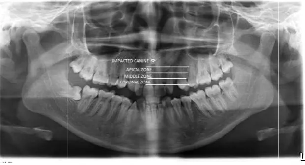
Two secondary objectives were also involved in our study. The first one was the evaluation of CCI index. This objective was studied by changing one of the index teeth of the Chausu’s technique (ipsilateral central incisor) to contralateral canine. Instead of measuring the mesiodistal diameter of the ipsilateral central incisor, the same parameter (d) of contralateral canines is measured and the ratio of widest mesiodistal diameter of the crown of impacted maxillary canines to that of contralateral canines (b/d) was evaluated. The significance of this change of the values was evaluated (Figure 3). Another secondary objective was the evaluation of vertical restriction. On the panoramic radiograph, the roots of the ipsilateral lateral incisors were divided arbitrarily into three equal zones and named as apical, middle and coronal zone. The crown tip of the impacted maxillary canine is located in any of these zones and the position of the canine is predicted. The prediction was compared with the actual position seen clinically or intra operatively (Figure 4). The statistical analysis was done on SPSS (Statistical Package for the Social Sciences) software
ver.12.0. Mean, median standard deviation of the
measurements was summarized. For comparing the reliability
of the techniques, accuracy, sensitivity, specificity and
likelihood ratios wereevaluated.
RESULTS
Out of 18 subjects included in this study, 6 (33.3percent) of them were males and 12 (66.7 percent) of them were females. Among the panoramic radiograph of 18 subjects, 5 (21.7 percent) were having bilateral canines, 13 were having unilateral (78.3 percent).
Thus 23 canines were included in this study. Thirteen canines were present on the right side (56.5 percent) and the rest (43.5 percent) were localized on the left quadrant. For evaluating the Katsnelsons technique, the angle formed between the long axis of the impacted maxillary canine and the occlusal plane (a) was measured for all the 23 cases. The angle varied from 19.50 to 840, having a mean value of 57.50 and standard deviation of 16.5. By using the angulation technique nine canines were predicted as palatal and 14 were predicted as buccaly placed. Out of 9 palatally predicted canines 4 of them were actually buccally placed. This technique shows 82.6 percent accuracy with 100 percent sensitivity and 61.1 percent specificity (Figure 5). While evaluating the Chausu’s technique, the widest mesiodistal dimensions of the crown of impacted maxillary canines (b) varied from 2.8 to 4.1 cm. Also with that the widest mesiodistal dimension of the ipsilateral central incisors varied from 2.5 to 4.6 cms (c). The ratio between these two values (also known as CII index) was calculated (b/c). The range of the ratio was between 1.0 to 1.50 with a mean value of 1.15 and standard deviation of 0.12. Based on the CII index values, 8 canines were predicted to be palatal out of them 3 were buccally placed. Thirteen canines were predicted buccaly and all of them were found buccally placed when compared with clinical records. This shows that the magnification has got an accuracy of 86.5 percent with 100 percent sensitivity and 77.8 percent specificity (Figure 6).
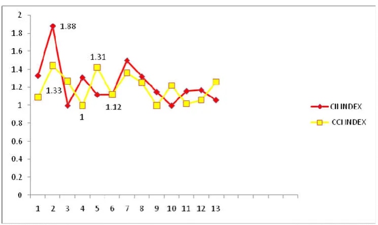
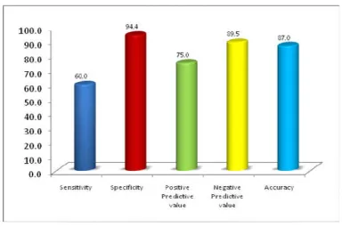
As a secondary objective the ratio between the widest mesiodistal diameter of the impacted maxillary canines (b) and that of the contralateral erupted canines (d) was calculated (known as CCI index). Since the contralateral canines have to be erupted ones, bilaterally impacted cases were excluded and the sample size diminished to 13. The mean value of 1.20 and standard deviation of 0.15 was present. The CII and CCI values were compared. There was wide difference in the corresponding values of the CII index which made it statistically insignificant (Figure 7). Based on the concept of vertical restriction, 23 canines were evaluated and found that 52.1 percent cases were located in middle zone, 42.63 percent cases were located in coronal zone and only 5.27 percent cases were seen on apical zone. According to this zonal distribution, 4 canines were predicted palatally and among them one was buccaly placed. Nineteen canines were predicted buccaly and two of them were palatally placed. The comparison was statistically analyzed and showed 87percent accuracy, 60 percent sensitivity and 94.4 percent specificity (Figure 8).
DISCUSSION
Figure 1. The lines and measurements drawn on panoramic radiograph for evaluating
Figure 2. The lines and measurements drawn on panoramic radiograph for evaluating Chausu’s technique
[image:3.595.164.439.439.582.2] [image:3.595.146.457.608.776.2]Figure 3
Figure
Figure 1. The lines and measurements drawn on panoramic radiograph for evaluating Katsnelson’s technique
The lines and measurements drawn on panoramic radiograph for evaluating Chausu’s technique
Figure 3. measurements taken for cci index
Figure 4. The zones drawn on the panoramic radiographs
Katsnelson’s technique
Figure 5. Clustered cylindrical representation of statistical summary of angulation technique.
Figure 6. Clustered cylindrical representation of statistical summary of CII index
[image:4.595.116.489.547.769.2]The dimensions of alveolar process of maxilla vary from coronal to apical region. The impacted canines most often represent the coronal portion and the bucco palatal dimension at that region is 11.2 +/- 2.1 mm [Vera et al., 2012]. The widest labio palatal width of the maxillary canine is about 8 mm. the impacted canine could be approached by removing the 2-3 mms of bone from palatally or buccally. This may even vary when we deals with rotated canines. Ideally the measurement of the depth of the bone removed for reaching the canine could have been measured for better results and the disparity could also be eliminated. In our study we have got 86% accuracy for Chausu’s technique which is more than that of the study by Wang et al. Also 100 % sensitivity for buccal canines was obtained in our study. Mudasar et al. [2016] compared the magnification technique and the angulation technique. He found that the values of palatal sensitivity and buccal sensitivity were 57% and 100% respectively for the technique of angulation. This when compared with our study stood as 100% sensitivity for buccaly placed canines. They considered magnification technique as more reliable than the angulation technique with the palatal and buccal sensitivity of impacted maxillary canine 71% and 100% respectively. We found that there was an overlap of values of the ratio when the contralateral canine was used as index tooth and hence the results had error. Other studies too have shown that CCI index is insignificant. While applying the concept of vertical restriction, we received 87% accuracy.
This accuracy was comparable with other studies including the research done by Wang et al. [2012]. However it cannot be denied that, our studies had certain limitations. Our sample size was only 18 so we could not involve an area based distribution in our study. Better significant changes in accuracy would have been achieved with increased sample size. The impacted maxillary canine present in the midway between buccal and palatal regions could be approached from both regions. The approach varies according to investigators. The evaluation of the depth of bone removal could have been included for such cases which could increase the significance for prediction. Another problem might have been occurred in this study was that the software used for measuring the values was not the inbuilt software provided with the OPG machine. The evaluation of differences of these two soft wares would have been done for better standardization and which needs to be studied later. For sure science is growing day by day. In future, researchers would come out with hundred percent reliable methods for localization of impacted maxillary canines and also we can expect that the CBCT will become economical and easily available.
Conclusion
[image:5.595.103.488.48.303.2]By comparing the two different techniques, simultaneously we were comparing two principles of radiographic localization (principle of magnification and principle of angulation).
Figure 8. Clustered cylindrical representation of statistical summary of prediction using vertical restriction
Table 1. Inclusion and exclusion criteria
INCLUSION CRITERIA EXCLUSION CRITERIA
1. Patients more than 12 years of age
with unerupted maxillary canine or canines needing orthodontic treatment.
2. Panoramic radiographs of acceptable diagnostic quality from the selected patients.
3. Patients referred from department of
Orthodontics for surgical removal or surgical exposure of the impacted maxillary canine.
1. Erupted canines.
2. Gross distortion of dental arches, as in craniofacial syndromes. 3. Rotated canines.
4. Patients contraindicated for radiation exposure.
5. Pathological lesions involving the impacted canine.
6. Extensively magnified panoramic radiographs.
[image:5.595.67.530.365.482.2]At the end we found that the principle of magnification is slightly more reliable. Also the buccaly placed canines were more predictable when compared to palatal ones.
List of abbreviations
OPG orthopantamogram
CT Computerised Tomogram
CBCT Cone Beam Computerised Tomogram
cm Centimeter
mA Milli ampere
kV Kilovoltage
REFERENCES
An S., Wang J., Li J., Cheng Q., Jiang CM., Wang YT., Huang YF., Yu WJ., Gou YC., Xiao L. 2013. Aug 21.Comparison of methods for localization of impacted maxillary canines
by panoramic radiographs. Dentomaxillofacial
Radiology.;42(8).
Chaushu S., Chaushu G., Becker A. 1999. Reliability of a method for the localization of displaced maxillary canines using a single panoramic radiograph. Clin Orthod Res. 2: 194-199.
Dachi SF., Howell FV. 1961. A survey of 3,874 routine full-mouth radiographs. II: A study of impacted teeth. Oral
Surg Oral Med Oral Pathol. 14:1165.
Katsnelson A., Flick WG., Susarla S., Tartakovsky JV., Miloro M. 2010. Use of panoramic x-ray to determine position of
impacted maxillary canines. Journal of Oral and
Maxillofacial Surgery. 31;68(5):996-1000.
Larsen HJ., Sørensen HB., Artmann L., Christensen IJ., Kjaer I. 2010. Sagittal, vertical and transversal dimensions of the maxillary complex in patients with ectopic maxillary
canines. Orthodontics & craniofacial research.
1;13(1):34-9.
Mudasar Ahad, Mohamad Imran, Azhar Khan, Muzafar Ahmad, Nowsheen Yaqoob,
Shayan Nazir. 2016. Evaluation of different radiographic methods for the localization of impacted maxillary canine – a comparative study. International Journal of
Contemporary Medical Research. 3(9):2589-2592
Vera C., De Kok IJ., Reinhold D., Limpiphipatanakorn P., Yap AK., Tyndall D., Cooper LF. 2012. Evaluation of buccal alveolar bone dimension of maxillary anterior and premolar teeth: a cone beam computed tomography investigation.
International Journal of Oral & Maxillofacial
Implants.1;27(6).