ISOLATION AND IDENTIFICATION OF ACTINOMYCETES ISOLATED FROM KARACHI SOIL AND
SCREENING OF ANTIMICROBIAL COMPOUNDS
*
Abdul Wahab
1, Shumaila
1
Department of Microbiology, University of Karachi, Karachi
2Department of Chemistr
ARTICLE INFO ABSTRACT
Twenty strains of actinomycetes were isolated from a soil sample in Karachi and screened for antimicrobial activity both by primary screening method (perpendicular st
method) and secondary screening method (agar well diffusion method) against several test organisms including bacteria and fungi. Out of twenty strains eight showed broad spectrum activity in primary screening. The secondary screen
microorganisms in primary screening. The strains were identified using various biochemical tests and their optimum growth conditions (temperature, pH and at different concentrations of NaCl)
determined. The most promising strain amongst all was SZ020 which was identified as
Pseudonocardia
Copyright © 2015 Abdul Wahab et al. This is an open access article distributed under the Creative Commons Att distribution, and reproduction in any medium, provided the original work is properly cited.
INTRODUCTION
Actinomycetes are slow growing, gram positive bacteria with high G+C ratio. They resemble to fungi because of their filamentous appearance and spore production property
(Waksman, 1940) and they resemble to bacteria because of
presence of peptidoglycan in their cell wall and possession of flagella (Mythili and Das, 2011). Their habitats include soil, sea sediments, invertebrates, near hydrothermal vents, sea floor, sea plants, ocean surface, in human and cattle bodies
(Rosebury, 1944). The genome size of act
from too large to too small. Actinomycetes play variety of roles like degradation of recalcitrant xenobiotic compounds, organic matter present in soil and waste release from agriculture field and urban community (Heur
nitrogen symbiotically in non-leguminous plants (
Silvester, 1993), produce agro active compounds which are
used to control phytopathogens (Doumbou
Most actinomycetes species have capability to synthesize biologically active secondary metabolites such as antibiotics, herbicides, antitumor, anticancer, pesticides, antiparasitic, immunomodulator, vitamins and enzyme inhibitors (
et al., 2004; Naikpatil and Rathod, 2011
compounds, the compounds which a
therapeutically and also commercially are the antibiotics.
*Corresponding author: Abdul Wahab,
Department of Microbiology, University of Karachi, Karachi
ISSN: 0975-833X
Vol.
Article History:
Received 22nd November, 2014
Received in revised form
09th December, 2014
Accepted 15th January, 2015
Published online 28th February,2015
Key words:
Soil actinomycetes, Antimicrobial compounds, Secondary metabolites,
Primary and secondary screening, Solvent extraction
RESEARCH ARTICLE
ISOLATION AND IDENTIFICATION OF ACTINOMYCETES ISOLATED FROM KARACHI SOIL AND
SCREENING OF ANTIMICROBIAL COMPOUNDS
Shumaila Shumaila
1, Syed Abdus Subhan
1, Syed Tariq Ali
Talat Yasmeen Mujahid
1Department of Microbiology, University of Karachi, Karachi- 75270
Department of Chemistry, University of Karachi, Karachi- 75270,
ABSTRACT
Twenty strains of actinomycetes were isolated from a soil sample in Karachi and screened for antimicrobial activity both by primary screening method (perpendicular st
method) and secondary screening method (agar well diffusion method) against several test organisms including bacteria and fungi. Out of twenty strains eight showed broad spectrum activity in primary screening. The secondary screening was done with those strains which gave activity against test microorganisms in primary screening. The strains were identified using various biochemical tests and their optimum growth conditions (temperature, pH and at different concentrations of NaCl)
determined. The most promising strain amongst all was SZ020 which was identified as
Pseudonocardia sp.
is an open access article distributed under the Creative Commons Attribution License, which distribution, and reproduction in any medium, provided the original work is properly cited.
Actinomycetes are slow growing, gram positive bacteria with high G+C ratio. They resemble to fungi because of their filamentous appearance and spore production property ) and they resemble to bacteria because of heir cell wall and possession of Their habitats include soil, sea sediments, invertebrates, near hydrothermal vents, sea floor, sea plants, ocean surface, in human and cattle bodies ). The genome size of actinomycetes varies from too large to too small. Actinomycetes play variety of roles like degradation of recalcitrant xenobiotic compounds, organic matter present in soil and waste release from
Heur et al., 1997), fix
leguminous plants (Benson and
), produce agro active compounds which are
Doumbou et al., 2001) etc.
Most actinomycetes species have capability to synthesize condary metabolites such as antibiotics, herbicides, antitumor, anticancer, pesticides, antiparasitic, immunomodulator, vitamins and enzyme inhibitors (Oskay
., 2004; Naikpatil and Rathod, 2011). Among these
compounds, the compounds which are important therapeutically and also commercially are the antibiotics.
Department of Microbiology, University of Karachi, Karachi- 75270 Pakistan.
About two-thirds of known antibiotics are produced by actinomycetes (Hozzein et al
antibiotics are produced by Streptomyces
2005). The Micromonospora is the runner up in the antibiotic production and produces one tenth of antibiotics (
As organisms are getting resist
we continuously need to search for the new compounds to which bacteria are not resistant for this purpose scientists are exploring different habitats where novel actinomycetes can be found. The goal of this research was to is
from soil, which was cultivated but dry and less granulated due to over use of synthetic fertilizers and screen them for antibacterial and especially antifungal activity.
MATERIALS AND METHODS
Sampling: soil sample was collected near
of an ornamental plant at the depth of 10
was collected in a dry and clean polythene bag and was air dried at ambient temperature for 9 days.
Culture media: Isolation of actinomycetes was done on oat
meal agar (Oat meal 20gm, Agar 15gm, Trace salt solution (FeSO4.7H2O 0.1gm, MnCl
0.1gm, distilled water 100ml) 1.0ml, distilled water 1000 ml, pH 7.2. Primary and secondary screening for the strains was done using nutrient agar. For the characterization purpose
International Journal of Current Research
Vol. 7, Issue, 02, pp.12760-12765, February, 2015
INTERNATIONAL
ISOLATION AND IDENTIFICATION OF ACTINOMYCETES ISOLATED FROM KARACHI SOIL AND
, Syed Tariq Ali
2and
75270, Pakistan
, Pakistan
Twenty strains of actinomycetes were isolated from a soil sample in Karachi and screened for antimicrobial activity both by primary screening method (perpendicular streak and spot inoculation method) and secondary screening method (agar well diffusion method) against several test organisms including bacteria and fungi. Out of twenty strains eight showed broad spectrum activity in primary ing was done with those strains which gave activity against test microorganisms in primary screening. The strains were identified using various biochemical tests and their optimum growth conditions (temperature, pH and at different concentrations of NaCl) were also determined. The most promising strain amongst all was SZ020 which was identified as
ribution License, which permits unrestricted use,
thirds of known antibiotics are produced by
et al., 2011). About 80% of these
Streptomyces (Kim and Garson,
is the runner up in the antibiotic production and produces one tenth of antibiotics (Lam, 2006). As organisms are getting resistance against preexisting drugs we continuously need to search for the new compounds to which bacteria are not resistant for this purpose scientists are exploring different habitats where novel actinomycetes can be found. The goal of this research was to isolate actinomycetes from soil, which was cultivated but dry and less granulated due to over use of synthetic fertilizers and screen them for antibacterial and especially antifungal activity.
MATERIALS AND METHODS
soil sample was collected near rhizospheric region of an ornamental plant at the depth of 10-12 cm this sample was collected in a dry and clean polythene bag and was air dried at ambient temperature for 9 days.
Isolation of actinomycetes was done on oat al 20gm, Agar 15gm, Trace salt solution O 0.1gm, MnCl2.4H2O 0.1gm, ZnSO4.7H2O
100ml) 1.0ml, distilled water 1000 ml, pH 7.2. Primary and secondary screening for the strains was done using nutrient agar. For the characterization purpose
yeast extract glucose agar, yeast extract malt extract agar, oat meal agar, inorganic salts starch agar, Jensen’s agar, nutrient agar, starch casein agar and Sabroud’s dextrose agar were used.
Test organisms: The bacteria test strains include Escherichia
coli, Bacillus subtilis, Staphylococcus aureus, Pseudomonas
aeruginosa, Proteus mirabilis, Micrococcus luteus,
Klebsiellapneumoniae, Salmonella typhi, Streptococcus
faecalis, Streptococcus pyogenes,
Corynebacteriumdiphtheriae, Corynebacterium Xerosis,
Corynebacteriumhofmannii, Methicillin-resistant
Staphylococcus aureus, Vancomycin resistant Enterococcus,
Shigella dysenteriae and Shigella flexneri, and the fungi test
strains include Candida albicans, Saccharomyces cerevisiae,
Aspergillus niger and Aspergillus terreus were used. The
bacterial strains were stored in the form of glycerol stock and fungal strains were store on the slants of potato dextrose agar. They were sub cultured as needed.
Isolation of Actinomycetes: One gram of air dried soil sample
was taken and added in the flask containing 100ml of sterile distilled water. The flask was shaken vigorously to release microorganisms from the soil particles then kept at smooth horizontal surface in still state for 15 minutes to allow the soil particles to get settle down. This suspension was 1:100 times diluted. Further dilutions were made up to 1:106. 100 µl from the last three dilutions (1:104, 1:105 and 1:106) was transferred onto the oat meal agar plate and spread with sterile spreader. The plates were incubated for 7 days at ambient temperature. The strains of actinomycetes were selected on the basis of colonial morphology. The microscopy of the isolates was performed and characteristics of mycelium and spores types and numbers per sporangium were noted. The actinomycetes strains were preserved on slants and as glycerol stock and screened for antimicrobial potential.
Primary screening methods
Perpendicular streak method: The actinomycetes strains were
streaked perpendicularly in the middle of the nutrient agar plate; the streaked plates were incubated at 37 ˚C for 3-4 days. After getting sufficient growth test organisms were streaked perpendicular to the producer actinomycetes strains and the plates were incubated at 37 ˚C for 24 hrs for bacteria and at ambient temperature for 4-5 days for unicellular fungi.
Spot inoculation method: The spots of actinomycetes strains
were inoculated on the nutrient agar. The plates were incubated at 37 ˚C for 3-4 days. When the spots of actinomycetes grown they were layered with soft agar of nutrient agar (for bacteria) and potato dextrose agar (for fungi) which was pre seeded with test organism, Plates were incubated at 37 ˚C for 24 hrs for bacteria and at ambient temperature for 4-5 days for single cell fungi. To check the activity of actinomycetes against filamentous fungi the actinomycetes strains were spot inoculated on potato dextrose agar and grown for 4-5 days at ambient temperature after that a disk of actively growing fungal mycelium was inoculated at some distance from actinomycetes spot and then plates were
incubated for 4 days at room temperature. The results were recorded by measuring zone of inhibition.
Secondary screening of actinomycetes
Crude extract: The producer strains which gave broad
spectrum activity in primary screening methods were further evaluated by secondary screening method. In doing this the producers were inoculated in 50ml of nutrient broth and incubated at 37 ˚C in shaking incubator for 10 days. After 10 days inoculated broth was centrifuged at 10000 rpm for 10 min. Supernatant was taken and again centrifuged twice at 5000 rpm for 15 min. The collected crude extract was kept at refrigerator temperature. Then crude extract was tested against test organisms by agar well diffusion.
Solvent extraction method for extracellular metabolites:
Solvents used for the extraction of extracellular metabolite were ethanol, methanol, ethyl acetate and chloroform. 5ml of crude extract was taken in sterile falcon tube and added with 5ml of solvent separately. Gently mix for 1 hr. Then it was centrifuged at 10000 rpm for 10 min. After centrifugation two phases were obtained the upper phase containing antimicrobial metabolites was collected in an Eppendorf and the activity of the extract was tested by agar well method.
Agar well diffusion method: The nutrient agar plate was
[image:2.595.314.552.467.754.2]layered with the soft nutrient agar pre seeded with the test organism when soft agar solidified the wells were dug with the help of sterile borer. The 100 µl of extract was added in the well. Sterile nutrient broth was also added in one well as a negative control. Then plates were kept in refrigerator for 15 min to allow the diffusion of extract and later on incubated at 37 ⁰C in inverted position for 24 hrs.
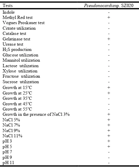
Table 1. Biochemical and physiological characterization of strain
Pseudonocardia sp. SZ020
Tests Pseudonocardiasp. SZ020
Indole -
Methyl Red test +
Vogues Proskauer test -
Citrate utilization -
Catalase test -
Gelatinase test +
Urease test -
H2S production -
Glucose utilization -
Mannitol utilization -
Lactose utilization -
Xylose utilization -
Fructose utilization -
Sucrose utilization -
Growth at 15˚C +
Growth at 25˚C +
Growth at 35˚C -
Growth at 45˚C -
Growth at 55˚C -
Growth in the presence of NaCl 3% +
NaCl 5% +
NaCl 7% +
NaCl 9% +
NaCl 11% +
pH 3 +
pH 5 +
pH 7 -
pH 9 pH 11
- - Key: +/ growth and -/no growth
Characterization of actinomycetes
Active strains were grown on different media for morphological characterization and biochemical characterization was performed using various tests. The active strains were also checked for their optimum growth conditions by growing on different temperatures (15˚C, 25˚C, 35˚C, 45˚C and 55˚C), pH (3, 5, 7, 9, and 11) and NaCl concentration (3%, 5%, 7%, 9% and 11%). the growth was measured in terms of optical density.
RESULTS AND DISCUSSION
Isolation of actinomycetes: The soil sample was collected
from depth as it has been found out that number of actinomycetes at depth is relatively higher than the soil surface
due to appropriate pH and water content in depth (Basavaraj
et al., 2010). The sample was air dried for one week at 30 0C
as this reduces the population of gram negative bacteria and other microorganisms (Jeffrey, 2010). The dry, pigmented, chalky, small to medium sized colonies which showed characteristic features of actinomycetes were selected and purified by streaking on nutrient agar. Total 20 strains were isolated. The culture of purified strains were maintained on slants and preserved in the glycerol stock also.
Screening of strains: The strains were assessed for their
antimicrobial potential. The primary screenings was performed by perpendicular streak method (Egorov, 1985) and spot inoculation method (Kumar et al., 2010). The actinomycetes strains were first tested by perpendicular streak method and it was found that six out of twenty strains showed activity against gram positive test organisms and only four strains showed activity against gram negative organisms. The results of spot inoculation method showed that eight isolates had potential to inhibit gram positive microorganisms and five isolates inhibited gram negative organisms. The difference between the sensitivity of gram negative and gram positive bacteria is due to their cell wall morphology. The gram negative organisms have outer layer which consists of lipopolysaccharide which decreases the cell wall permeability and in this way interrupts the entrance of lipophilic molecules in cell. Cell wall of gram negative organisms also contains porins which serve as selective barrier for the passage of hydrophilic molecules. The gram positive organisms are more sensitive to antimicrobial compounds because they do not have outer layer and their cell wall is more permeable to these compounds (Scherrer and Gerhardt, 1971). Strains Faenia
sp.SZ014 and Pseudonocardia sp. SZ020 showed broad spectrum activity. The broad spectrum activity is may be due to the production of more than one antimicrobial compound that make actinomycetes effective against both gram negative and gram positive organisms (Gurung et al., 2009), these five isolates were selected for secondary screening. Secondary screening of active isolates was performed by agar well method (Pandey et al., 2004).
The secondary screening results were conclusive. Those strains which gave activity in primary screening did not exhibit antimicrobial potential in secondary method except
Pseudonocardia sp. SZ020 (only against some
microorganisms). The reason behind this is may be the difference in the morphology of actinomycetes when grow on solid medium (filamentous mycelia) and in liquid broth (fragementing mycelia). Also many of the actinomycetes are poor fermenter (Pandey et al., 2004). It may also be possible that the active compounds release by actinomycetes become inactive or chemically modified in broth or bind to the component of liquid medium (Gurung et al., 2009).
One of the reasons is may be the failure of diffusion of inhibitory compound in aqueous environment. This observation was reported by Rosenfield and Zobell, 1947. They reported that antimicrobial compounds are closely bound to the outer surface of cell and release into solid medium slowly. Lemos et al. (1985) reported that bound antibiotics excrete in the environment slowly and continually and prevent the colonization of competitors in its surrounding (Hosnyet al.,
2011). For the isolation of antimicrobial metabolites from
broth different solvents including methanol, ethanol, chloroform and ethyl acetate were used. This procedure is referred as solvent extraction method (Khan and Patel, 2011). Only the chloroform and ethyl acetate extract of strain
Pseudonocardia (SZ020) showed activity against MRSA and
M. lutues. Other solvent did not extract the antimicrobial
metabolite from fermented broth. The reasons for the failure of metabolite extraction by using solvents could be the presence of polar functional group in metabolite which make metabolite insoluble in solvent and soluble in water, inadequate shaking of the mixture, and the use of inappropriate solvents (Gurung
et al., 2009). The strains showing activity against bacteria were also tested against Candida albicans, Saccharomyces
cerevisea, Aspergillus niger and Aspergillus tereus. Spot
inoculation and perpendicular streak methods were used to check activity against Candida albicansand Saccharomyces
cerevisae. The activity against Aspergillus niger and
Aspergillus tereus was tested by the method used by
Prapagdee et al. (2008). Only three isolates exhibited
antifungal activity against C. albicans and S. cerevisea and one isolate exhibited activity against Aspergillus niger. The potential of inhibiting fungi is attributed to the production of secondary antifungal compounds and extracellular hydrolytic enzymes (Prapagdee et al., 2008). The growth kinetics of the most active and broad spectrum activity showing strain
Pseudonocardia sp. SZ020 was also determined by following
the method mentioned by Khan and Patel (2011). The strain started to produce antibiotic at day 3 but the activity of the extract was not too strong because of various reasons as described above. This strain completed its life cycle in 6 days.
At present we need to find out such isolates which secret novel antimicrobial compounds as the preexisting drugs have been failed due to the development of resistance among the microorganisms. The prevalence of increasing resistance is mainly due to the dissemination of existing resistant pathogenic strains or due to emergence of peculiar resistive pathogens (World Health Organization, 2001). The present study is also a little contribution towards this need, the isolate
Pseudonocardia sp. SZ020 showed broad spectrum activity
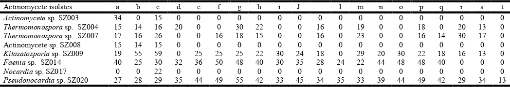
Table 2. Antimicrobial activity of actinomycetes strains by spot inoculation method (zone of inhibition in mm)
Actinomycete isolates a b c d e f g h i J l m n o p q r s t
Actinomycete sp. SZ003 34 0 15 0 0 0 0 0 0 0 0 0 0 0 0 0 0 0 0 0
Thermomonospora sp. SZ004 15 14 16 20 0 0 30 22 0 0 16 0 19 0 0 18 0 20 13 0
Thermomonospora sp. SZ007 17 16 26 0 0 16 18 15 0 0 16 0 23 0 0 16 14 30 17 0
Actinomycete sp. SZ008 15 14 15 0 0 0 0 0 0 0 0 0 0 0 0 0 0 0 0 0
Kitasatosporia sp. SZ009 19 55 59 0 25 25 25 22 30 24 18 0 29 20 30 22 18 16 13 0
Faenia sp. SZ014 40 25 30 32 36 50 48 40 30 35 28 24 22 44 48 48 40 0 0 0
Nocardia sp. SZ017 0 0 22 0 0 0 0 0 0 0 0 0 0 0 0 0 0 0 0 0
Pseudonocardia sp. SZ020 27 28 29 35 44 49 55 42 33 45 34 35 33 39 44 49 42 29 34 13 Test organisms
a: B. subtilis; b: S. aureus; c: M. luteus; d: S. faecalis; e: S. pyogenes; f: C. diphtheriae; g: C. xerosis; h: C. hofmannii; i: MRSA; j: VRE; k: E. coli; l: P. aeruginosa; m: P. mirabilis; n: K. pneumoniae; o: S. typhi; p: S. dysenteriae; q: S. flexneri; r: C. albicans; s: S. cerevisiae; t: A. niger
Fig. 1. Growth kinetics ofPseudonocardia sp. SZ020
The further studies related to the determination of
the antimicrobial compounds released by Pseudonocardia
SZ020 is currently going on.
Acknowledgement
This research project was funded by Dean Faculty of Science, University of Karachi grant DFS / 2011-2012 given to Dr. Abdul Wahab.
REFERENCES
Basavaraj, N. K., Chandrashekhara, S. et al.,
and morphological characterization of antibiotic producing actinomycetes, Tropical Journal of Pharmaceutical
Research, 9(3): 231-236
Benson, D. R. and Silvester, W. B., 1993. Biology of
Strains, actinomycetes symbionts of actinorhizal plants,
Microbiological Reviews, 57(2): 293-319
Doumbou, C. L., Salove, M. K. H. et al., 2001. Actinomycetes, promising tools to control plant diseases and to promote plant growth, Phytoprotection, 82(3):
85-Egorov, N. S. 1985, Antibiotics, a scientific approach, Mir publishers, Moscow
Gurung, T. D., Sherpa, C. et al., 2009. Isolation and characterization of antibacterial actinomycetes
samples of Kalapatthar; Mount Everest region,
Journal of Science and Technology, 10: 173
Heuer, H., Krsek, M., et al., 1997. Analysis of Actinomycete Communities by Specific Amplification of Genes Encoding 16S rRNA and Gel-Electrophoretic Separation in Denaturing Gradients, Applied and Environmental
Microbiology, 63(8): 3233–3241
Hosny, A.E.D.M.S., Sheir, D.H., et al., 2011. Production of antimicrobial agent from marine bacteria isolated from Mediterranean, Australian Journal of Basic and Applied
Sciences, 5(5): 121-128
Hozzein, W. N., Rabie, W. et al., 2011. Screening the Egyptian desert actinomycetes as candidates for new antimicrobial compounds and identification of a new desert
strain, African Journal of Biotechnology
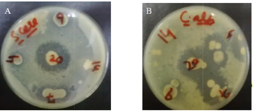
[image:5.595.91.514.57.245.2]2301
Figure 2. Antifungal activity of Thermomonospora Pseudonocardia
The further studies related to the determination of structure of
Pseudonocardia sp.
This research project was funded by Dean Faculty of Science, 2012 given to Dr.
et al., 2010. Isolation
and morphological characterization of antibiotic producing
Tropical Journal of Pharmaceutical
Benson, D. R. and Silvester, W. B., 1993. Biology of Frankia
symbionts of actinorhizal plants, 319
2001. Actinomycetes, eases and to promote
-102
Egorov, N. S. 1985, Antibiotics, a scientific approach, Mir
2009. Isolation and characterization of antibacterial actinomycetes from soil samples of Kalapatthar; Mount Everest region, Nepal
, 10: 173-182
1997. Analysis of Actinomycete Communities by Specific Amplification of Genes Encoding Electrophoretic Separation in
Applied and Environmental
2011. Production of antimicrobial agent from marine bacteria isolated from
l of Basic and Applied
2011. Screening the Egyptian desert actinomycetes as candidates for new antimicrobial compounds and identification of a new desert Streptomyces
echnology, 10(12):
2295-Khan, J. A. and Patel, A. S. 2011. Extraction and purification of antimicrobial metabolites from actinomycetes spp. Isolated from soil sample
Pharmaceutical Research and Development
Kim, T. K. and Garson, M. J. 2005. Marine actinomycetes related to the ‘Salinospora’ group from the Great Barrier Reef sponge Pseudoceratinaclavata.
Microbiology 7:509-518
Kumar, N., Singh, R. K. et al.,
soil actinomycetes as source of antibiotics active against bacteria, International Journal of Microbiology Research
2(2): 12-16
Lam, K. S. 2006. Discovery of novel metabolites from marine actinomycetes, Current Opinion in Microbiology,
251
Lemos, M. L., Toranzo A. E. and Barja J. L, 1985. Antibiotic activity of epiphytic bacteria isolated from intertidal seaweeds. Journal of Microbial Ecology
Mythili, B. and Das, M. P. A. 2011. Studies on antimicrobial activity of Streptomyces spp.
soil, Research Journal of Agricultural Sciences
106
Naikpatil, S. V. and Rathod, J. L. 2011. Antimicrobial and cytotoxic activity of actinomycetes from Karwar coast, west coast of India, World Journal of Scien
Technology, 1(1): 07-10
Oskay, M., Tamer, A. U. et al.,
some actinomycetes isolated from farming soils of Turkey,
African Journal of Biotechnology
Pandey, B., Ghimire, P. et al.,
antibacterial activity of the actinomycetes isolated from the Khumbu Region of Nepal,
23: 44-53
Prapagdee, B., Kuekulvong, C.
potential of extracellular metabolites produced by
Streptomyces hygroscopicus
fungi, International Journal of Biological Sciences,
330-337
Rosebury, T. 1944. The parasitic actinomycetes
filamentous microorganisms of the mouth: a review of their characteristics and relationships, of the bacteriology of the
Thermomonospora sp. SZ004, Thermomonospora sp. SZ007, Kitasatosporia
Pseudonocardia sp. SZ020 against (A) S. cerevisiae (B) C. albicans
Khan, J. A. and Patel, A. S. 2011. Extraction and purification of antimicrobial metabolites from actinomycetes spp. Isolated from soil sample, International Journal of
Pharmaceutical Research and Development, 3(10): 63-71
Kim, T. K. and Garson, M. J. 2005. Marine actinomycetes related to the ‘Salinospora’ group from the Great Barrier Reef sponge Pseudoceratinaclavata. Environmental
2010. Isolation and screening of as source of antibiotics active against
International Journal of Microbiology Research,
Lam, K. S. 2006. Discovery of novel metabolites from marine
Current Opinion in Microbiology,
9:245-Lemos, M. L., Toranzo A. E. and Barja J. L, 1985. Antibiotic activity of epiphytic bacteria isolated from intertidal
Journal of Microbial Ecology, 11: 149-163
Mythili, B. and Das, M. P. A. 2011. Studies on antimicrobial spp. isolates from tea plantation
Research Journal of Agricultural Sciences, 2(1):
104-Naikpatil, S. V. and Rathod, J. L. 2011. Antimicrobial and cytotoxic activity of actinomycetes from Karwar coast,
World Journal of Science and
et al., 2004. Antibacterial activity of
some actinomycetes isolated from farming soils of Turkey,
African Journal of Biotechnology, 3 (9): 441-446
et al., 2004. Studies on the
antibacterial activity of the actinomycetes isolated from the
, Journal of Biological Sciences,
Prapagdee, B., Kuekulvong, C. et al., 2008. Antifungal potential of extracellular metabolites produced by
icus against phytopathogenic
International Journal of Biological Sciences, 4(5):
Rosebury, T. 1944. The parasitic actinomycetes and other filamentous microorganisms of the mouth: a review of their characteristics and relationships, of the bacteriology of the
actinomycosis, of salivary calculus in man, Microbiology
and Molecular Biology Reviews, 8(3):189-223
Rosenfeld, W.D. and C.E. Zobell. 1947. Antibiotic production by marine microorganisms. Journal of Bacteriology, l54:393-398
Scherrer, R. and Gerhardt, P. 1971, Molecular sieving by Bacillus megatherium cell wall and protoplast, Journal of
Bacteriology, 107: 718-735
Waksman, S. A. 1940. On the classification of actinomycetes, Journal of Bacteriology, 39(5): 549-558
WHO, 2001, WHO Global strategy for containment of antimi crobial resistance