202
https://doi.org/10.1107/S2056989019000458 Acta Cryst.(2019). E75, 202–207research communications
Received 8 January 2019 Accepted 9 January 2019
Edited by M. Zeller, Purdue University, USA
Keywords:piperazines; crystal structure; mol-ecular conformation; hydrogen bonding; supra-molecular assembly.
CCDC references:1889708; 1889709; 1889710
Supporting information:this article has supporting information at journals.iucr.org/e
Three closely related
1-[(1,3-benzodioxol-5-yl)-methyl]-4-(halobenzoyl)piperazines: similar
molecular structures but different intermolecular
interactions
Ninganayaka Mahesha,aBelakavadi K. Sagar,aHemmige S. Yathirajan,a* Tetsundo Furuya,bTomoyuki Haraguchi,b Takashiro Akitsuband Christopher Glidewellc
aDepartment of Studies in Chemistry, University of Mysore, Manasagangotri, Mysuru-570 006, India,bDepartment of
Chemistry, Faculty of Science, Tokyo University of Science, 1-3 Kagurazaka, Shinjuku-ku, Tokyo 162-8601, Japan, and cSchool of Chemistry, University of St Andrews, St Andrews, Fife KY16 9ST, UK. *Correspondence e-mail:
yathirajan@hotmail.com
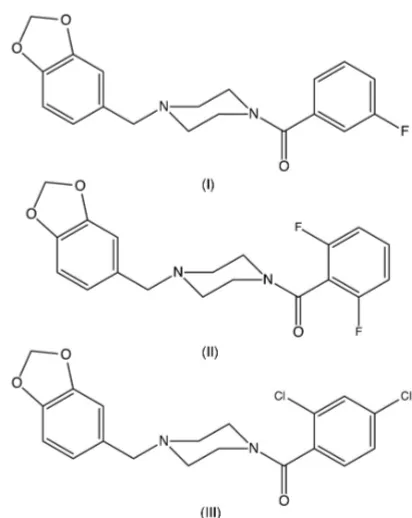
In each of the compounds 1-[(1,3-benzodioxol-5-yl)methyl]-4-(3-fluorobenzoyl)-piperazine, C19H19FN2O3 (I),
1-[(1,3-benzodioxol-5-yl)methyl]-4-(2,6-difluoro-benzoyl)piperazine, C19H18F2N2O3(II), and
1-[(1,3-benzodioxol-5-yl)methyl]-4-(2,4-dichlorobenzoyl)piperazine, C19H19Cl2N2O3 (III), the piperazine rings
adopt a chair conformation with the (1,3-benzodioxol-5-yl)methyl substituent occupying an equatorial site: the five-membered rings are all slightly folded across the O O line leading to envelope conformations. The dihedral angle between the planar amidic fragment and the haloaryl ring is 62.97 (5)in (I) but
77.72 (12)and 75.50 (5)in (II) and (III), respectively. Despite their similarity in constitution and conformation, the supramolecular interactions in (I)–(III) differ: in (I), a combination of C—H O and C—H (arene) hydrogen bonds links the molecules into a three-dimensional framework structure, but there are no hydrogen bonds of any sort in either (II) or (III), although the structure of (III) contains a short Cl Cl contact between inversion-related pairs of molecules.
1. Chemical context
1-[(1,3-Benzodioxol-5-yl)methyl]piperazine is an important intermediate for the synthesis (Dunctonet al., 2006; Hamid & Williams, 2007) of piribedil, 1-[(1,3-benzodioxol-5-yl)methyl]-4-(pyrimidin-2-yl)piperazine, which is used in the treatment of Parkinson’s disease, particularly in the reduction of tremor (Rondot & Ziegler, 1992; Millan et al., 2001). The synthetic routes to piribedil reported hitherto have utilized either palladium-catalysed (Duncton et al., 2006) or ruthenium-catalysed (Hamid & Williams, 2007) processes, requiring extensive purification procedures to ensure that the final product is free of heavy metals. With this in mind, we have now synthesized a series ofN-aroyl analogues (I)–(III) (Figs. 1–3) using a metal-free procedure involving a straightforward coupling reaction between 1-[(1,3-benzodioxol-5-yl)methyl]-piperazine and a carboxylic acid, promoted by 1-(3-dimeth-ylaminopropyl)-3-ethylcarbodimide as the dehydrating agent, and we report here the molecular and supramolecular struc-tures of compounds (I)–(III).
2. Structural commentary
In each of (I)–(III), the five-membered ring is slightly non-planar: while the atoms O11, C7A, C3A and O13 are co-planar, as expected, the atom C12 is slightly displaced from this plane by 0.150 (2), 0.099 (6) and 0.210 (2) A˚ in (I)–(III), respectively, giving an envelope conformation in each case, with the ring folded across the line O11 O13. The piperazine rings all adopt chair conformations with the substituent at atom N1 in an equatorial site, while the atoms of the amide fragment (C3, N4, C5, C47, O47 and C41) are coplanar. The only significant conformational difference between the mol-ecules in (I)–(III) lies in the dihedral angle between the amide unit and the adjacent aryl ring (C41–C46), 62.97 (5)in (I) but
77.72 (12) and 75.50 (5) in (II) and (III), respectively. The
molecules of (I)–(III) exhibit no internal symmetry and hence they are all conformationally chiral, but the space groups (Table 2) confirm that equal numbers of the two conforma-tional enantiomorphs are present in each crystal.
3. Supramolecular features
Despite their similar molecular constitutions and conforma-tions, compounds (I)–(III) all exhibit different types of direction-specific intermolecular interactions. In the crystal structure of compound (I), a combination of one C—H O hydrogen bond and two C—H (arene) hydrogen bonds (Table 1) links the molecules into a three-dimensional framework structure, whose formation can readily be analysed in terms of simple sub-structures (Ferguson et al., 1998a,b; Gregson et al., 2000). The C—H O hydrogen bond links molecules related by the 21screw axis along (0.25,y, 0.25) to
form a C(5) (Etter, 1990; Etteret al., 1990; Bernstein et al.,
1995) chain running parallel to the [010] direction. In addition, the C—H (arene) hydrogen bond having atom C5 as the donor links molecules related by the 21screw axis along (0.75,
y, 0.25) into a second chain running parallel to [010] and, together, these two interactions generate a sheet lying parallel to (001) (Fig. 4). The second C—H (arene) hydrogen bond, having atom C45 as the donor, links molecules related
research communications
Acta Cryst.(2019). E75, 202–207 Maheshaet al. C
19H19FN2O3, C19H18F2N2O3and C19H18Cl2N2O3
203
Figure 2
[image:2.610.65.271.79.338.2] [image:2.610.313.564.107.181.2]The molecular structure of compound (II) showing the atom-labelling scheme. Displacement ellipsoids are drawn at the 50% probability level.
Figure 3
[image:2.610.315.564.313.407.2] [image:2.610.316.566.454.561.2]The molecular structure of compound (III) showing the atom-labelling scheme. Displacement ellipsoids are drawn at the 50% probability level.
Figure 1
The molecular structure of compound (I) showing the atom-labelling scheme. Displacement ellipsoids are drawn at the 50% probability level.
Table 1
Hydrogen-bond geometry (A˚ ,) for (I).
Cg1 represents the centroid of the C3A, C14, C15, C16, C17, C7Aring.
D—H A D—H H A D A D—H A
C42—H42 O47i 0.95 2.34 3.273 (2) 168 C5—H5A Cg1ii 0.99 2.76 3.7310 (18) 168
C45—H45 Cg1iii 0.95 2.90 3.7470 (18) 149
Symmetry codes: (i) xþ1 2;y
1 2;zþ
1
2; (ii) xþ 3 2;yþ
1 2;zþ
1 2; (iii) x1
2;yþ 3 2;zþ
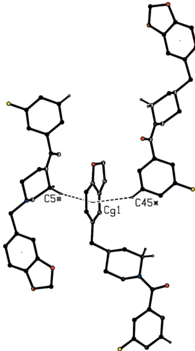
[image:2.610.314.568.614.718.2]by then-glide plane aty= 0.75 into a chain running parallel to the [101] direction (Fig. 5), and chains of this type link the (001) sheets into a continuous three-dimensional structure. It is interesting to note that both C—H (arene) hydrogen bonds utilize the same ring as the acceptor, with one donor approaching each face of this ring (Fig. 6), with the angle H5i Cg1 H45ii= 152, whereCg1 represents the centroid of the ring (C3A, C14, C15, C16, C17, C7A) and the symmetry codes are (i)3
2x, 1 2+y,
1
2z) and (ii) ( 1 2+x,
3 2y,
1 2+z).
Hence, the two molecules providing the donor atoms here are related by inversion across (1, 1/2, 0). In this structure, the atoms of type O11 in the molecules at (x,y,z) and (2x, 1y, z) are separated by a distance of only 2.7888 (18) A˚ . At the same time, the atoms C12 and H12 at (x,y,z) are distant from O11 at (2x, 1y,z) by 2.66 and 3.008 (2) A˚ , respectively,
with an associated C—H O angle of 101; the H O
distance is too long and the C—H O angle is too small for this contact to be regarded as a hydrogen bond, but the short O O distance here is perhaps associated with this ‘failed’ hydrogen bond involving atom C12.
In contrast to the three-dimensional supramolecular assembly in (I) generated by three hydrogen bonds, the only direction-specific intermolecular interaction in (II) is a single C—H O contact, in which theD–-H Aangle is only 123
so that this cannot be regarded as structurally significant (Woodet al., 2009). The only direction-specific intermolecular interactions in (III) are a C—Cl (ring) contact involving the 1,3-dioxolane ring, but since this ring is not aromatic, this contact cannot be regarded as structurally significant; and a short Cl Cl contact between inversion-related pairs of molecules. For the atoms of type Cl44 in the molecules at (x,y,
204
Maheshaet al. C19H19FN2O3, C19H18F2N2O3and C19H18Cl2N2O3 Acta Cryst.(2019). E75, 202–207
[image:3.610.74.520.265.703.2]research communications
Figure 4
[image:3.610.67.271.267.699.2] [image:3.610.344.519.267.705.2]Part of the crystal structure of compound (I) showing the formation of a sheet lying parallel to (001) and built from C—H O and C— H (arene) hydrogen bonds, which are drawn as dashed lines. For the sake of clarity, the H atoms bonded to the C atoms not involved in the motifs shown have been omitted.
Figure 5
z) and (x, y, 2 z), the Cl Clidistance is 3.3963 (7) A˚ with an associated C—Cl Cliangle of 137.68 (5)[symmetry
code: (i)x,y, 2z]. For C—Cl Cl angles of 90 and 180, values of 1.78 and 1.58 A˚ have been suggested (Nyburg & Faerman, 1985) for the major and minor van der Waals radii: on this basis, a value of around 1.68 A˚ would seem appropriate to a C—Cl Cl angle close to 135, so that the observed
Cl Cl contact distance in (III) is not exceptional, and is probably therefore of no structural significance. Thus for both (II) and (III), the molecular packing depends solely on mol-ecular shape and van der Waals forces.
4. Database survey
It is of interest briefly to compare the supramolecular assembly found here for compounds (I)–(III) with that observed in some related compounds. In 1-[(1,3-benzodioxol-5-yl)methyl]-4-(pyrimidin-2-yl)piperazine (piribedil), the molecules are linked into sheets by three independent C—
H hydrogen bonds (Wu et al., 2013), and in 1-(2-iodo-benzoyl)-4-(pyrimidin-2-yl)piperazine, the molecules are linked by a combination of C—H O and C—H
hydrogen bonds to form a three-dimensional structure which is augmented by – stacking interactions and N I inter-actions (Mahesha et al., 2019). The amidic compound N -(4-chlorophenyl)-4-(pyrimidin-2-yl)piperazine-1-carboxamide crystallizes withZ0
= 2 in space groupP21/c, and the molecules
are linked by two independent N—H O hydrogen bonds to form chains of C2
2(8) type, although these are described as
C(4) in the original report (Li, 2011). Finally, we note the structures of three salts derived by monoprotonation of the starting material 1-[(1,3-benzodioxol-5-yl)methyl]piperazine used in the synthesis of compounds (I)–(III): protonation occurs at the unsubstituted N atom of the piperazine unit in each of the picrate (Kavitha et al., 2014a), 4-nitrobenzoate (Kavithaet al., 2014b) and 4-chlorobenzoate (Kavithaet al., 2014c) salts, although the schematic diagrams given for the two carboxylate salts depict protonation at the substituted N atom.
5. Synthesis and crystallization
1-[(1,3-Benzodioxol-5-yl)methyl]piperazine was purchased from Sigma–Aldrich and used as received. For the synthesis of compounds (I)–(III), 1-(3-dimethylaminopropyl)-3-ethyl-carbodimide (207 mg, 1.08 mmol), 1-hydroxybenzotriazole (121.6 mg, 0.9 mmol) and triethylamine (0.5 ml, 3.7 mmol) were added to solutions of the appropriately substituted benzoic acid [3-fluorobenzoic acid for (I), 2,6-difluorobenzoic acid for (II) or 2,4-dichlorobenzoic acid for (III)] (0.9 mmol) inN,N-dimethylformamide (5 ml) and the resulting mixtures were then stirred at 273 K for 20 min. A solution of 1-[(1,3-benzodioxol-5-yl)methyl]piperazine (200 mg, 0.9 mmol) in
N,N-dimethylformamide (5 ml) was then added to each mixture and stirring was continued overnight at ambient temperature. When the reactions were complete as confirmed using thin-layer chromatography, an excess of water was added to each of the mixtures, which were then exhaustively extracted using ethyl acetate. Each of the organic fractions was then washed successively with aqueous hydrochloric acid (1 mol dm3), then with a saturated aqueous solution of sodium hydrogencarbonate, and finally with brine. The organic fractions were then dried over anhydrous sodium sulfate and concentrated under reduced pressure. Slow evaporation of these solutions, at ambient temperature and in the presence of air, gave crystals of compounds (I)–(III) suitable for single-crystal X-ray diffraction: m.p. (I) 383– 386 K, (II) 373 K, (III) 394–396 K.
6. Refinement
Crystal data, data collection and structure refinement details are summarized in Table 2. All H atoms were located in difference maps, and they were subsequently treated as riding atoms in geometrically idealized positions with C—H distances 0.95 A˚ (aromatic) or 0.99 A˚ (CH2) and with
research communications
Acta Cryst.(2019). E75, 202–207 Maheshaet al. C
[image:4.610.72.262.72.414.2]19H19FN2O3, C19H18F2N2O3and C19H18Cl2N2O3
205
Figure 6
Part of the crystal structure of compound (I) showing the two C— H (arene) hydrogen bonds with a common aryl acceptor. The hydrogen bonds are drawn as dashed lines and, for the sake of clarity, the unit-cell outline and the H atoms bonded to the C atoms not involved in the motifs shown have been omitted. The atoms marked with an asterisk (*) or a hash (#) are at the symmetry positions (1
2+x, 3 2y,
1 2+z)
and (3 2x,
1 2+y,
1
Uiso(H) = 1.2Ueq(C). For compound (I), fifteen bad outlier
reflections were omitted from the data set. For compound (II), the correct orientation of the structure with respect to the polar axis direction could not be established because of the lack of significant resonant scattering: thus calculation of the Flackxparameter (Flack, 1983) using using 1369 quotients of the type [(I+)(I)]/[(I+) + (I)] (Parsonset al., 2013) gave a value 0.3 (10), which must be regarded as indeterminate (Flack & Bernardinelli, 2000), despite the 93% coverage of Friedel pairs, while the value of the Hooftyparameter (Hooft
et al., 2008),y=0.2 (6), is likewise indeterminate.
Acknowledgements
NM is grateful to the University of Mysore for research facilities.
Funding information
HSY is grateful to the UGC, New Delhi for the award of a BSR Faculty Fellowship for three years. BKS thanks the UGC for the award of a Rajeev Gandhi Fellowship.
References
Bernstein, J., Davis, R. E., Shimoni, L. & Chang, N.-L. (1995).Angew. Chem. Int. Ed. Engl.34, 1555–1573.
Bruker (2004).APEX2. Bruker AXS Inc., Madison, Wisconsin, USA. Bruker (2013).SAINT, Bruker AXS Inc., Madison, Wisconsin, USA. Bruker (2015). SADABS, Bruker AXS Inc., Madison, Wisconsin,
USA.
Duncton, M. A. J., Roffey, J. R. A., Hamlyn, R. J. & Adams, D. R. (2006).Tetrahedron Lett.47, 2549–2552.
Etter, M. C. (1990).Acc. Chem. Res.23, 120–126.
Etter, M. C., MacDonald, J. C. & Bernstein, J. (1990).Acta Cryst.B46, 256–262.
Ferguson, G., Glidewell, C., Gregson, R. M. & Meehan, P. R. (1998a). Acta Cryst.B54, 129–138.
Ferguson, G., Glidewell, C., Gregson, R. M. & Meehan, P. R. (1998b). Acta Cryst.B54, 139–150.
Flack, H. D. (1983).Acta Cryst.A39, 876–881.
Flack, H. D. & Bernardinelli, G. (2000).J. Appl. Cryst.33, 1143–1148. Gregson, R. M., Glidewell, C., Ferguson, G. & Lough, A. J. (2000).
Acta Cryst.B56, 39–57.
Hamid, M. H. S. A. & Williams, J. M. J. (2007).Tetrahedron Lett.48, 8263–8265.
Hooft, R. W. W., Straver, L. H. & Spek, A. L. (2008).J. Appl. Cryst. 41, 96–103.
Kavitha, C. N., Kaur, M., Anderson, B. J., Jasinski, J. P. & Yathirajan, H. S. (2014a).Acta Cryst.E70, o208–o209.
Kavitha, C. N., Kaur, M., Anderson, B. J., Jasinski, J. P. & Yathirajan, H. S. (2014b).Acta Cryst.E70, o270–o271.
206
Maheshaet al. C19H19FN2O3, C19H18F2N2O3and C19H18Cl2N2O3 Acta Cryst.(2019). E75, 202–207
[image:5.610.48.563.91.422.2]research communications
Table 2
Experimental details.
(I) (II) (III)
Crystal data
Chemical formula C19H19FN2O3 C19H18F2N2O3 C19H18Cl2N2O3
Mr 342.36 360.35 393.25
Crystal system, space group Monoclinic,P21/n Orthorhombic,Pca21 Monoclinic,P21/n
Temperature (K) 173 173 173
a,b,c(A˚ ) 12.2358 (16), 10.3185 (14), 14.2310 (19)
14.2762 (9), 15.9821 (10), 7.3753 (5)
12.2889 (14), 12.3034 (14), 13.3667 (15)
,,() 90, 111.199 (2), 90 90, 90, 90 90, 116.295 (1), 90
V(A˚3) 1675.2 (4) 1682.78 (19) 1811.9 (4)
Z 4 4 4
Radiation type MoK MoK MoK
(mm1) 0.10 0.11 0.38
Crystal size (mm) 0.480.290.28 0.910.350.17 0.490.480.38
Data collection
Diffractometer Bruker APEXII CCD Bruker APEXII CCD Bruker APEXII CCD
Absorption correction Multi-scan (SADABS; Bruker, 2015)
Multi-scan (SADABS; Bruker, 2015)
Multi-scan (SADABS; Bruker, 2015)
Tmin,Tmax 0.813, 0.972 0.587, 0.981 0.776, 0.867
No. of measured, independent and observed [I> 2(I)] reflections
8635, 3674, 2975 9016, 3743, 3449 9718, 4054, 3545
Rint 0.021 0.057 0.017
(sin/ )max(A˚
1) 0.651 0.650 0.648
Refinement
R[F2> 2(F2)],wR(F2),S 0.040, 0.114, 1.10 0.054, 0.155, 1.16 0.031, 0.087, 1.04
No. of reflections 3674 3743 4054
No. of parameters 226 235 235
No. of restraints 0 1 0
H-atom treatment H-atom parameters constrained H-atom parameters constrained H-atom parameters constrained
max,min(e A˚ 3
) 0.24,0.18 0.17,0.22 0.37,0.38
Absolute structure – Flackxdetermined using 1369
quotients [(I+)(I)]/[(I+)+(I)] (Parsonset al., 2013)
–
Kavitha, C. N., Kaur, M., Anderson, B. J., Jasinski, J. P. & Yathirajan, H. S. (2014c).Acta Cryst.E70, o283–o284.
Li, Y.-F. (2011).Acta Cryst.E67, o2575.
Mahesha, N., Yathirajan, H. S., Furuya, T., Akitsu, T. & Glidewell, C. (2019).Acta Cryst.E75, 129–133.
Millan, M. J., Cussac, D., Milligan, G., Carr, C., Audinot, V., Gobert, A., Lejeune, F., Rivet, J.-M., Brocco, M., Duqueyroix, D., Nicolas, J.-P., Boutin, J. A. & Newman-Tancredi, A. (2001).J. Pharmacol. Exp. Ther.297, 876–887.
Nyburg, S. C. & Faerman, C. H. (1985).Acta Cryst.B41, 274–279.
Parsons, S., Flack, H. D. & Wagner, T. (2013).Acta Cryst.B69, 249– 259.
Rondot, P. & Ziegler, M. (1992).J. Neurol.239, S28–S34. Sheldrick, G. M. (2015).Acta Cryst.C71, 3–8.
Spek, A. L. (2009).Acta Cryst.D65, 148–155.
Wood, P. A., Allen, F. H. & Pidcock, E. (2009).CrystEngComm,11, 1563–1571.
Wu, C., Li, J., Wei, H., Hang, Y. & Jiang, Y. (2013).Acta Cryst.E69, o1140.
research communications
Acta Cryst.(2019). E75, 202–207 Maheshaet al. C
supporting information
sup-1 Acta Cryst. (2019). E75, 202-207
supporting information
Acta Cryst. (2019). E75, 202-207 [https://doi.org/10.1107/S2056989019000458]
Three closely related
1-[(1,3-benzodioxol-5-yl)methyl]-4-(halobenzoyl)-piperazines: similar molecular structures but different intermolecular
interactions
Ninganayaka Mahesha, Belakavadi K. Sagar, Hemmige S. Yathirajan, Tetsundo Furuya, Tomoyuki
Haraguchi, Takashiro Akitsu and Christopher Glidewell
Computing details
For all structures, data collection: APEX2 (Bruker, 2004); cell refinement: SAINT (Bruker, 2013); data reduction: SAINT
(Bruker, 2013). Program(s) used to solve structure: SHELXS97 (Sheldrick 2015) for (I); SHELXS97 (Sheldrick, 2015) for
(II), (III). Program(s) used to refine structure: SHELXL2014 (Sheldrick,2015) for (I); SHELXL2014 (Sheldrick, 2015) for
(II), (III). For all structures, molecular graphics: PLATON (Spek, 2009); software used to prepare material for
publication: SHELXL2014 and PLATON (Spek, 2009).
1-[(1,3-Benzodioxol-5-yl)methyl]-4-(3-fluorobenzoyl)piperazine (I)
Crystal data
C19H19FN2O3
Mr = 342.36
Monoclinic, P21/n
a = 12.2358 (16) Å b = 10.3185 (14) Å c = 14.2310 (19) Å β = 111.199 (2)° V = 1675.2 (4) Å3
Z = 4
F(000) = 720 Dx = 1.357 Mg m−3
Mo Kα radiation, λ = 0.71073 Å Cell parameters from 3689 reflections θ = 1.9–27.6°
µ = 0.10 mm−1
T = 173 K Block, colourless 0.48 × 0.29 × 0.28 mm
Data collection
Bruker APEXII CCD diffractometer
Radiation source: fine focus sealed tube Graphite monochromator
Detector resolution: 0.3333 pixels mm-1
φ and ω scans
Absorption correction: multi-scan (SADABS; Bruker, 2015) Tmin = 0.813, Tmax = 0.972
8635 measured reflections 3674 independent reflections 2975 reflections with I > 2σ(I) Rint = 0.021
θmax = 27.6°, θmin = 1.9°
h = −10→15 k = −13→13 l = −18→9
Refinement
Refinement on F2
Least-squares matrix: full R[F2 > 2σ(F2)] = 0.040
wR(F2) = 0.114
supporting information
sup-2 Acta Cryst. (2019). E75, 202-207
Hydrogen site location: inferred from neighbouring sites
H-atom parameters constrained
w = 1/[σ2(F
o2) + (0.0476P)2 + 0.4713P]
where P = (Fo2 + 2Fc2)/3
(Δ/σ)max < 0.001
Δρmax = 0.24 e Å−3
Δρmin = −0.18 e Å−3
Special details
Geometry. All esds (except the esd in the dihedral angle between two l.s. planes) are estimated using the full covariance matrix. The cell esds are taken into account individually in the estimation of esds in distances, angles and torsion angles; correlations between esds in cell parameters are only used when they are defined by crystal symmetry. An approximate (isotropic) treatment of cell esds is used for estimating esds involving l.s. planes.
Fractional atomic coordinates and isotropic or equivalent isotropic displacement parameters (Å2)
x y z Uiso*/Ueq
supporting information
sup-3 Acta Cryst. (2019). E75, 202-207
C42 0.22151 (13) 0.57722 (14) 0.38544 (11) 0.0333 (3) H42 0.2306 0.5049 0.3476 0.040* C43 0.16209 (14) 0.56607 (15) 0.45098 (12) 0.0367 (3) F43 0.11374 (10) 0.44972 (10) 0.45775 (9) 0.0590 (3) C44 0.14867 (13) 0.66616 (16) 0.50952 (12) 0.0377 (4) H44 0.1079 0.6542 0.5544 0.045* C45 0.19660 (13) 0.78531 (16) 0.50088 (12) 0.0381 (4) H45 0.1896 0.8563 0.5409 0.046* C46 0.25459 (13) 0.80141 (14) 0.43428 (12) 0.0345 (3) H46 0.2857 0.8839 0.4280 0.041*
Atomic displacement parameters (Å2)
U11 U22 U33 U12 U13 U23
N1 0.0328 (6) 0.0277 (6) 0.0323 (6) 0.0018 (5) 0.0140 (5) 0.0033 (5) C2 0.0307 (7) 0.0341 (8) 0.0338 (8) 0.0021 (6) 0.0113 (6) 0.0106 (6) C3 0.0349 (8) 0.0444 (9) 0.0275 (7) 0.0076 (6) 0.0114 (6) 0.0080 (6) N4 0.0373 (7) 0.0345 (7) 0.0283 (6) 0.0068 (5) 0.0145 (5) 0.0068 (5) C5 0.0388 (8) 0.0300 (7) 0.0317 (7) 0.0015 (6) 0.0157 (6) 0.0061 (6) C6 0.0350 (8) 0.0311 (7) 0.0285 (7) −0.0005 (6) 0.0121 (6) 0.0025 (6) C11 0.0408 (8) 0.0276 (7) 0.0427 (9) 0.0007 (6) 0.0160 (7) 0.0009 (6) O11 0.0512 (7) 0.0504 (7) 0.0447 (7) −0.0015 (5) 0.0275 (6) −0.0093 (5) C12 0.0400 (9) 0.0524 (10) 0.0469 (10) 0.0013 (7) 0.0209 (8) −0.0054 (8) O13 0.0426 (6) 0.0511 (7) 0.0478 (7) −0.0086 (5) 0.0231 (6) −0.0146 (5) C3A 0.0325 (7) 0.0279 (7) 0.0318 (7) 0.0059 (6) 0.0082 (6) −0.0006 (6) C14 0.0367 (8) 0.0306 (7) 0.0290 (7) 0.0048 (6) 0.0113 (6) −0.0022 (6) C15 0.0351 (8) 0.0273 (7) 0.0344 (8) 0.0064 (6) 0.0108 (6) 0.0013 (6) C16 0.0358 (8) 0.0344 (8) 0.0391 (8) 0.0030 (6) 0.0066 (7) −0.0068 (6) C17 0.0465 (9) 0.0388 (8) 0.0324 (8) 0.0077 (7) 0.0117 (7) −0.0073 (6) C7A 0.0401 (8) 0.0330 (8) 0.0330 (8) 0.0109 (6) 0.0160 (7) 0.0023 (6) C47 0.0369 (8) 0.0260 (7) 0.0370 (8) 0.0018 (6) 0.0150 (6) 0.0059 (6) O47 0.0624 (8) 0.0576 (8) 0.0686 (9) 0.0303 (6) 0.0401 (7) 0.0385 (7) C41 0.0263 (7) 0.0280 (7) 0.0303 (7) 0.0019 (5) 0.0071 (6) 0.0036 (5) C42 0.0400 (8) 0.0268 (7) 0.0329 (7) −0.0001 (6) 0.0128 (7) 0.0002 (6) C43 0.0383 (8) 0.0311 (8) 0.0398 (8) −0.0047 (6) 0.0130 (7) 0.0068 (6) F43 0.0759 (8) 0.0378 (6) 0.0757 (7) −0.0142 (5) 0.0424 (6) 0.0061 (5) C44 0.0342 (8) 0.0466 (9) 0.0337 (8) 0.0027 (7) 0.0141 (7) 0.0058 (7) C45 0.0368 (8) 0.0382 (8) 0.0387 (8) 0.0020 (6) 0.0127 (7) −0.0064 (7) C46 0.0332 (8) 0.0270 (7) 0.0431 (8) −0.0020 (6) 0.0135 (7) −0.0006 (6)
Geometric parameters (Å, º)
supporting information
sup-4 Acta Cryst. (2019). E75, 202-207
C3—N4 1.4619 (18) C16—C17 1.402 (2) C3—H3A 0.9900 C16—H16 0.9500 C3—H3B 0.9900 C17—C7A 1.363 (2) N4—C47 1.3423 (19) C17—H17 0.9500 N4—C5 1.4692 (17) C47—O47 1.2283 (18) C5—C6 1.5156 (19) C47—C41 1.507 (2) C5—H5A 0.9900 C41—C46 1.390 (2) C5—H5B 0.9900 C41—C42 1.3960 (19) C6—H6A 0.9900 C42—C43 1.379 (2) C6—H6B 0.9900 C42—H42 0.9500 C11—C15 1.510 (2) C43—F43 1.3572 (17) C11—H11A 0.9900 C43—C44 1.374 (2) C11—H11B 0.9900 C44—C45 1.387 (2) O11—C7A 1.3777 (19) C44—H44 0.9500 O11—C12 1.431 (2) C45—C46 1.384 (2) C12—O13 1.4268 (19) C45—H45 0.9500 C12—H12A 0.9900 C46—H46 0.9500 C12—H12B 0.9900
supporting information
sup-5 Acta Cryst. (2019). E75, 202-207
H6A—C6—H6B 108.1 F43—C43—C44 118.11 (14) N1—C11—C15 111.97 (12) F43—C43—C42 118.15 (14) N1—C11—H11A 109.2 C44—C43—C42 123.73 (14) C15—C11—H11A 109.2 C43—C44—C45 117.76 (14) N1—C11—H11B 109.2 C43—C44—H44 121.1 C15—C11—H11B 109.2 C45—C44—H44 121.1 H11A—C11—H11B 107.9 C46—C45—C44 120.34 (14) C7A—O11—C12 105.29 (11) C46—C45—H45 119.8 O13—C12—O11 108.48 (13) C44—C45—H45 119.8 O13—C12—H12A 110.0 C45—C46—C41 120.77 (14) O11—C12—H12A 110.0 C45—C46—H46 119.6 O13—C12—H12B 110.0 C41—C46—H46 119.6 O11—C12—H12B 110.0
C6—N1—C2—C3 −60.96 (15) C16—C17—C7A—C3A −0.4 (2) C11—N1—C2—C3 175.17 (12) C12—O11—C7A—C17 174.64 (16) N1—C2—C3—N4 57.23 (15) C12—O11—C7A—C3A −6.53 (16) C2—C3—N4—C47 133.38 (15) C14—C3A—C7A—C17 0.5 (2) C2—C3—N4—C5 −55.00 (16) O13—C3A—C7A—C17 179.30 (14) C47—N4—C5—C6 −133.39 (14) C14—C3A—C7A—O11 −178.38 (13) C3—N4—C5—C6 54.54 (16) O13—C3A—C7A—O11 0.39 (17) C11—N1—C6—C5 −175.10 (11) C3—N4—C47—O47 171.03 (16) C2—N1—C6—C5 60.95 (15) C5—N4—C47—O47 0.0 (2) N4—C5—C6—N1 −56.63 (15) C3—N4—C47—C41 −8.9 (2) C6—N1—C11—C15 71.31 (15) C5—N4—C47—C41 −179.92 (13) C2—N1—C11—C15 −165.70 (12) O47—C47—C41—C46 −56.5 (2) C7A—O11—C12—O13 10.19 (17) N4—C47—C41—C46 123.39 (16) O11—C12—O13—C3A −9.99 (17) O47—C47—C41—C42 116.97 (17) C12—O13—C3A—C14 −175.37 (15) N4—C47—C41—C42 −63.11 (19) C12—O13—C3A—C7A 5.95 (17) C46—C41—C42—C43 1.1 (2) O13—C3A—C14—C15 −178.36 (14) C47—C41—C42—C43 −172.33 (13) C7A—C3A—C14—C15 0.2 (2) C41—C42—C43—F43 178.02 (14) C3A—C14—C15—C16 −0.9 (2) C41—C42—C43—C44 −1.7 (2) C3A—C14—C15—C11 176.73 (13) F43—C43—C44—C45 −178.92 (14) N1—C11—C15—C16 −138.33 (14) C42—C43—C44—C45 0.8 (2) N1—C11—C15—C14 44.02 (18) C43—C44—C45—C46 0.7 (2) C14—C15—C16—C17 1.1 (2) C44—C45—C46—C41 −1.3 (2) C11—C15—C16—C17 −176.55 (14) C42—C41—C46—C45 0.4 (2) C15—C16—C17—C7A −0.4 (2) C47—C41—C46—C45 174.01 (13) C16—C17—C7A—O11 178.28 (14)
Hydrogen-bond geometry (Å, º)
Cg1 represents the centroid of the C3A, C14, C15, C16, C17, C7A ring.
D—H···A D—H H···A D···A D—H···A
supporting information
sup-6 Acta Cryst. (2019). E75, 202-207
C5—H5A···Cg1ii 0.99 2.76 3.7310 (18) 168
C45—H45···Cg1iii 0.95 2.90 3.7470 (18) 149
Symmetry codes: (i) −x+1/2, y−1/2, −z+1/2; (ii) −x+3/2, y+1/2, −z+1/2; (iii) x−1/2, −y+3/2, z+1/2.
1-[(1,3-Benzodioxol-5-yl)methyl]-4-(2,6-difluorobenzoyl)piperazine (II)
Crystal data
C19H18F2N2O3
Mr = 360.35
Orthorhombic, Pca21
a = 14.2762 (9) Å b = 15.9821 (10) Å c = 7.3753 (5) Å V = 1682.78 (19) Å3
Z = 4
F(000) = 752
Dx = 1.422 Mg m−3
Mo Kα radiation, λ = 0.71073 Å Cell parameters from 3743 reflections θ = 1.9–27.5°
µ = 0.11 mm−1
T = 173 K Needle, colourless 0.91 × 0.35 × 0.17 mm
Data collection
Bruker APEXII CCD diffractometer
Radiation source: fine focus sealed tube Graphite monochromator
Detector resolution: 0.3333 pixels mm-1
φ and ω scans
Absorption correction: multi-scan (SADABS; Bruker, 2015) Tmin = 0.587, Tmax = 0.981
9016 measured reflections 3743 independent reflections 3449 reflections with I > 2σ(I) Rint = 0.057
θmax = 27.5°, θmin = 1.9°
h = −14→18 k = −16→20 l = −9→9
Refinement
Refinement on F2
Least-squares matrix: full R[F2 > 2σ(F2)] = 0.054
wR(F2) = 0.155
S = 1.16 3743 reflections 235 parameters 1 restraint
Hydrogen site location: inferred from neighbouring sites
H-atom parameters constrained w = 1/[σ2(F
o2) + (0.096P)2]
where P = (Fo2 + 2Fc2)/3
(Δ/σ)max < 0.001
Δρmax = 0.17 e Å−3
Δρmin = −0.21 e Å−3
Absolute structure: Flack x determined using 1369 quotients [(I+)-(I-)]/[(I+)+(I-)] (Parsons et
al., 2013)
Special details
Geometry. All esds (except the esd in the dihedral angle between two l.s. planes) are estimated using the full covariance matrix. The cell esds are taken into account individually in the estimation of esds in distances, angles and torsion angles; correlations between esds in cell parameters are only used when they are defined by crystal symmetry. An approximate (isotropic) treatment of cell esds is used for estimating esds involving l.s. planes.
Fractional atomic coordinates and isotropic or equivalent isotropic displacement parameters (Å2)
x y z Uiso*/Ueq
supporting information
sup-7 Acta Cryst. (2019). E75, 202-207
H3A 0.3686 0.8328 0.3249 0.036* H3B 0.2777 0.7762 0.3590 0.036* N4 0.37766 (17) 0.76146 (14) 0.5509 (4) 0.0265 (5) C5 0.3658 (2) 0.67513 (15) 0.6138 (4) 0.0247 (5) H5A 0.2984 0.6633 0.6317 0.030* H5B 0.3979 0.6679 0.7317 0.030* C6 0.40582 (18) 0.61456 (18) 0.4776 (4) 0.0243 (6) H6A 0.4742 0.6236 0.4659 0.029* H6B 0.3953 0.5564 0.5193 0.029* C11 0.3880 (2) 0.5620 (2) 0.1720 (4) 0.0320 (7) H11A 0.4569 0.5628 0.1566 0.038* H11B 0.3592 0.5740 0.0526 0.038* O11 0.26117 (18) 0.25056 (13) 0.4272 (4) 0.0445 (6) C12 0.1638 (3) 0.2617 (2) 0.3896 (6) 0.0426 (8) H12A 0.1431 0.2208 0.2971 0.051* H12B 0.1266 0.2526 0.5011 0.051* O13 0.15012 (16) 0.34454 (15) 0.3247 (4) 0.0436 (6) C3A 0.2382 (2) 0.37818 (18) 0.3049 (5) 0.0295 (6) C14 0.2621 (2) 0.45542 (17) 0.2400 (4) 0.0299 (6) H14 0.2157 0.4937 0.1993 0.036* C15 0.35761 (19) 0.47614 (18) 0.2356 (4) 0.0276 (6) C16 0.4231 (2) 0.4189 (2) 0.2983 (4) 0.0309 (6) H16 0.4875 0.4339 0.2961 0.037* C17 0.3983 (2) 0.34019 (19) 0.3646 (5) 0.0336 (7) H17 0.4438 0.3015 0.4067 0.040* C7A 0.3045 (2) 0.32183 (18) 0.3656 (4) 0.0317 (6) C47 0.4015 (2) 0.82057 (17) 0.6718 (4) 0.0262 (6) O47 0.41835 (17) 0.80559 (13) 0.8317 (3) 0.0366 (5) C41 0.4064 (2) 0.91022 (16) 0.6064 (5) 0.0277 (6) C42 0.3281 (2) 0.96071 (19) 0.5945 (6) 0.0384 (8) F42 0.24359 (14) 0.92331 (12) 0.6222 (4) 0.0620 (8) C43 0.3316 (3) 1.0447 (2) 0.5555 (7) 0.0476 (9) H43 0.2760 1.0772 0.5492 0.057* C44 0.4181 (3) 1.0804 (2) 0.5258 (5) 0.0432 (9) H44 0.4221 1.1383 0.4980 0.052* C45 0.4992 (2) 1.03346 (19) 0.5357 (5) 0.0375 (8) H45 0.5587 1.0584 0.5158 0.045* C46 0.4913 (2) 0.94955 (18) 0.5754 (5) 0.0303 (6) F46 0.56966 (12) 0.90206 (13) 0.5843 (4) 0.0464 (6)
Atomic displacement parameters (Å2)
U11 U22 U33 U12 U13 U23
supporting information
sup-8 Acta Cryst. (2019). E75, 202-207
C6 0.0267 (12) 0.0220 (13) 0.0241 (14) 0.0012 (10) 0.0000 (11) 0.0044 (11) C11 0.0400 (15) 0.0328 (15) 0.0232 (14) 0.0002 (13) 0.0051 (12) −0.0003 (12) O11 0.0469 (14) 0.0305 (11) 0.0563 (17) 0.0005 (10) −0.0008 (12) 0.0081 (11) C12 0.0465 (19) 0.0387 (17) 0.043 (2) −0.0081 (14) −0.0024 (15) −0.0013 (15) O13 0.0332 (11) 0.0427 (13) 0.0547 (16) −0.0046 (10) −0.0009 (11) 0.0050 (12) C3A 0.0282 (13) 0.0343 (14) 0.0260 (14) 0.0040 (12) −0.0012 (11) −0.0043 (12) C14 0.0338 (13) 0.0308 (13) 0.0252 (14) 0.0072 (12) −0.0048 (12) −0.0008 (13) C15 0.0371 (14) 0.0266 (13) 0.0191 (12) 0.0031 (11) 0.0018 (11) −0.0044 (11) C16 0.0283 (12) 0.0336 (16) 0.0307 (15) 0.0059 (11) 0.0018 (12) −0.0063 (13) C17 0.0374 (14) 0.0270 (14) 0.0365 (17) 0.0093 (12) −0.0025 (13) −0.0011 (13) C7A 0.0420 (15) 0.0242 (13) 0.0289 (16) 0.0036 (12) 0.0004 (13) −0.0027 (12) C47 0.0293 (11) 0.0188 (12) 0.0304 (15) −0.0014 (10) −0.0014 (11) 0.0043 (11) O47 0.0560 (13) 0.0233 (10) 0.0305 (13) −0.0055 (9) −0.0064 (10) 0.0021 (9) C41 0.0365 (14) 0.0183 (12) 0.0284 (15) −0.0031 (11) −0.0002 (12) 0.0023 (11) C42 0.0344 (14) 0.0295 (16) 0.051 (2) −0.0016 (12) 0.0038 (14) 0.0095 (15) F42 0.0323 (9) 0.0451 (11) 0.109 (2) −0.0006 (10) 0.0056 (12) 0.0293 (14) C43 0.0502 (19) 0.0283 (16) 0.064 (3) 0.0091 (14) 0.0094 (18) 0.0128 (16) C44 0.068 (2) 0.0187 (14) 0.043 (2) −0.0038 (14) 0.0079 (17) 0.0061 (13) C45 0.0461 (17) 0.0282 (14) 0.038 (2) −0.0127 (14) 0.0074 (13) 0.0019 (14) C46 0.0331 (14) 0.0265 (13) 0.0313 (17) −0.0021 (11) 0.0026 (12) 0.0003 (13) F46 0.0330 (9) 0.0404 (11) 0.0658 (16) 0.0006 (8) 0.0054 (10) 0.0060 (11)
Geometric parameters (Å, º)
supporting information
sup-9 Acta Cryst. (2019). E75, 202-207
C6—N1—C2 107.9 (2) H12A—C12—H12B 108.4 C6—N1—C11 111.2 (2) C3A—O13—C12 105.9 (2) C2—N1—C11 111.8 (2) C14—C3A—O13 128.2 (3) N1—C2—C3 110.1 (3) C14—C3A—C7A 122.1 (3) N1—C2—H2A 109.6 O13—C3A—C7A 109.7 (3) C3—C2—H2A 109.6 C3A—C14—C15 117.6 (3) N1—C2—H2B 109.6 C3A—C14—H14 121.2 C3—C2—H2B 109.6 C15—C14—H14 121.2 H2A—C2—H2B 108.2 C16—C15—C14 119.4 (3) N4—C3—C2 111.0 (2) C16—C15—C11 120.5 (3) N4—C3—H3A 109.4 C14—C15—C11 120.0 (3) C2—C3—H3A 109.4 C15—C16—C17 122.6 (3) N4—C3—H3B 109.4 C15—C16—H16 118.7 C2—C3—H3B 109.4 C17—C16—H16 118.7 H3A—C3—H3B 108.0 C7A—C17—C16 116.3 (3) C47—N4—C5 118.8 (3) C7A—C17—H17 121.8 C47—N4—C3 125.6 (2) C16—C17—H17 121.8 C5—N4—C3 114.9 (2) C17—C7A—O11 128.3 (3) N4—C5—C6 110.5 (2) C17—C7A—C3A 121.9 (3) N4—C5—H5A 109.6 O11—C7A—C3A 109.8 (3) C6—C5—H5A 109.6 O47—C47—N4 123.4 (3) N4—C5—H5B 109.6 O47—C47—C41 118.8 (3) C6—C5—H5B 109.6 N4—C47—C41 117.8 (3) H5A—C5—H5B 108.1 C42—C41—C46 115.6 (2) N1—C6—C5 109.5 (2) C42—C41—C47 122.4 (2) N1—C6—H6A 109.8 C46—C41—C47 121.6 (3) C5—C6—H6A 109.8 F42—C42—C43 119.5 (3) N1—C6—H6B 109.8 F42—C42—C41 116.8 (2) C5—C6—H6B 109.8 C43—C42—C41 123.7 (3) H6A—C6—H6B 108.2 C42—C43—C44 118.0 (3) N1—C11—C15 111.4 (2) C42—C43—H43 121.0 N1—C11—H11A 109.3 C44—C43—H43 121.0 C15—C11—H11A 109.3 C43—C44—C45 121.2 (3) N1—C11—H11B 109.3 C43—C44—H44 119.4 C15—C11—H11B 109.3 C45—C44—H44 119.4 H11A—C11—H11B 108.0 C46—C45—C44 118.1 (3) C7A—O11—C12 105.7 (2) C46—C45—H45 120.9 O13—C12—O11 108.4 (3) C44—C45—H45 120.9 O13—C12—H12A 110.0 F46—C46—C45 119.2 (3) O11—C12—H12A 110.0 F46—C46—C41 117.4 (2) O13—C12—H12B 110.0 C45—C46—C41 123.3 (3) O11—C12—H12B 110.0
supporting information
sup-10 Acta Cryst. (2019). E75, 202-207
C2—C3—N4—C5 −48.1 (3) O13—C3A—C7A—O11 −0.4 (4) C47—N4—C5—C6 −140.1 (3) C5—N4—C47—O47 3.6 (4) C3—N4—C5—C6 49.5 (3) C3—N4—C47—O47 173.0 (3) C2—N1—C6—C5 65.0 (3) C5—N4—C47—C41 −175.7 (2) C11—N1—C6—C5 −172.1 (2) C3—N4—C47—C41 −6.3 (4) N4—C5—C6—N1 −57.3 (3) O47—C47—C41—C42 −95.9 (4) C6—N1—C11—C15 62.1 (3) N4—C47—C41—C42 83.4 (4) C2—N1—C11—C15 −177.2 (2) O47—C47—C41—C46 76.3 (4) C7A—O11—C12—O13 −6.7 (4) N4—C47—C41—C46 −104.4 (4) O11—C12—O13—C3A 6.5 (4) C46—C41—C42—F42 179.2 (3) C12—O13—C3A—C14 177.4 (3) C47—C41—C42—F42 −8.2 (5) C12—O13—C3A—C7A −3.9 (4) C46—C41—C42—C43 −0.2 (6) O13—C3A—C14—C15 179.0 (3) C47—C41—C42—C43 172.4 (4) C7A—C3A—C14—C15 0.4 (5) F42—C42—C43—C44 −179.1 (4) C3A—C14—C15—C16 −0.8 (4) C41—C42—C43—C44 0.3 (7) C3A—C14—C15—C11 −177.6 (3) C42—C43—C44—C45 −0.3 (7) N1—C11—C15—C16 −107.8 (3) C43—C44—C45—C46 0.3 (6) N1—C11—C15—C14 69.0 (4) C44—C45—C46—F46 179.4 (3) C14—C15—C16—C17 0.8 (5) C44—C45—C46—C41 −0.3 (5) C11—C15—C16—C17 177.6 (3) C42—C41—C46—F46 −179.5 (3) C15—C16—C17—C7A −0.3 (5) C47—C41—C46—F46 7.8 (5) C16—C17—C7A—O11 −178.2 (3) C42—C41—C46—C45 0.3 (5) C16—C17—C7A—C3A −0.2 (5) C47—C41—C46—C45 −172.4 (3) C12—O11—C7A—C17 −177.4 (4)
Hydrogen-bond geometry (Å, º)
D—H···A D—H H···A D···A D—H···A
C45—H45···O47i 0.95 2.58 3.204 (4) 123
Symmetry code: (i) −x+1, −y+2, z−1/2.
1-[(1,3-Benzodioxol-5-yl)methyl]-4-(2,4-dichlorobenzoyl)piperazine (III)
Crystal data
C19H18Cl2N2O3
Mr = 393.25
Monoclinic, P21/n
a = 12.2889 (14) Å b = 12.3034 (14) Å c = 13.3667 (15) Å β = 116.295 (1)° V = 1811.9 (4) Å3
Z = 4
F(000) = 816 Dx = 1.442 Mg m−3
Mo Kα radiation, λ = 0.71073 Å Cell parameters from 4054 reflections θ = 2.4–27.4°
µ = 0.38 mm−1
T = 173 K Block, colourless 0.49 × 0.48 × 0.38 mm
Data collection
Bruker APEXII CCD diffractometer
Radiation source: fine focus sealed tube Graphite monochromator
Detector resolution: 0.3333 pixels mm-1
φ and ω scans
Absorption correction: multi-scan (SADABS; Bruker, 2015) Tmin = 0.776, Tmax = 0.867
supporting information
sup-11 Acta Cryst. (2019). E75, 202-207
4054 independent reflections 3545 reflections with I > 2σ(I) Rint = 0.017
θmax = 27.4°, θmin = 2.4°
h = −15→11 k = −15→15 l = −17→17
Refinement
Refinement on F2
Least-squares matrix: full R[F2 > 2σ(F2)] = 0.031
wR(F2) = 0.087
S = 1.04 4054 reflections 235 parameters 0 restraints
Hydrogen site location: inferred from neighbouring sites
H-atom parameters constrained w = 1/[σ2(F
o2) + (0.043P)2 + 0.6034P]
where P = (Fo2 + 2Fc2)/3
(Δ/σ)max = 0.001
Δρmax = 0.37 e Å−3
Δρmin = −0.37 e Å−3
Special details
Geometry. All esds (except the esd in the dihedral angle between two l.s. planes) are estimated using the full covariance matrix. The cell esds are taken into account individually in the estimation of esds in distances, angles and torsion angles; correlations between esds in cell parameters are only used when they are defined by crystal symmetry. An approximate (isotropic) treatment of cell esds is used for estimating esds involving l.s. planes.
Fractional atomic coordinates and isotropic or equivalent isotropic displacement parameters (Å2)
x y z Uiso*/Ueq
supporting information
sup-12 Acta Cryst. (2019). E75, 202-207
C16 0.75523 (12) 0.15362 (12) 0.54436 (12) 0.0316 (3) H16 0.7471 0.0773 0.5330 0.038* C17 0.81427 (12) 0.21356 (12) 0.49382 (11) 0.0311 (3) H17 0.8479 0.1795 0.4501 0.037* C7A 0.82108 (11) 0.32314 (12) 0.51047 (11) 0.0269 (3) C47 0.15968 (12) 0.18798 (11) 0.55740 (11) 0.0273 (3) O47 0.08510 (9) 0.19648 (11) 0.45924 (8) 0.0426 (3) C41 0.11656 (11) 0.15520 (11) 0.64264 (10) 0.0240 (3) C42 0.09914 (11) 0.22960 (10) 0.71238 (10) 0.0236 (3) Cl42 0.13921 (3) 0.36508 (3) 0.71225 (3) 0.03486 (11) C43 0.04921 (11) 0.19988 (11) 0.78313 (11) 0.0261 (3) H43 0.0384 0.2518 0.8306 0.031* C44 0.01549 (11) 0.09273 (12) 0.78272 (11) 0.0276 (3) Cl44 −0.04771 (4) 0.05479 (4) 0.87079 (3) 0.04291 (12) C45 0.02990 (12) 0.01613 (11) 0.71408 (12) 0.0310 (3) H45 0.0052 −0.0570 0.7143 0.037* C46 0.08095 (12) 0.04794 (11) 0.64495 (12) 0.0291 (3) H46 0.0920 −0.0044 0.5980 0.035*
Atomic displacement parameters (Å2)
U11 U22 U33 U12 U13 U23
supporting information
sup-13 Acta Cryst. (2019). E75, 202-207
Geometric parameters (Å, º)
N1—C6 1.4540 (19) O13—C3A 1.3787 (17) N1—C2 1.4600 (17) C3A—C14 1.3701 (19) N1—C11 1.4665 (17) C3A—C7A 1.3839 (19) C2—C3 1.512 (2) C14—C15 1.404 (2) C2—H2A 0.9900 C14—H14 0.9500 C2—H2B 0.9900 C15—C16 1.389 (2) C3—N4 1.4660 (17) C16—C17 1.400 (2) C3—H3A 0.9900 C16—H16 0.9500 C3—H3B 0.9900 C17—C7A 1.363 (2) N4—C47 1.3462 (17) C17—H17 0.9500 N4—C5 1.4658 (17) C47—O47 1.2276 (17) C5—C6 1.518 (2) C47—C41 1.5088 (17) C5—H5A 0.9900 C41—C42 1.3890 (18) C5—H5B 0.9900 C41—C46 1.3950 (19) C6—H6A 0.9900 C42—C43 1.3852 (17) C6—H6B 0.9900 C42—Cl42 1.7383 (13) C11—C15 1.5138 (19) C43—C44 1.381 (2) C11—H11A 0.9900 C43—H43 0.9500 C11—H11B 0.9900 C44—C45 1.380 (2) O11—C7A 1.3817 (17) C44—Cl44 1.7370 (13) O11—C12 1.4242 (19) C45—C46 1.384 (2) C12—O13 1.4249 (17) C45—H45 0.9500 C12—H12A 0.9900 C46—H46 0.9500 C12—H12B 0.9900
supporting information
sup-14 Acta Cryst. (2019). E75, 202-207
N4—C5—H5B 109.6 O47—C47—C41 118.97 (12) C6—C5—H5B 109.6 N4—C47—C41 117.62 (11) H5A—C5—H5B 108.1 C42—C41—C46 117.74 (12) N1—C6—C5 110.42 (12) C42—C41—C47 122.74 (12) N1—C6—H6A 109.6 C46—C41—C47 119.22 (12) C5—C6—H6A 109.6 C43—C42—C41 122.06 (12) N1—C6—H6B 109.6 C43—C42—Cl42 117.72 (10) C5—C6—H6B 109.6 C41—C42—Cl42 120.22 (10) H6A—C6—H6B 108.1 C44—C43—C42 118.17 (12) N1—C11—C15 111.53 (11) C44—C43—H43 120.9 N1—C11—H11A 109.3 C42—C43—H43 120.9 C15—C11—H11A 109.3 C45—C44—C43 121.85 (12) N1—C11—H11B 109.3 C45—C44—Cl44 119.64 (11) C15—C11—H11B 109.3 C43—C44—Cl44 118.50 (11) H11A—C11—H11B 108.0 C44—C45—C46 118.71 (13) C7A—O11—C12 105.00 (10) C44—C45—H45 120.6 O11—C12—O13 108.54 (12) C46—C45—H45 120.6 O11—C12—H12A 110.0 C45—C46—C41 121.47 (13) O13—C12—H12A 110.0 C45—C46—H46 119.3 O11—C12—H12B 110.0 C41—C46—H46 119.3 O13—C12—H12B 110.0
supporting information
sup-15 Acta Cryst. (2019). E75, 202-207
![Figure 5Part of the crystal structure of compound (I) showing the formation of achain running parallel to [101] and built from C—H� � ��(arene) hydrogenbonds, which are drawn as dashed lines](https://thumb-us.123doks.com/thumbv2/123dok_us/9023426.398883/3.610.67.271.267.699/figure-crystal-structure-compound-formation-running-parallel-hydrogenbonds.webp)