Andrew F. Bent
A Thesis Submitted for the Degree of PhD at the
University of St Andrews
2016
Full metadata for this item is available in St Andrews Research Repository
at:
http://research-repository.st-andrews.ac.uk/
Please use this identifier to cite or link to this item:
http://hdl.handle.net/10023/10404
This item is protected by original copyright
Structural and Biochemical Studies on the
Biosynthetic Pathways of Cyanobactins
A thesis submitted in fulfilment for the degree of
Doctor of Philosophy
March 2016
Andrew F. Bent
II. Abstract
Cyclic peptides have potential as scaffolds for novel pharmaceuticals, however their chemical synthesis can be challenging and as such natural sources are often explored. Several species of cyanobacteria produce a family of cyclic peptides, the cyanobactins, through the ribosomal synthesis of precursor peptides and post-translational tailoring. The patellamides, a member of the cyanobactin family, are cyclic octapeptides containing D-stereo centres and heterocyclised amino acids. A single gene cluster, patA
- patG, contains the genes for the expression of the precursor peptide and the
enzymes responsible for post-translational modifications including a heterocyclase, protease, macrocyclase and oxidase. Biochemical and structural analysis on the patellamide and related cyanobactin pathways has been carried out.
The crystal structure of PatF, a proposed prenyl transferase, has been determined, highlighting that it is likely evolutionary inactive due to changes to key residues when compared to active homologues. This is in agreement with the knowledge that no naturally prenylated patellamides have been discovered to date.
The crystal structure of the macrocyclase domain of PatG has been determined in complex with a substrate analogue peptide. The structure, together with biochemical analysis has allowed a mechanism of macrocyclisation to be proposed, confirming the requirement of a specific substrate conformation to enable macrocyclisation.
Using isolated enzymes from the patellamide and related pathways, a small scale library of macrocycles made up of diverse sequences has been created in vitro and characterised by mass spectrometry and in certain cases NMR. In order to further enhance diversity, macrocycles containing unnatural amino acids have been created using three approaches; SeCys derived precursor peptides, intein-mediated peptide ligation and pEVOL amber codon technology.
III. Declaration
I, Andrew F. Bent, hereby certify that this thesis, which is approximately 50,000 words in length, has been written by me, and that it is the record of work carried out by me, or principally by myself in collaboration with others as acknowledged, and that it has not been submitted in any previous application for a higher degree.
I was admitted as a research student in September, 2011 and as a candidate for the degree of Doctor of Philosophy in August, 2015; the higher study for which this is a record was carried out in the University of St Andrews between 2011 and 2015.
Date …... Signature of candidate …...
I hereby certify that the candidate has fulfilled the conditions of the Resolution and Regulations appropriate for the degree of Doctor of Philosophy in the University of St Andrews and that the candidate is qualified to submit this thesis in application for that degree.
IV. Copyright Declaration
In submitting this thesis to the University of St Andrews I understand that I am giving permission for it to be made available for use in accordance with the regulations of the University Library for the time being in force, subject to any copyright vested in the work not being affected thereby. I also understand that the title and the abstract will be published, and that a copy of the work may be made and supplied to any bona fide library or research worker, that my thesis will be electronically accessible for personal or research use unless exempt by award of an embargo as requested below, and that the library has the right to migrate my thesis into new electronic forms as required to ensure continued access to the thesis. I have obtained any third-party copyright permissions that may be required in order to allow such access and migration, or have requested the appropriate embargo below.
The following is an agreed request by candidate and supervisor regarding the publication of this thesis:
Access to all of printed copy but embargo of all electronic publication of thesis for a period of one year on the following grounds:
Publication would be commercially damaging to the researcher, or to the supervisor, or the University
Date …... Signature of candidate …...
V. Contents Page
I. Title Page ... i
II. Abstract ... ii
III. Declaration ...iii
IV. Copyright Declaration ... iv
V. Contents Page ... v
VI. List of Figures and Tables ... xii
VII. Abbreviations ... xix
VIII. Acknowledgements ... xxii
IX. Publications ... xxiii
IX.1 Journal Articles Published ... xxiii
IX.2 Patents Filed ... xxiv
1. Introduction ... 1
1.1 Cyclic Peptides, RiPPs and Cyanobactins ... 1
1.1.1 Cyclic Peptides ... 1
1.1.2 Ribosomally Synthesised and Post-translationally Modified Peptides (RiPPs) ... 2
1.1.3 Cyanobactins ... 6
1.2 Patellamide Biosynthesis ... 8
1.2.1 Heterocyclisation ... 14
1.2.2 N-terminal Core Peptide Cleavage ... 22
1.2.3 C-terminal Core Peptide Cleavage and Macrocyclisation ... 26
1.2.4 Epimerisation ... 28
1.2.5 Oxidation ... 30
1.2.6 Prenylation ... 31
1.2.7 Non-defined Patellamide Proteins ... 33
2. Structural and Biochemical Studies of PatF and Homologues ... 37
2.1 Introduction ... 37
2.2 Materials and Methods ... 39
2.2.1 DNA Cloning ... 39
2.2.2 PatF Expression and Purification ... 41
2.2.3 LynF Expression and Purification ... 42
2.2.4 PatF Crystallography ... 43
2.2.5 PatF Additive Screening ... 44
2.2.6 PatF Seeding ... 44
2.2.7 PatF Data Collection and Structure Solution ... 44
2.2.8 Homology Model Building ... 45
2.2.9 Structural Alignments ... 45
2.2.10 Biochemical Studies of Pat and LynF ... 45
2.3 Results ... 46
2.3.1 PatF Expression, Purification and Crystallisation ... 46
2.3.2 PatF Additive Screening ... 48
2.3.3 PatF Seeding ... 49
2.3.4 Selenomethionine-labelled PatF ... 51
2.3.5 Crystal Structure of PatF ... 58
2.3.6 Homology Models... 59
2.3.7 Structural Alignments ... 62
2.3.8 LynF Expression and Purification ... 64
2.3.9 Biochemical Studies of PatF and LynF ... 66
2.3.10 Active Site Mutants of PatF ... 68
2.4 Discussion ... 69
2.5 Conclusions and Future Work ... 71
3. Structural and Biochemical Studies of PatGmac ... 73
3.1 Introduction ... 73
3.2.1 DNA Cloning ... 76
3.2.2 Expression and Purification ... 76
3.2.3 Crystallography ... 77
3.2.4 Data Collection and Structure Solution ... 77
3.2.5 Biochemical Studies ... 78
3.3 Results ... 79
3.3.1 Expression and Purification ... 79
3.3.2 Crystallography ... 80
3.3.3 Crystal Structure of PatGmac H618A in Complex with VPAPIPFPAYDG Peptide ... 83
3.3.4 Biochemical Studies on PatGmac ... 86
3.4 Discussion ... 88
3.5 Conclusions and Future Work ... 91
4. In vitro Biosynthesis of Patellamides ... 93
4.1 Introduction ... 93
4.2 Materials and Methods ... 95
4.2.1 DNA Cloning ... 95
4.2.2 PatE Expression and Purification ... 96
4.2.3 Enzyme Expression and Purification ... 98
4.2.4 Heterocyclisation ... 99
4.2.5 N-terminal Core Peptide Cleavage ... 99
4.2.6 C-terminal Core Peptide Cleavage and Macrocyclisation
...
1004.2.7 Cyclic Peptide Purification ... 100
4.2.8 Mass Fragmentation ... 100
4.2.9 NMR Spectroscopy ... 101
4.3 Results ... 102
4.3.1 DNA Cloning ... 102
4.3.2 PatE Expression and Purification ... 102
4.3.4 Heterocyclisation ... 105
4.3.5 N-terminal Core Peptide Cleavage ... 106
4.3.6 C-terminal Core Peptide Cleavage and Macrocyclisation ... 107
4.3.7 Cyclic Peptide Purification ... 109
4.3.8 Mass Fragmentation ... 110
4.3.9 NMR Spectroscopy ... 112
4.3.10 Further Compounds ... 113
4.4 Discussion ... 120
4.5 Conclusions and Future Work ... 123
5. Selenazoline Incorporation into Macrocyclic Peptides ... 125
5.1 Introduction ... 125
5.2 Materials and Methods ... 127
5.2.1 Expression and Purification ... 127
5.2.2 Heterocyclisation ... 127
5.2.3 N-terminal Cleavage ... 127
5.2.4 Macrocyclisation, Purification and Characterisation ... 128
5.3 Results ... 129
5.3.1 Expression and Purification ... 129
5.3.2 Heterocyclisation ... 130
5.3.3 N-terminal Cleavage ... 130
5.3.4 Macrocyclisation ... 131
5.3.5 Cyclic Peptide Purification ... 133
5.3.6 Mass Fragmentation ... 133
5.4 Discussion ... 136
5.5 Conclusions and Future work... 137
6. Structural and Biochemical Studies on Cyanobactin Oxidases ... 139
6.1 Introduction ... 139
6.2.1 DNA Cloning ... 144
6.2.2 PatGox Expression and Purification... 145
6.2.3 Homologue Expression Trials ... 146
6.2.4 ArtGox Expression, Purification and Crystallisation ... 146
6.2.5 CyaGox (PCC 7425) Expression, Purification and Crystallisation ... 147
6.2.6 Phasing Trials ... 148
6.2.7 Oxidase Activity Assays ... 148
6.2.8 Structural Predictions ... 148
6.3 Results ... 149
6.3.1 PatGox Expression and Purification... 149
6.3.2 Homologue Expression Trials ... 151
6.3.3 ArtGox Expression and Purification ... 152
6.3.4 CyaGox (PCC 7425) Expression and Purification ... 153
6.3.5 ArtGox Crystallisation ... 154
6.3.6 CyaGox (PCC 7425) Crystallisation ... 155
6.3.7 CyaGox (PCC 7425) Phasing Trials ... 158
6.3.8 Oxidase Activity Assays ... 161
6.3.9 Structure Predictions ... 164
6.4 Discussion ... 166
6.5 Conclusions and Future Work ... 167
7. Unnatural Amino Acid Incorporation into Cyclic Peptides ... 169
7.1 Introduction ... 169
7.1.1 Intein Technology ... 169
7.1.2 pEVOL Amber Stop Codon Technology ... 173
7.2 Intein-mediated Approach ... 176
7.2.1 Materials and Methods ... 176
7.2.1.1 Construct Design and DNA Cloning ... 176
7.2.1.3 C-terminal Peptide Production ... 179
7.2.1.4 Thiol-mediated Cleavage ... 180
7.2.1.5 Peptide Ligation ... 180
7.2.1.6 One Step Thiol Cleavage/Peptide Ligation ... 181
7.2.1.7 In vitro Processing ... 181
7.2.1.8 Mass Fragmentation ... 181
7.2.2 Results... 182
7.2.2.1 Expression and Purification ... 182
7.2.2.2 C-terminal Peptide Production ... 183
7.2.2.3 Thiol-mediated Cleavage ... 184
7.2.2.4 Peptide Ligation ... 186
7.2.2.5 One Step Thiol Cleavage/Peptide Ligation ... 186
7.2.2.6 In Vitro Processing ... 188
7.2.2.7 Mass Fragmentation ... 191
7.2.3 Discussion ... 192
7.3 pEVOL approach ... 194
7.3.1 Materials and Methods ... 194
7.3.1.1 Construct Design and DNA Cloning ... 194
7.3.1.2 Expression and Purification ... 195
7.3.1.3 In vitro Processing ... 195
7.3.1.4 Mass Fragmentation ... 195
7.3.2 Results... 196
7.3.2.1 Expression and Purification ... 196
7.3.2.2 In vitro Processing ... 197
7.3.2.3 Mass Fragmentation ... 198
7.3.3 Discussion ... 199
7.4 Conclusions and Future Work ... 200
9. Appendices ... 215 Appendix A – Media and Buffer Compositions ... 215 Appendix B – Sequence Alignment of Members of the PatF Family ... 222 Appendix C – Sequence Alignment of Members of the PatG Macrocyclase
VI. List of Figures and Tables
VI.1 List of Figures
Figure 1.1: Chemical Structures of Cyclic Peptides ... 1
Figure 1.2: Chemical Structures of RiPPs ... 3
Figure 1.3: Schematic of RiPP Biosynthesis ... 4
Figure 1.4 Structure of NisB in Complex with Substrate NisA ... 5
Figure 1.5: Chemical Structures of Cyanobactins ... 7
Figure 1.6: Chemical Structures of Patellamides ... 8
Figure 1.7: P-glycoprotein in Complex with Cyclic Peptide ... 10
Figure 1.8: Chemical Synthesis of Patellamides ... 11
Figure 1.9: Schematic of Patellamide Gene Cluster ... 12
Figure 1.10: Sequence Schematic of PatE Precursor Peptide ... 13
Figure 1.11: PatD Catalysed Heterocyclisation of PatE ... 14
Figure 1.12: PatD Reaction at a Molecular Level... 15
Figure 1.13: PatD, TruD and LynD Sequence Alignment ... 16
Figure 1.14: Crystal Structure of TruD Heterocyclase Enzyme... 17
Figure 1.15: Crystal Structure of LynD Heterocyclase Enzyme in Complex with PatE and ATP... 18
Figure 1.16: ATP Molecular Machine Mechanism for PatD ... 19
Figure 1.17: Kinase Mechanism of Heterocyclisation ... 20
Figure 1.18: Adenylation Mechanism of Heterocyclisation ... 20
Figure 1.19: Nucleotide Binding to LynD ... 21
Figure 1.20: PatA Protease Activity ... 22
Figure 1.21: PatA Protease Mechanism ... 23
Figure 1.22: PatA Protease Domain Structure ... 23
Figure 1.23: PatA Structural Alignment with Subtilisin ... 24
Figure 1.25: Peptide Macrocyclisation ... 27
Figure 1.26: Epimerisation in Patellamides ... 28
Figure 1.27: Epimerisation Mechanism ... 28
Figure 1.28: PatA and PatG DUF Alignment ... 29
Figure 1.29: PatG DUF Structure ... 30
Figure 1.30: Thiazole Structure... 30
Figure 1.31: Cyanobactin Prenylation ... 32
Figure 1.32: Prenylation ... 32
Figure 1.33: PatB Homologue Alignment ... 33
Figure 1.34: PatC Homologue Alignment ... 34
Figure 2.1: PatF Family Prenylation ... 37
Figure 2.2: Schematic for PatF Constructs ... 39
Figure 2.3: Schematic for Primer Design for Site-directed Mutagenesis ... 40
Figure 2.4: Schematic for LynF Constructs ... 41
Figure 2.5: Crystallography of PatF ... 47
Figure 2.6: Purification of PatF ... 48
Figure 2.7: Phase Diagram for Crystal Growth ... 50
Figure 2.8: Crystals of PatF from Seeding Experiments ... 51
Figure 2.9: Friedel’s Law ... 52
Figure 2.10: Purification of SeMet PatF ... 53
Figure 2.11: Crystallisation of SeMet PatF ... 54
Figure 2.12: SeMet PatF Fluorescence Scan ... 54
Figure 2.13: SHELX Output Statistics ... 55
Figure 2.14: Density Modification Map ... 56
Figure 2.15: The Crystal Structure of PatF ... 60
Figure 2.16: Homology Models of LynF and TruF1 ... 61
Figure 2.17: Structures of Prenyl Transferases ... 62
Figure 2.18: Structural Alignment of PatF Met136 ... 63
Figure 2.19: Structural Alignment of PatF His125 ... 64
Figure 2.21: LynF Purification (pSUMO) ... 66
Figure 2.22: LC-ESI MS Spectrum for BocTyr Reactions ... 67
Figure 2.23: SDS-PAGE of PatF Mutants ... 68
Figure 2.24: Partial Sequence Alignment of PatF ... 70
Figure 3.1: Sequence Alignment of PatGmac and Ak1 ... 74
Figure 3.2: The Structure of PatGmac ... 75
Figure 3.3: Purification of PatGmac H618A ... 79
Figure 3.4: Crystallisation of PatGmac H618A in Complex with VPAPIPFPAYDG ... 81
Figure 3.5: PatGmac Electron Density Representations ... 81
Figure 3.6: PatGmac Binding to VPAPIPFPAYDG peptide ... 84
Figure 3.7: The Crystal Structure of PatGmac H618A in Complex with Peptide ‘VPAPIPFPAYDG’ ... 85
Figure 3.8: Mass Spectrometry Analysis of Peptide VGAGIGFPAYDG after Reaction with Native and Mutant PatGmac ... 87
Figure 3.9: PatGmac in Complex with Peptide VPAPIPFPAYDG ... 89
Figure 3.10: Proposed PatGmac Mechanism ... 90
Figure 4.1: PatE Protein Sequences ... 95
Figure 4.2: PatE DNA Cloning ... 102
Figure 4.3: Purification of PatE2 Precursor Peptide ... 103
Figure 4.4: Enzyme Purification ... 104
Figure 4.5: MS Analysis of Heterocyclisation ... 105
Figure 4.6: Purification of PatE2 from TruD ... 106
Figure 4.7: MS Analysis of N-terminal Cleavage ... 107
Figure 4.8: MS Analysis of Macrocyclisation ... 108
Figure 4.9: HPLC Purification of Macrocycle ... 109
Figure 4.10: Mass Fragmentation of cyclo[ITACThnITFCThn] ... 110
Figure 4.11: Mass Fragmentation of cyclo[ITOxnACThnITOxnFCThn] ... 111
Figure 4.12: 1H-NMR Spectrum of cyclo[ITACThnITFCThn] ... 112
Figure 4.13: 1H-NMR Spectrum of cyclo[IMACThnIMACThn] ... 119
Figure 5.1: Selenium Containing Heterocycles ... 125
Figure 5.2: Chemical Structures of Selenazole Macrocycles ... 126
Figure 5.3: Purification of PatE2K-SeCys ... 129
Figure 5.4: Heterocyclisation of PatE2K-SeCys ... 130
Figure 5.5: N-terminal Cleavage of PatE2K-SeCys ... 131
Figure 5.6: Macrocyclisation of PatE2K-SeCys ... 132
Figure 5.7: Chemical Structures of Selenazoline Containing Cyclic Peptides ... 132
Figure 5.8: SeCys Macrocycle Purification ... 133
Figure 5.9: Mass Fragmentation of Single Selenazoline Containing Macrocycle ... 134
Figure 5.10: Mass Fragmentation of cyclo[ITA(SeCys)SenITF(SeCys)Sen] ... 135
Figure 6.1: Cyanobactin Oxidation ... 139
Figure 6.2: PatGox Homologue Sequence Alignment ... 140
Figure 6.3: Lissoclinamide Oxidation ... 141
Figure 6.4: BcerB Utilisation in TOMMs pathway ... 141
Figure 6.5: BcerB and PatGox Oxidation Reactions ... 142
Figure 6.6: PatGox and BcerB Sequence Alignment ... 143
Figure 6.7: Schematic for Oxidase Protein Constructs ... 144
Figure 6.8: SDS-PAGE Analysis of PatGox Expression Trials ... 149
Figure 6.9: SDS-PAGE Analysis of PatGox Non-Bound Material ... 150
Figure 6.10: Gel Filtration of PatGox ... 151
Figure 6.11: SDS-PAGE Analysis of Oxidase Homologues ... 152
Figure 6.12: Purification of ArtGox ... 153
Figure 6.13: Purification of CyaGox ... 154
Figure 6.14: Crystallisation of ArtGox ... 154
Figure 6.15: Crystallisation of CyaGox ... 155
Figure 6.16: Optimisation and Analysis of CyaGox Crystals ... 156
Figure 6.17: Co-crystallisation of CyaGox ... 157
Figure 6.18: SDS-PAGE of CyaGox Crystals ... 158
Figure 6.19: Robetta Molecular Model ... 160
Figure 6.21: MS of cyclo[IMACThzIMACThz] ... 162
Figure 6.22: LC/MS/MS Analysis of cyclo[IMACThzIMACThz] ... 162
Figure 6.23: Oxidation of cyclo[ATACThnITFCThn] ... 163
Figure 6.24: Oxidation of cyclo[ITAAITFCThn] ... 163
Figure 6.25: Homology Models of Oxidase Proteins ... 165
Figure 7.1: Intein Protein Splicing ... 170
Figure 7.2: Intein-mediated Protein Ligation (IPL) ... 171
Figure 7.3: Intein Cleavage Thiols ... 172
Figure 7.4: Semi Synthetic PatE Production ... 172
Figure 7.5: Unnatural Amino Acid Incorporation ... 173
Figure 7.6: Genetically Encoded Unnatural Amino Acids ... 174
Figure 7.7: p-Benzoyl-L-Phenylalanine ... 175
Figure 7.8: PatE Intein Fusion PCR Schematic ... 176
Figure 7.9: PatE-intein Construct Schematic ... 178
Figure 7.10: Chemical Structures of Synthetic Peptides ... 180
Figure 7.11: Gel Filtration and SDS-PAGE Analysis of PatE-intein ... 182
Figure 7.12: SDS-PAGE and Gel Filtration Analysis of CKITAPITWPAYDGELEHHHHHH ... 183
Figure 7.13: MALDI MS of CKITAPITWPAYDGELEHHHHHH ... 184
Figure 7.14: Gel Filtration of PatE-Large-MESNA ... 184
Figure 7.15: MSMS of Activated PatE Constructs ... 185
Figure 7:16: MS of Ligated PatE-Large-CIS[Bpa]CAYDGE Peptide ... 186
Figure 7.17: MALDI MS of PatE-Intein-GLEA Ligation with CKITAPITWPAYDGELEHHHHHH ... 187
Figure 7:18: MALDI MS of PatE-Intein-Large with CIS[Bpa]CAYDGE ... 187
Figure 7.19: MALDI MS of PatE-Intein-GLEA Ligation with CKI[Thi]ACI[Hyp]APAYDG ... 188
Figure 7.20: MALDI MS of PatE-Large-CIS[Bpa]CAYDGE and TruD ... 189
Figure 7.21: MALDI MS of PatE-GLEA-CKI[Thi]ACI[Hyp]APAYDG and TruD ... 189
Figure 7.23: Chemical Structure of cyclo[I[Thi]ACThnI[Hyp]AP] ... 190
Figure 7.24: LC/MS/MS of cyclo[I[Thi]ACThnI[Hyp]AP] ... 191
Figure 7.25: Schematic of PatE Amber Stop ... 194
Figure 7.26: Purification of Bpa-containing PatE ... 196
Figure 7.27: MS of TruD-treated PatE-ITA[Bpa]ITAC ... 197
Figure 7.28: Chemical Structure of cyclo[ITA[Bpa]ITACThn] ... 197
Figure 7.29: MALDI TOF MS of cyclo[ITA[Bpa]ITACThn] ... 198
Figure 7.30: LC/MS/MS Analysis of Bpa-containing Macrocycle ... 198
VI.1 List of Tables
Table 1.1: PatA Protease and Homologue Recognition Sites ... 24Table 2.1: PatF Primer Sequences ... 40
Table 2.2: LynF Primer Sequences ... 41
Table 2.3: Data Collection Statistics for Native Dataset ... 47
Table 2.4: Data Collection and Refinement Statistics for SeMet PatF ... 57
Table 2.5: MolProbity Statistical Output for PatF Structure ... 58
Table 3.1: Data Collection and Refinement Statistics of PatGmac H618A in Complex with VPAPIPFPAYDG Peptide ... 82
Table 3.2: MolProbity Statistical Output for PatGmac Structure in Complex with VPAPIPFPAYDG Peptide ... 83
Table 3.3: Relative Ion Counts of Linear Cleaved and Macrocyclised Peptide ... 86
Table 4.1: PatE Primer Sequences ... 97
Table 4.2: Protein Purification Yields ... 104
Table 4.3: In vitro Biosynthesis Derived Cyclic Peptides ... 113
Table 6.1: PatGox Primer Sequences ... 144
Table 6.2: PatGox Homologues ... 145
Table 6.3: Data Collection Statistics of CyaGox in Complex with PatE-C50P ... 156
Table 6.5: CyaGox Molecular Replacement Trials ... 159
Table 6.6: Phyre2 Structural Predictions ... 164
Table 7.1: PatE-intein Mutagenesis Primers ... 177
VII. Abbreviations
ADP Adenosine diphosphate AMP Adenosine monophosphate ATP Adenosine triphosphate AU Asymmetric unit
BME -mercaptoethanol
-OG n-octyl-β-D-glucoside CCD Charge coupled device
CCP4 Collaborative Computing Project Number 4 DMAPP Dimethylallyl pyrophosphate
DMATS Dimethylallyl tryptophan synthase DMSO Dimethyl sulfoxide
DNA Deoxyribonucleic acid DNAse Deoxyribonuclease I DTT Dithiothreitol
FMN Flavin mononucleotide
HPLC High-performance liquid chromatography IPTG Isopropylthio-β-D-galactoside
LAPs Linear azol(in)e-containing peptides LB Luria-Bertani (growth medium) LC Liquid chromatography
MALDI Matrix-assisted laser desorption ionisation MBP Maltose-binding protein
MME Monomethyl ether MR Molecular replacement MS Mass spectrometry MW Molecular weight
OD Optical density
PAGE Polyacrylamide gel electrophoresis PCR Polymerase chain reaction
PDB Protein Data Bank PEG Polyethylene glycol PPi Pyrophosphate
RiPPs Ribosomally synthesized and post-translationally modified peptides RNA Ribonucleic acid
SAD Single-wavelength anomalous dispersion SAR Structure-activity relationship
SDM Site-directed mutagenesis SDS Sodium dodecylsulfate
SEC Size-exclusion chromatography SUMO Small ubiquitin-like modifier TCEP Tris(2-carboxyethyl)phosphine TEV Tobacco Etch Virus
TLS Translation, libration and screw-rotation TOF Time of flight
TOMMS Thiazole/oxazole-modified microcins tRNA Transfer ribonucleic acid
Standard Amino Acids
Alanine A Ala Leucine L Leu Arginine R Arg Lysine K Lys Asparagine N Asn Methionine M Met Aspartic acid D Asp Phenylalanine F Phe Cysteine C Cys Proline P Pro Glutamic acid E Glu Serine S Ser Glutamine Q Gln Threonine T Thr Glycine G Gly Tryptophan W Trp Histidine H His Tyrosine Y Tyr Isoleucine I Ile Valine V Val
Non-Standard Amino Acids
Benzophenylalanine Bpa Selenocysteine SeCys Selenomethionine SeMet Hydroxyproline Hyp Thienylalanine Thi
Heterocyclised Amino Acids
Oxazolines (Serine) SOxn Oxazoles (Serine) SOxz Oxazolines (Threonine) TOxn Oxazoles (Threonine) TOxz Thiazolines (Cysteine) CThn Thiazoles (Cystine) CThz Selenazolines SeCysSen Selenazoles SeCysSez
Nucleic Acids
VIII. Acknowledgements
First and foremost my thanks go to Prof Jim Naismith for allowing me the fantastic opportunity to study for my PhD and for his guidance and support throughout. I would like to thank Dr Jesko Koehnke, Mr Greg Mann and Dr Emilia Oueis and the numerous project students for making the entire patellamide project viable by collaborative discussion. Thanks to Prof. Marcel Jaspars and his laboratory (University of Aberdeen) for continued collaboration, in particular the transfer of DNA constructs and synthetic peptides. My thanks go to the entire Naismith laboratory in particular Dr Huanting Liu, Dr Judith Reeks and Mr Fraser Duthie for their help in getting me started with molecular biology and protein purification. A massive thank you must go to BSRC Mass Spectrometry team of Dr Catherine Botting, Dr Sally Shirran and Dr Matt Fuszard for their fast and efficient service which has been crucial in many of my studies. Thanks to Prof Nick Westwood for useful discussion, Dr Uli Schwarz-Linek for NMR analysis, Prof Tom Muir (Princeton University) and his group for helpful tips with intein ligation and to Prof Satish Nair (University of Illinois) for the kind sharing of the unpublished PagF structure.
IX. Publications
IX.1 Journal Articles Published
J. Koehnke*, A. Bent*, W.E. Houssen*, D. Zollman, F. Morawitz, S. Shirran, J. Vendome, A.F. Nneoyiegbe, L. Trembleau, C.H. Botting, M.C.M. Smith, M. Jaspars & J. H Naismith, The mechanism of patellamide
macrocyclization revealed by the characterization of the PatG macrocyclase domain, Nature Structural &
Molecular Biology, 19, 767-773, doi: 10.1038/nsmb.2340 (2012).
J. Koehnke, F. Morawitz, A.F. Bent, W.E. Houssen, S.L. Shirran, M.A. Fuszard, I.A. Smellie, C.H. Botting, M.C.M. Smith, M. Jaspars & J. H Naismith, An enzymatic route to selenazolines, ChemBioChem, 14, 564-567, doi: 10.1002/cbic.201300037 (2013).
A.F. Bent, J. Koehnke, W.E. Houssen, M.C.M. Smith, M. Jaspars & J. H Naismith, Structure of PatF from
Prochloron didemni, Acta. Cryst. F, 69, 618-623, doi: 10.1107/S1744309113012931 (2013).
J. Koehnke*, A.F. Bent*, D. Zollman, K. Smith, W.E. Houssen, X. Zhu, G. Mann, T. Lebl, R. Scharff, S. Shirran, C.H. Botting, M. Jaspars, U. Schwarz-Linek & J. H Naismith, The cyanobactin heterocyclase
enzyme operates via an adenylation mechanism and is processive with a defined order of reaction. Ange.
Chem. Int. Edt. 52, 13991-13996 doi: 10.1002/anie.201306302 (2013)
W.E. Houssen*, A.F. Bent*, A.R. McEwan, N. Pieiller, J. Tabudravu, A. Raab, J. Koehnke, G. Mann, R.I. Adaba, L. Thomas, U. Hawas, M.C.M. Smith, J.H. Naismith and M. Jaspars - An Efficient Method for the in
vitro Production of Azol(in)e-based Cyclic Peptides. Ange. Chem. Int. Edt. 53, 14171-14174 doi:
10.1002/anie.201408082 (2014)
G. Mann, J. Koehnke, A.F. Bent, R. Graham, W.E. Houssen, M. Jaspars and J.H. Naismith, Structural
studies of the cyanobactin DUF domains. Acta. Cryst. F 70, 1597-1603, doi: 10.1107/S2053230X1402425X (2014).
J. Koehnke*, A.F. Bent*, W.E. Houssen*, G. Mann*, M. Jaspars* and J.H. Naismith*, The structural
biology of cyanobactin synthesis. Curr. Opin. Struc. Biol. 29, 112-121, doi: 10.1016/j.sbi.2014.10.006
J. Koehnke*, G. Mann*, A.F. Bent*, H. Ludewig, S. Shirran, C. Botting, T. Lebl, W. Houssen, M. Jaspars, and J.H. Naismith, Structural analysis of leader peptide binding enables leader-free cyanobactin
processing. Nature Chem. Biol. 11, 558-563, doi: 10.1038/nchembio.1841 (2015)
* Authors contributed equally.
Copies of all published papers are enclosed at the rear of this thesis.
IX.2 Patents Filed
1. Introduction
1.1 Cyclic Peptides, RiPPs and Cyanobactins
1.1.1 Cyclic Peptides
Cyclic peptides have long been of interest to the biotechnology and pharmaceutical industries as novel therapeutics. They already have diverse applications in biology including antibiotics (e.g. Daptomycin [1],[2]Figure 1.1 A), immunosuppressants (e.g. Cyclosporine A[3]Figure 1.1 B), anti-cancer drugs (e.g. Somatostatin[4]Figure 1.1 C), and also as various probes and tools in biotechnology[5](Figure 1.1).
[image:27.595.114.483.379.685.2]Cyclic peptides possess several properties that make them interesting pharmaceutical entities. They are considerably more stable (less prone to breakdown) than linear peptides due to their conformational rigidity and their lack of N- and C- termini means they are less prone to degradation by exo-proteases. They have increased bioavailability as the absence of the charged termini facilitates their movement across lipid membranes[6]. In some cases, stability is further increased by the presence of internal bonding between side chains e.g. disulfide bonds between cysteine residues [7]. As drug molecules, cyclic peptides often occur beyond the molecular weight limits defined by the Lipinski “rule of five” guidelines for small molecule compounds [8], however their size and increased chemical diversity can offer improvements in potency and specificity[9]. The use of peptides also bridges the chemical space gap between small molecules and biologics (e.g. antibodies).
The chemical synthesis of cyclic peptides is particularly challenging due to cost and low yields [10] and achieving diversity is difficult due to limitations in chemical composition [11]. As a result, there is interest in examining the biosynthesis of natural cyclic peptides and harnessing the potential for utilising these processes to generate novel diverse compounds [12] [13]
Many cyclic peptide molecules used in therapies today are derived from natural sources, e.g. Cyclosporine A from the fungi Tolypocladium inflatum [3], Daptomycin
(Streptomyces roseosporus) [1] [2], or derivatised from a natural source e.g. the antibiotic Gramicidin S (Figure 1.1 D), a cyclic derivative of Gramicidin (Bacillus brevis) [14]. Marine organisms are one such natural source which provide a significant numbers of cyclic peptides [15].
1.1.2 Ribosomally Synthesised and Post-translationally Modified Peptides (RiPPS)
precursor peptide and matured through a range of post translational modifications [16]. RiPPs cover a diverse range of natural products and can be sub-characterised into over twenty groups; exemplars include lanthipeptides [17], bottromycins [18], microcins [19], linear azol(in)e-containing peptide (LAPs) [20], amatoxins and phallotoxins [21], cyanobactins [22] and cyclotides [23] (Figure 1.2). Across the RiPP family, a range of biological activities have been established including antibiotics [18] [24], toxins [21] and anti-HIV activity [25].
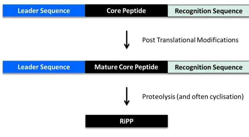
[image:29.595.120.482.244.711.2]All RiPPs have a common general biosynthesis with a precursor peptide ribosomally synthesised containing a leader peptide (or in rarer cases a follower peptide) prior to one or multiple core peptides and in some cases is also followed by a C-terminal enzyme recognition sequence (Figure 1.3) [16]. The full length peptide undergoes post translational modifications with enzymes that interact with the leader sequence and then subsequently modify the core peptide. This process therefore uncouples recognition from catalysis, potentially to allow hypervariable core peptide sequences to be modified by the same set of enzymes. Following modification, the core peptide is proteolytically excised to yield the mature RiPP [16]. In several subcategories of RiPPs the peptide is cyclised to form a macrocycle (e.g. cyanobactins [22] and cyclotides [23]).
Figure 1.3: Schematic of RiPP Biosynthesis. General schematic of RiPP biosynthesis with a ribosomal precursor peptide tailored by enzymatic post-translational modifications. The enzymes act through recognition of the N-terminal leader and in some cases C-terminal recognition sequences. These sequences are subsequently cleaved leaving the mature compound. Figure adapted from Arnison et al (2013) [16]
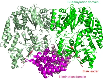
[image:30.595.96.494.344.563.2]dehydrobutyrine respectively in the precursor peptide NisA, one of the early steps in the biosynthesis of the complex molecule NisinA (Figure 1.2 A) [31]. The dehydration is known to occur via the glutamylation of the serine and threonine side chains followed by a glutamate elimination step [32]. The study determined the crystal structure of the NisB in complex with the precursor peptide NisA showing the distinct peptide binding and glutamylation sites (Figure 1.4) [30]. The distinct binding and catalytic sites is an uncommon feature of enzymes in general but is highly prevalent among the RiPP enzymes [16].
Figure 1.4 Structure of NisB in Complex with Substrate NisA. Dimer structure of NisB in complex with NisA with glutamylation domain represented in green, elimination domain in magenta and the NisA leader sequence in red (secondary molecule shown in paler equivalent colours) [30]. All protein structure figures in this thesis were created in Pymol unless otherwise stated [33].
1.1.3 Cyanobactins
The cyanobactins, a member of the RiPPs, are a superfamily of cyclic peptides (macrocycles) produced by both free-living and symbiotic cyanobacteria [22]. The cyanobactins are derived from a precursor peptide which is post-translationally tailored by a range of enzymatic reactions to form cyclic peptides containing modifications such as D-stereo centres, heterocyclised amino acids, disulfide bonds and prenylation. The size of these peptides ranges from six to at least twenty amino acids [22], [34]. The post-translation modifications are carried out by a range of enzymes including proteases, heterocyclases, oxidases and prenyl transferases. Bioinformatic comparisons of the related cyanobactin pathways show a high degree of homology between the related enzymes [35].
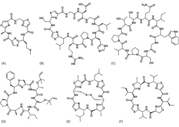
Figure 1.5 represents the diverse range of cyanobactins including tenuecyclamide (N. spongiaeforme) [36], anacyclamide (Anabaena sp.) [34], trichamide (T. erythraeum)
[37] and trunkamide [38], ulithiacyclamide [39] and patellamide [40] (all Prochloron
sp.). The cyanobactins are of particular interest as they have been shown to have
diverse biological effects including anticancer (e.g. Ulithiacyclamide A [41], [42]) and trunkamide A [43]), anti-parasitics (e.g. Venturamide A [44]) as well as reversal of multi-drug resistance (e.g. Dendroamide A [45]). In addition, the cyanobactins are produced using a defined set of tailoring enzymes and so are attractive for bioengineering [46].
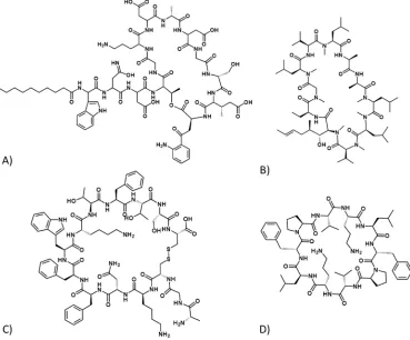
Figure 1.5: Chemical Structures of Cyanobactins. The chemical structures of cyanobactins from the following families (A) Tenuecyclamide, (B) Anacyclamide, (C) Trichamide, (D) Trunkamide, (E) Ulithiacyclamide and (F) Patellamide. (Adapted from Sivonen et al. [22])
[image:33.595.116.478.81.338.2]1.2 Patellamide Biosynthesis
The patellamides are a cyanobactin family produced by Prochloron sp., cyanobacteria that exist symbiotically with the marine sea squirt Lissoclinum patella [47]. L. patella, along with its obligate symbionts, is commonly found in tropical oceans, such as near the Palau Islands or at the Great Barrier Reef, both in the Pacific Ocean[40] [48].
The patellamides are macrocyclised octapeptides containing heterocyclised residues (Thr/Ser and Cys) giving rise to oxazolines and thiazolines, the latter of which can be further oxidised to thiazoles [49] (Figure 1.6). Additionally, D-stereocentres are found adjacent to the thiazoles.
Figure 1.6: Chemical Structure of Patellamides. The chemical structures of (A) Patellamide A and (B) Patellamide D.
The patellamides show considerable chemical diversity and as such their activity is dependent on their cyclic nature, the heterocycles present, and the amino acids side chains which make up the macrocycle. Variation in any of these properties, even subtle changes, can have large consequences on their function [49]. Lissoclinamide 4, a related cyanobactin, shows an increase of two orders of magnitude in cytotoxicity against T24 bladder carcinoma cells compared to lissoclinamide 5, despite varying only by the presence of a thiazoline as opposed to a thiazole, its oxidised analogue. [42] [54].
Although the patellamides function for the organism is unknown, they could potentially be produced as a defence mechanism to protect its environment from other bacterial species. The patellamides have been tested for a range of biological activities and been found to be cytotoxic to leukaemia cells and also having the ability to reverse multiple drug resistance [40][55]. To date the basis of patellamide mode of action is not well defined but there is data to suggest that proteins associated with DNA, RNA and protein synthesis are all targets [49]. Patellamide D is however known to target the ATP-binding cassette multidrug efflux pump P-glycoprotein (P-gp) [55] which regulates the distribution and bioavailability of drugs [56]. Inhibition of this protein could play a role in cancer therapies where chemotherapeutic resistance has occurred. Previous structural studies on the mouse P-gp protein have identified the binding sites for cyclic peptide inhibitors containing heterocycles and it is likely that Patellamide D interacts in a similar manner [57] [58]. The inhibitors are bound in the transmembrane portion of the protein (Figure 1.7).
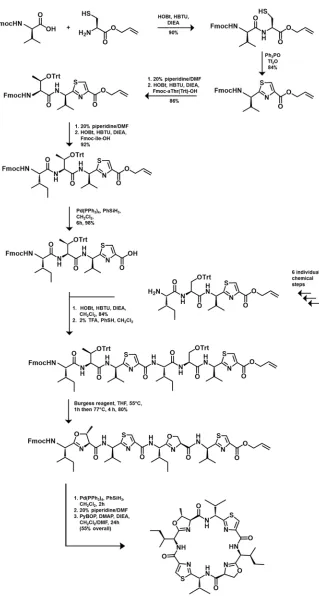
(mirror image with exception of serine/threonine) which allows for some of the initial starting materials to be used for both the initial Ser and Thr containing 4-mers. As a result of this, it is likely that when more diversity is required in the patellamide, the synthesis would require additional steps and reduce the efficiency of the yield further. As such, the chemical synthesis is a plausible yet lengthy strategy for producing patellamides and patellamide-derived novel compounds.
Patellamides are biosynthesised ribosomally as a precursor peptide which undergoes several post-translational modifications through a range of enzymatic (and potentially non-enzymatic) reactions. A single gene cluster (Figure 1.9) of Prochloron sp. has been identified which encodes both the precursor peptide (PatE) and the tailoring enzymes (PatA, B, C, D, F, G) [47]. Studies of the gene cluster when heterologously expressed in
E. coli have shown that PatA, D, E, F and G are essential for patellamide production and
their absence leads to no detectable products. The absence of PatB and PatC however still results in patellamide products [47][61][62].
Figure 1.9: Schematic of Patellamide Gene Cluster. The gene cluster responsible for encoding the seven proteins of the patellamide biosynthetic pathway with seven genes patA-G.
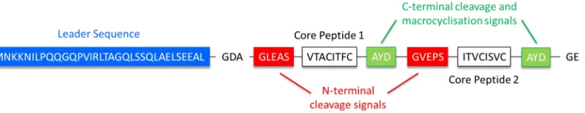
Figure 1.10: Sequence Schematic of the PatE Precursor Peptide. The PatE precursor peptide highlighting the leader sequence (blue), the N-terminal cleavage recognition signals (red), the variable core peptides (white) and the C-terminal cleavage signals (green).
PatA (77 kDa) consists of an N-terminal subtilisin-like protease domain and a C-terminal domain of unknown function (DUF). The protease domain is responsible for N-terminal cleavage of the core peptides [63]. PatB (9 kDa) and PatC (7 kDa) currently have no defined function [61]. PatD (89 kDa) is a three domain protein responsible for heterocyclisation of specific amino acids within the core peptides [64]. PatF (36 kDa) currently has no defined function but equivalent enzymes in related pathways have shown that the PatF family is responsible for the prenylation of specific amino acids within the core peptide [65]. Finally, PatG (131 kDa) consists of three domains, an N-terminal oxidoreductase domain, a subtilisin-like protease domain and a C-N-terminal DUF [63] [66]. The oxidoreductase domain is likely to catalyse the oxidation of thiazolines to thiazoles, however this has yet to be confirmed, while the protease domain carries out C-terminal cleavage and macrocyclisation of each core peptide [63]. In addition, the epimerisation of two C positions adjacent to thiazolines also occurs
and may be an enzymatic or a non-enzymatic process.
[image:39.595.99.522.106.188.2]1.2.1 Heterocyclisation
Note: A selection of the work in this section has been carried out by the Naismith group including myself. This was achieved during the PhD research period but not within the scope of this thesis. All work has been published and references for this work are noted with an asterisk.
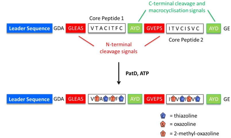
PatD is an ATP-dependent heterocyclase enzyme which catalyses the formation of oxazolines and thiazolines from threonine/serine and cysteine residues respectively (Figure 1.11, 1.12). Each heterocyclisation event results in the loss of one water molecule which can be observed by a reduction in mass of 18 Da [64]. The binding of the PatE precursor peptide to the PatD is mediated through the 37 amino acid leader sequence of the PatE and removal of this sequence abolishes the majority of the heterocyclase activity [64]. The C-terminal cysteine of the core peptide will still process without the leader sequence as long as the C-terminal recognition sequence remains present [67]* [68].
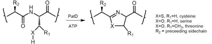
[image:40.595.90.492.434.675.2]Figure 1.12: PatD Reaction at a Molecular Level. Chemical structure schematic of the substrate amino acids cysteine, serine and threonine and their heterocyclic products upon reaction with PatD and ATP.
Within the cyanobactin family there are variations in the type of activity each heterocyclase adopts. TruD, an analogue from the trunkamide pathway, and LynD, an analogue from the aestuaramide pathway, are reported to only heterocyclise cysteine residues and both leave threonine and serine residues in their native state [64] [69]*. This is intriguing considering the high homology of TruD and LynD to PatD; 88 % and 76 % identity respectively (Figure 1.13). The variation between the three enzymes may be a result of chemical selectivity, however one study on PatD and TruD has suggested that this is primarily down to regiospecificity [64]. Two positions within the core peptide (positions 1 and 5 from C-terminus) were found to be more liable to heterocyclisation by TruD, while PatD could act on at least five positions (positions 1, 3, 4, 5 and 7). The order in which the heterocyclisations within the precursor peptide occur has been determined using the heterocyclase TruD and was found to process from the C-terminus backwards [67]*.
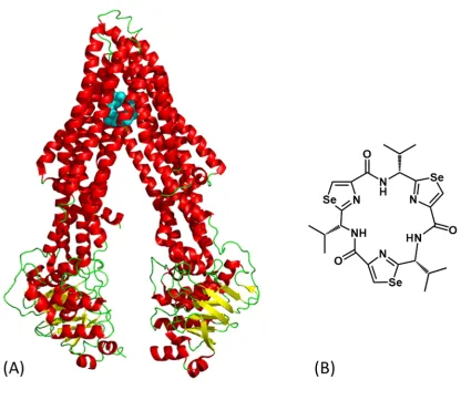
Figure 1.14: Crystal Structure of TruD Heterocyclase Enzyme. TruD crystal structure to 2.95 Å resolution (PDB entry: 4BS9). (A) The asymmetric unit of domains one, two and three coloured magenta, cyan and green respectively. The zinc ion is shown as a grey sphere (B) The dimer representation of TruD which is biologically relevant showing the anti-parallel dimer interface occurring between domains one and two (symmetry molecule in darker equivalent colours) [67]*.
The PatE leader sequence had been proposed to contain a helical structure from residues 13 - 26 [75]which would resemble that of the precursor peptide for microcin B17, where the helix plays a crucial role in peptide:protein binding [76],[77]. These original studies however were carried out in organic solvent and a more recent study has determined using NMR analysis of a 15N labelled PatE in aqueous buffer that there is no degree of secondary structure in the leader sequence [67]*.
secondary molecule essentially bringing the two domains together and confirming the biological relevance of the dimer. The conformational change which occurs upon leader sequence binding results in the activation of the heterocyclase, subsequently allowing complete processing of the core peptide.
[image:44.595.97.499.203.529.2]The catalytic reaction of PatD is known to be ATP-dependent although the mechanism of this reaction remains unclear. McIntosh et al. (2010) initially proposed that heterocyclisation is driven by ATP through a molecular machine basis where there is no direct interaction between the heterocyclisation chemistry and ATP breakdown [79] (Figure 1.16).
Figure 1.16: ATP Molecular Machine Mechanism for PatD: Proposed mechanism for PatD ATP depletion with ATP binding at a distinct site from the PatE causing turnover. Figure adapted from McIntosh et al (2010) [79].
Studies on the related RiPP biosynthetic pathway of thiazole/oxazole-modified microcins (TOMMs) then suggested that heterocyclisation occurs through a kinase mechanism where the peptide backbone carbonyl oxygen is phosphorylated by ATP [80] [81]. This then drives an O-elimination reaction which generates the final oxazoline/thiazoline (Figure 1.17). This releases H2O, ADP and phosphate, the latter of
Figure 1.17 – Kinase Mechanism of Heterocyclisation. Proposed mechanism for heterocycle formation in the TOMMS pathway by BalhC/BalhD/BalhF with ATP phosphorylating the carbonyl oxygen followed by an O-elimination with the release of ADP, PO4- and H2O. X = O or S. (Figured adapted from Dunbar et al., 2012, [80])
Next, a study on the TruD protein showed by an NMR approach that heterocyclisation potentially occurs via an adenylation mechanism through production of AMP and pyrophosphate from ATP during turnover and failed to observe ADP [67]* (Figure 1.18). In addition, the crystal structure identified an adenylase fold in the second domain correlating with the NMR data (Figure 1.14).
Figure 1.18: Adenylation Mechanism of Heterocyclisation. Proposed mechanism for thiazoline formation by TruD with ATP adenylating the carbonyl oxygen followed by an O-elimination with the release of AMP, PPi and H2O. (Figure adapted from Koehnke et al, 2013, [67]*)
The adenylation mechanism appeared to be further backed up by a study on the E.coli YcaO domain, a close homologue of BalhD, which was, in the absence of substrate, shown to also hydrolyse ATP to AMP and PPi [82]
adenylation mechanism [78]*. The binding of ATP in the active site (where in the structure ATP has actually hydrolysed to ADP and PO4-) found that the nucleotide and
the alpha phosphate were buried in a pocket whilst the gamma phosphate was exposed (Figure 1.19) [78]*. This would therefore favour a kinase mechanism through nucleophilic attack of the beta-gamma bond. Thus the two sets of data are conflicting.
Figure 1.19: Nucleotide Binding to LynD. Structural binding of ADP and PO4- to LynD showing structural shielding of the adenosine ring and the alpha phosphate by side chains of the enzyme whilst the beta and gamma phosphates are exposed. The phosphates are co-ordinated by three Mg2+ ions (grey spheres). (Figure adapted from Koehnke et al, 2015, [78]*)
1.2.2 N-terminal Core Peptide Cleavage
The terminal subtilisin-like protease domain of PatA (PatApr) catalyses the N-terminal cleavage of the modified core peptide residue, resulting in the removal of the PatE precursor peptide leader sequence (Figure 1.20). This step must proceed after heterocyclisation due to the requirement of the leader sequence to bind PatD to enable heterocyclase activity [79].
Figure 1.20: PatA Protease Activity. Schematic for the action of PatApr which cleaves at the N-terminal of the modified core peptide (white box) upon recognition of the sequence ‘G (L/V) E (A/P) S’ (red box).
intermediate. The tetrahedral intermediate collapses, breaking the peptide bond and the C-terminal peptide is then released and protonated by His58 thus leaving an acyl-enzyme intermediate. Finally, upon water addition a second tetrahedral intermediate is formed and its subsequent collapse releases the N-terminal peptide and returns the enzyme to its native state, ready to catalyse the next reaction (Figure 1.21).
Figure 1.21: PatA Protease Mechanism. Schematic for the mechanism of action of PatApr showing classic serine protease characteristics.
The crystal structure of PatApr has been determined and is consistent with known subtilisin like protease domains (Figure 1.22, 1.23) [66][87]
Figure 1.23: PatA Structural Alignment with Subtilisin. Structural alignment of the protease domain from PatA (red, PDB code: 3ZXX) with subtilisin (cyan, PDB code: 2ZRQ) shows a high degree of structural homology.
The cleavage by PatApr is a relatively slow reaction (up to 48 hours) when compared to heterocyclisation by PatD (two hours) [63],[79]. This may be intentional to ensure that heterocyclisation can be driven to completion before removal of the leader sequence which reduces heterocyclisation activity [67]*. In all related cyanobactin pathways, a subtilisin-like protease domain (homologous to PatApr) is always present (Figure 1.24) however the N-terminal cleavage recognition sites on the precursor peptide upon which they act can vary quite significantly (Table 1.1).
Table 1.1: PatA Protease and Homologue Recognition Sites. Variation of N-terminal cleavage sites within the cyanobactin precursor peptides which are recognised by the PatApr family of enzymes.
Protease Organism N-terminal Cleavage Site 1 N-terminal Cleavage Site 2
PatApr Prochloron sp. GLEAS GVEPS
PagApr Planktothrix agardhii GLTPH -
LynApr Lyngbya aestuarii GVDAS -
TruApr Prochloron sp GVDAS -
MicApr Microcystis aeruginosa GMDAS GADAS
[image:50.595.206.389.83.290.2]1.2.3 C-terminal Core Peptide Cleavage and Macrocyclisation
The C-terminal cleavage of the modified core peptide is catalysed by the PatG subtilisin-like protease domain (PatGmac). This process occurs after both heterocyclisation (PatD) and N-terminal cleavage (PatApr) because the leader sequence must be removed to allow the N-terminus of the core peptide to bond directly with its C-terminus [64]. PatGmac, like PatApr, contains a classic subtilisin triad and the active site is comprised of residues Asp548, His618 and Ser783 [84], [85]. However, unlike PatApr, PatGmac has macrocyclase activity. At a molecular level, it has been reported that PatGmac recognises the cleavage signal ‘AYD’ in the P1’ – P3’ positions (and possibly Gly in the P4’ position), [88] whilst in many related cyanobactin pathways the C-terminal cleavage signal is ‘SYD’. There is also the requirement for a heterocycle (thiazoline/thiazole, oxazoline/oxazole) or a proline in the final position of the modified core peptide, the residue which also constitutes the P1 position of the protease substrate [88].
Macrocyclisation is a common feature in both non-ribosomal and ribosomal peptide biosynthetic pathways. For non-ribosomal peptides macrocyclisation is generally carried out by thioesterase (TE) domains containing the same Asp-His-Ser catalytic triad as PatGmac and PatApr [89]. The substrate initially binds to a peptidyl carrier protein (PCP) as a thioester, followed by the transfer to the active site serine of a TE domain, forming an acyl-enzyme intermediate [90]. (Figure 1.25 A). The N-terminal amine of the peptide then attacks the ester carbonyl, displacing the serine and forming the macrocycle.
cleavage recognition residues from the active site, the N-terminal amine of the modified core peptide attacks the acyl-enzyme intermediate, displacing the serine and yielding the macrocycle in a similar mechanism to the TE domain [63], [88]. (Figure 1.25 B). The macrocyclisation process has been found to be particularly slow with only one reaction per enzyme per day [63]. This may be to allow time for the N-terminal of the peptide to orientate itself for the nucleophilic attack of the acyl-enzyme intermediate.
Figure 1.25: Peptide Macrocyclisation. Schematic of (A) peptide macrocyclisation via thioesterase intermediate formation and (B) the proposed peptide macrocyclisation by PatGmac showing N-terminal attack of the acyl-enzyme intermediate.
[image:53.595.102.503.239.495.2]1.2.4 Epimerisation
In the patellamides, two of the amino acids, adjacent to the thiazoles, are epimerised to D-stereoisomers (Figure 1.26). The process of this epimerisation is likely to occur
after heterocyclisation and before oxidation of the thiazoline as the -carbon before a thiazoline is more liable to epimerisation due to the adjacent imine bond [94] (Figure 1.27). The pKaof the -carbon when adjacent to a thiazoline will be approximately 30
however when adjacent to a thiazole it will be higher at approximately 40 (estimated using JChem). Epimerisation post-oxidation is possible but disrupting the newly formed, highly stable, aromatic heterocycle makes this less likely.
Figure 1.26: Epimerisation in Patellamides. Chemical structures of A) Patellamide A and B) Patellamide B with the epimerised amino acids adjacent to the thiazoles highlighted in magenta.
There are currently two hypotheses for how the epimerisation occurs; either it is controlled enzymatically or it can occur spontaneously [94]. Bioinformatics analysis of the patellamide gene cluster however shows no evidence of an epimerase enzyme [47]. As there are two epimerisation events, it is possible that these are catalysed individually by the related DUF domains of PatA and PatG (51 % homologous to each other, Figure 1.28), however as they show no sequence homology to any known epimerases, these would represent a novel class.
Figure 1.28: PatA and PatG DUF Alignment. Sequence alignment of the C-terminal domains of unknown function from PatA and PatG.
Although a spontaneous epimerisation remains an option, occurring under these harsh conditions would be unlikely in nature.
Figure 1.29: PatG DUF Structure. Secondary structure representation of the C-terminal DUF domain from PatG (alpha helices – red, beta sheets – yellow) (PDB code: 4UVQ, [95])
1.2.5 Oxidation
Heterocyclisation of threonine/serine and cysteine residues results in the formation of oxazolines and thiazolines respectively as described previously. In final natural products the thiazolines have been further oxidised to form thiazoles (Figure 1.30) [49]. Although at present there is no definitive evidence that the N-terminal domain of PatG carries out this oxidation, it would appear to be highly likely as sequence analysis of this domain shows a high degree of conservation with other oxidases [47], and most cyanobactin pathways also contain a homologous oxidase domain either within the PatG equivalent or as a stand-alone protein [34].
It is also currently unknown at what stage oxidation occurs, whether before or after macrocyclisation of the modified core peptide. An interesting observation is the lack of oxazoles within the patellamides [49], suggesting that the PatG oxidase domain is specific in oxidising only thiazolines. This is in contrast to other cyanobactins where oxazoles and thiazoles occur together (e.g. tenuecyclamides [36], Figure 1.5 A). This may be a result of either substrate recognition (only binds thiazolines) or down to chemistry (can only oxidise thiazolines). At this stage it is also unclear which co-factors are required for oxidase activity, although FMN is required for similar thiazoline oxidases such as in the TOMMS, bleomycin and epothilone biosynthetic pathways [96] [97].
1.2.6 Prenylation
Figure 1.31: Cyanobactin Prenylation. Chemical structures of cyanobactins containing prenylated side chains (magenta). (A) Prenylagaramide B exhibiting forward O-prenylation on a tyrosine residue (B) Trunkamide A containing reverse O-prenylation on both threonine and serine residues [65] (C) an Aestuaramide showing forward C-prenylation on a tyrosine residue [102].
Figure 1.32: Prenylation. Chemical structures of (A) a prenyl group, (B) O-linked forward prenylation (C) O-linked reverse prenylation and (D) Dimethylallyl pyrophosphate (DMAPP).
To date the function of PatF has not been confirmed however studies on related cyanobactin pathways have shown that the PatF family of enzymes is responsible for the prenylation of specific amino acids within the core peptide [65], [103]. TruF1 from the trunkamide pathway (41 % homologous to PatF [62]) catalyses the addition of prenyl groups to the hydroxyl group of threonine and serine residues in the macrocycle [103], while studies on LynF (44 % homologous to PatF) from the Aestuaramide pathway (Lyngbya aestuarii) show that it catalyses prenylation on the hydroxyl of tyrosine residues which is followed by a Claisen rearrangement to yield forward C-prenylation ortho to the hydroxyl group on the aromatic ring [65][104][102].
residues within the core peptide or that PatD, unlike TruD, heterocyclises threonine and serine residues early in the biosynthesis process and as a result these residues would no longer be amenable to prenylation.
Interestingly, although these proteins have some degree of sequence homology among themselves, they show no sequence homology to other known prenyl transferases. The PatF family of enzymes therefore represent a novel subclass of prenyl transferases unique to the cyanobactins.
1.2.7 Non-defined Patellamide Proteins
The PatB and PatC proteins have been determined to be non-essential in the production of patellamides [47],[61], yet these proteins are highly conserved across cyanobactin pathways (Figures 1.33, 1.34). Nevertheless, their protein sequences do not offer any insights into their potential function [34]. There can be several hypotheses for the function in which they may be involved from offering the host a form of resistance against the patellamide or to regulators of the pathway.
1.3 Aims
This thesis is concerned with the ribosomal biosynthesis of the cyanobactin cyclic peptide superfamily, with particularly emphasis on the patellamides.
The study of the prenylase family of enzymes from cyanobactins (PatF family) will be explored through structural and biochemical methods to obtain an understanding of prenylation on the final macrocyclic peptides.
The PatG macrocyclase domain will be explored in structural and biochemical studies to determine substrate binding and rationalise the mechanism of macrocyclisation.
The oxidation step in patellamide biosynthesis has not been characterised to date, but is believed to be catalysed by the N-terminal oxidoreductase domain of PatG. This protein and its homologues will be explored both structurally and biochemically to confirm this hypothesis and determine its associated mechanism.
Through isolation of the proteins (or their individual domains) from the patellamide (or related) biosynthetic pathway(s) the reconstitution of patellamide production in vitro will be pursued the process used to create a diverse range of compounds including known and unknown amino acid sequences, and also in varying peptide length.
2. Structural and Biochemical Studies of PatF and
Homologues
2.1 Introduction
PatF has previously been reported as essential in the production of patellamides on the basis of studies knocking out the gene encoding for it which results in no detectable patellamide products [47]. The biochemical role of PatF in patellamide production has yet to be confirmed; however related pathways have revealed the PatF family to be prenyl transferases. LynF, from Lyngbya aestuarii [65], [104] and PagF from Planktothrix agardhii (unpublished, Nair et al.) have both been shown to prenylate tyrosine residues, while TruF1 from the trunkamide pathway (Prochloron sp.) prenylates serine and threonine residues [103] (Figure 2.1 A). At present no patellamide natural products have been discovered where prenylation is evident so it is unknown which residue(s) PatF acts on (if any).
It is clear that there are significant differences in the mechanisms associated with the closely related prenylases. LynF catalyses the reverse O-prenylation of tyrosine residues which is followed by spontaneous Claisen rearrangement resulting in forward C-prenylation of the tyrosine aromatic ring ortho to the hydroxyl group (Figure 2.1 B) [65][102][104]. PagF carries out forward prenylation directly on the hydroxyl group with no rearrangement (unpublished, personal communication, S. Nair - University of Illinois). Finally, TruF1 also reverse prenylates on oxygen, this time in threonine and serine residues.
2.2 Materials and Methods
2.2.1 DNA Cloning
The gene encoding full length PatF was synthesised in the pJexpress 411 plasmid using optimised codons for E. coli (DNA 2.0)[105]. The plasmid consists of full length PatF with an N-terminal His6-tag and additional Arg and Ser residues at the C-terminus (a
cloning artefact) (Figure 2.2 A).
A Tobacco Etch Virus (TEV) protease recognition site was subsequently introduced between the His6-tag and the PatF protein (Figure 2.2 B) using the site-directed
mutagenesis (SDM) technique of Liu & Naismith [106] (Figure 2.3). Polymerase chain reactions (PCR) were carried out on the original plasmid using KOD polymerase (Novagen) with specifically designed mutagenesis primers (Life technologies, Table 2.1) to insert the nucleotide sequence for TEV recognition site. The template DNA (methylated) was digested using DPN-1 enzyme (20 units) leaving only the newly synthesised (mutated) plasmid. This plasmid was used to transform DH5 E. coli cells. Single colonies were picked and grown in 10 ml Luria-Bertani (LB) media at 37 °C, 200 rpm overnight. The cultures were harvested (4,000 x g) and the plasmid DNA was extracted using the Qiagen mini-prep kit. Purified plasmid DNA was sequenced (GATC Biotech) to confirm the presence of the desired mutation(s).
Figure 2.3: Schematic for Primer Design for Site-directed Mutagenesis. Primer design using non-overlapping and non-overlapping regions for (A) insertion and (B) point mutations. INS indicates the location of insertions, and triangles indicate the locations of mutations in the primer sequences. Figure adapted from Liu & Naismith (2008) [106].
Point mutations of the active site (M136K and H125D/G127R/M136K) were created using the mutagenesis technique using primers shown in Table 2.1. (Note: The triple mutant was made from a single PCR experiment using the single mutant, M136K, as the template.)
5’ PatFTEV gaaaacctgtattttcaggacttgatcgaccgtctgcag
3’ PatFTEV gtcctgaaaatacaggttttcgtggtgatggtgatgatgcat
5' PatF H125DG127R attggtgtggatctgcgtagcaagttggaggacagcagcgtc
3' PatF H125DG127R caacttgctacgcagatccacaccaatggtgttattgataat
5' PatF M136K agcagcgtcaaactgtacattcacatcaaaccggaa
3' PatF M136K aatgtacagtttgacgctgctgtcctccaacttgct
Table 2.1: PatF Primer Sequences. Mutagenesis oligonucleotide sequences for PatF mutagenesis experiments.
The LynF gene was subsequently PCR amplified using specifically designed primers to yield NcoI and XhoI restriction sites on the 5’ and 3’ ends respectively (Table 2.2). The amplified DNA was then digested with NcoI and XhoI restriction enzymes and ligated into the pSUMO plasmid (a gift from C.D. Owen, University of St Andrews) consisting of an N-terminal His6-tag, SUMO (Small Ubiquitin-like Modifier) protein to aid solubility
and TEV protease site (Figure 2.4 B).
Figure 2.4:Schematic for LynF Constructs. (A) LynF construct with N-terminal His6-tag and TEV protease recognition site. (B) LynF SUMO construct with N-terminal His6-tag, SUMO domain and TEV protease recognition site.
5’ LynFNcoI ggcgccatggcgattgcaaaccgtgtaccgtac
3’ LynFXhoI tttctcgagttagccgaagctacgacggta
Table 2.2:LynF Primer Sequences. Oligonucleotide sequences for LynF cloning experiments.
2.2.2 PatF Expression and Purification
PatF was expressed from the pJexpress 411 plasmid using BL21 (DE3) E. coli cells grown on auto-induction medium using the Studier method [107] (see Appendix A.1 for media and buffer composition). The cultures were grown at 20 °C, 250 rpm for 48 hours before harvesting by centrifugation.
(L)-Selenomethionine-labelled PatF was expressed in BL21 (DE3) E. coli cells, cultures of
![Figure 1.2 Chemical Structures of RiPPs. Chemical structures of (A) The lanthipeptide NisinA [24], (B) The LAP](https://thumb-us.123doks.com/thumbv2/123dok_us/8983123.394774/29.595.120.482.244.711/figure-chemical-structures-ripps-chemical-structures-lanthipeptide-nisina.webp)