ISSN Online: 2151-1942 ISSN Print: 2151-1934
DOI: 10.4236/jct.2019.109063 Sep. 19, 2019 747 Journal of Cancer Therapy
Dunaliella salina
and
Haloferax volcanii
Synergistically Attenuate Skin Cancer
in Vitro
Oren Raz1, Ahmad Fahham1, Nona Kuchina2, Zvi Bentwich1,3, Guy Cohen1*
1The Skin Research Institute, The Dead-Sea & Arava Science Center, Masada, Israel 2Clinic Lenom Ltd., Rishon Lezion, Israel
3Department of Microbiology, Immunology and Genetics, Center for Emerging and Tropical Diseases and AIDS, Ben Gurion
University of the Negev, Beer Sheba, Israel
Abstract
Skin cancer, including both melanoma and non-melanoma, is the most common type of malignancy, which causes substantial morbidities and mor-talities. Although the significant increase in the understanding of skin cancer formation and the development of novel personalized drug regimens have occurred, new treatment options are always of need. The use of natural com-pounds to alleviate the symptoms or even to prevent and treat cancer has long been proposed. Specifically, the use of marine-based organisms as a source for cancer cure and remedy is being evaluated extensively. The objec-tive of the current study was to assess the ability of the green microalgae Du-naliella salina, the Dead-Sea-derived Haloferax volcanii, and its combinations to treat skin cancer in vitro. The results demonstrate the Dunaliella and Ha-loferax can reduce sarcoma and basal cell carcinoma cellular growth. Impor-tantly, their combination acts synergistically in a caspase-3 independent manner. Moreover, a synergistic action was found when evaluated sarcoma cell invasion rate, which was completely blocked at pharmacological relevant amounts of the compounds. Collectively, the results demonstrate that the combination of Haloferax volcanii and Dunaliella salina can be used as a new treatment for skin cancer. The specific mechanism of action and further in vivo validation studies are of need.
Keywords
Skin Cancer, Sarcoma, Dunaliella salina, Haloferax volcanii
1. Introduction
In the last decade, the reported incidence of melanoma and non-melanoma skin How to cite this paper: Raz, O., Fahham,
A., Kuchina, N., Bentwich, Z. and Cohen, G. (2019) Dunaliella salina and Haloferax volcanii Synergistically Attenuate Skin Cancer in Vitro. Journal of Cancer Thera-py, 10, 747-754.
https://doi.org/10.4236/jct.2019.109063
Received: August 21, 2019 Accepted: September 16, 2019 Published: September 19, 2019 Copyright © 2019 by author(s) and Scientific Research Publishing Inc. This work is licensed under the Creative Commons Attribution International License (CC BY 4.0).
http://creativecommons.org/licenses/by/4.0/
DOI: 10.4236/jct.2019.109063 748 Journal of Cancer Therapy
cancer has been consistently growing worldwide [1] [2]. These have been pri-marily ascribed to genetic predisposition and increased exposure to environ-mental factors, such as solar radiation, and in particular to ultraviolet (UV) range. The latter induces direct damage to macromolecules within the cells, in-cluding proteins, membranes, and DNA, and regards as the major risk factors for skin cancers formation [3]. UVB-induced carcinogenesis is related to UV absorption by the cell’s DNA, which results in DNA breakdown, and production of mutagenic dimeric photoproducts, namely cyclobutane-pyrimidine dimers (CPDs) and 6-4 photoproducts (6-4PPs) [4]. Both genetic and environmental factors converge eventually to an imbalance between proliferation and differen-tiation states of the cells and alter their ability to migrate and escape the immune system [5].
Squamous cell carcinoma (SCC) is one of the most common life-threatening cancers worldwide [6]. This malignancy also exhibits high recurrence rate fol-lowing therapy. Thus, the use of SCC in screening assays to novel treatments is superior to other skin cancer models. Skin sarcomas comprise a heterogeneous group of malignant mesenchymal tumors that originated in the dermis or sub-cutis [7]. Recreant studies have provided a better understanding of the pathoge-nesis at the molecular level, identifying a new therapeutic target, typically resulting in a good prognosis. However, if surgical removal is incomplete or without suffi-cient excisional margin, distant metastases are rare but extremely lethal [8].
Herbal- and marine-based natural compounds have long been used as a source for cure and remedies [9]. Several active compounds were previously harnessed to alleviate symptoms of cancer, adverse chemotherapy effect, or even as part of the treatment regimen [10].
Dunaliella salina is a green microalga that had been reported to possess several health beneficial effects [11] [12]. In addition to its importance as a nu-tritional source, studies have found neuromodulator [13], antibacterial [14], re-duce cardiac aging [15] and even anti-cancer properties [16]. These observations were attributed to several active compounds, such as phytosterols, glycerol, ca-rotene, and second metabolites. Isolated from the Dead Sea, Haloferax volcanii (formerly Halobacterium volcanii) flourishes in high salinity and has emerged as an important archaeal model system for life in extreme conditions [17]. Howev-er, the possibility to harness this organism as a source of novel natural medicinal compound has not been explored.
In the current study, we investigated the therapeutic properties of Dunaliella and Haloferax volcanii. The results indicate that their combination acts syner-gistically and can be used as a novel treatment option for skin cancer.
2. Materials and Methods
DOI: 10.4236/jct.2019.109063 749 Journal of Cancer Therapy
2.1. Cell Culture
Human skin sarcoma cell line (WS1-CLS) was purchase from CLS Cell Lines Service GmbH. The cells were grown in RPMI 1640 medium supplemented with 2 mM L-glutamine and 10% fetal bovine serum, and 1% (v/v) penicil-lin/streptomycin and maintained at 37˚C in a humidified 5% CO2 incubator.
SCC cell lines were purchased from ATCC and grown similarly in DMEM (dulbecco’s modified eagle medium).
2.2. Cytotoxicity Assay
The ability of the compounds to reduced cancer cell viability was evaluated by an MTT assay, as previously reported, with minor modifications [4]. Briefly, the cells were incubated with 3-(4,5-dimethylthiazol-2-yl)-2,5-diphenyltetrazolium bromide (MTT) (0.5 mg/ml) in PBS for 1 hr at 37˚C. The medium was then as-pirated, and isopropanol was added to solubilize the colored crystals. The ab-sorbance at 570 nm was measured in an ELISA reader.
2.3. Determination of Apoptosis (Caspase-3 Activity Assay)
Following treatment, the cells were exposed to caspase-3 substrate solution (10 µM Caspase 3 substrate II-Fluorogenic (Calbiochem), 0.02% Triton X-100, and 10 mM DTT). The enzyme’s fluorescent product was measured kinetically (20 times at 2-min intervals) using the Thermo Scientific Fluoroskan Ascent™ mi-croplate reader (Ex. 355 nm, Em. 460 nm) [5].
2.4. Invasion Assay
The cancer cell lines were treated without or with the maximal dose of Dunaliel-la salina and Haloferax volcanii that did not reduce the cell’s viability. After 24 hr, the cells were harvested and 50,000 cells were labeled with Calcein-AM for 1 hr and mounted into the invasion chamber (Trevigen), in serum-free conditions. The invasive rate of the tumor cells was determined fluorescently (excitation 485 nm; emission 520; Tecan modular fluorescence system), following the manufac-turer’s instructions.
2.5. Statistical Analysis
Results are given as mean ± SD. Statistical analyses were performed using single factor ANOVA. P < 0.05 is considered significant. All experiments were per-formed in 4 repetition.
3. Results
result-DOI: 10.4236/jct.2019.109063 750 Journal of Cancer Therapy
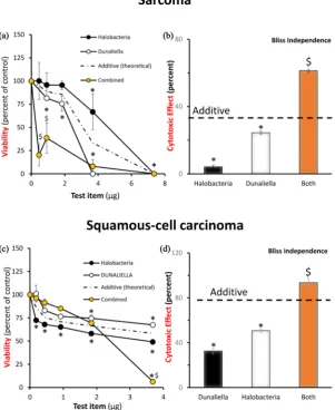
ing in a significant cytotoxic effect (Figure 1(a) & Figure 1(b)). Of note, the combined effect was also higher than double of each individual compound.
Similarly, when the SCC cells were exposed to the compounds, a dose depen-dent reduction was observed. Haloferax volcanii treatment was more potent, but Dunaliella was more effective, resulting in 100% cytotoxic effect at high concentrations. Importantly, a mild but significant synergic action was observed (Figure 1(c) & Figure 1(d)).
[image:4.595.225.527.286.655.2]Next, the ability of the compounds to reduce the ability of the human sarcoma cells and SCC for invasion and migration was evaluated. Thus, the cells were harvested and mounted and treated with one selected non-toxic concentrations of the compounds or combination. As shown in Figure 2, similar synergistic ac-tion was seen for sarcoma cells. However, no added value was observed in SCC (data not shown).
Figure 1. Haloferax volcanii and Dunaliella synergistic act against human skin can-cer cells. (a) Sarcoma cells were treated w/o or with increasing concentrations of
Halofe-rax volcanii, Dunaliella, or both. 24 hr later, the impact on sarcoma cell viability was
DOI: 10.4236/jct.2019.109063 751 Journal of Cancer Therapy
Figure 2. Haloferax volcanii and Dunaliella synergistic reduce human skin cancer cells invasion. The sarcoma cells were harvested and 50,000 cells were mounted in the invasion chamber with Haloferax volcanii, Dunaliella or both, according to the manufac-turer’s instructions. Inhibition of invasion rate is depicted. n = 4, *p < 0.05 in comparison to control; $ indicates synergy.
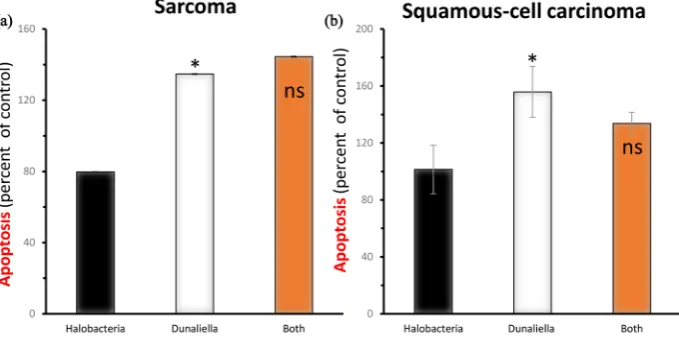
To gain insight into the molecular mechanism underlining the effect of the compound, the hypothesis that the induction of programmed cell death was investigated. In Figure 3, a small but significant enhancement of apoptosis by Dunaliella in both cancer cell lines demonstrates. However, the
supplementa-tion of Haloferax volcanii did not show any further increase in caspase-3 acti-vation.
4. Discussion
The current study was aimed at elucidating the impact of Dunaliella and Halo-ferax volcanii on human skin cancer. The results clearly show synergistic action in two independent models.
The increased prevalence of skin cancer in the last years have been linked to environmental stress, such as UV. Like other forms of cancer, two main aspects defined their harmful potential: the ability to fast increase in mass and their mi-gration capacity, to forms metastasis [5]. Here we show that the combination of Dunaliella and Haloferax volcanii can reduce both. However, the active com-pound(s) should be elucidated prior to drug development.
DOI: 10.4236/jct.2019.109063 752 Journal of Cancer Therapy
Figure 3. The impact of the compound on induction of apoptosis. The ability of the compounds to induce programmed cell death was evaluated by caspase-3 activity assay. n = 4, *p < 0.05 in comparison to control; $ indicates synergy.
caspase-3 actively in both skin cancer cell lines. Another interesting study re-ported once more on the antiproliferative action of Dunaliella [23]; however, that group attributed the antiproliferative action of Dunaliella on skin carci-noma cells to its high β-carotene content. They have also reported that the growth conditions, and in particular stressful culture can increase the potency of the extract with correlation to carotene amount. Interestingly, the use of Dunaliella to even treat radiation damage (such in chemotherapy) have also been reported [24] as well as to reduce chemical induced-cancer formation by 20-methylcholanthrene [25].
Not enough is known on the possible medicinal properties of Haloferax volcanii. This organism can survive at high salinity and was isolated originally at the Dead Sea [26] [27]. In the current study, we have shown that when com-bined with Dunaliella, synergistic action is noticeable. However, this action is not due to induction of apoptosis, as caspase-3 activity remains unchanged by Haloferax. Interestingly, Sikkandar et al. have found a high content of caroteno-ids that correlated with their ability to reduce HepG2 hepatic cancer cell viability
[28]. However, further research is needed to ascertain the mechanism of action (MOA) of both extracts and their synergistic action.
5. Conclusion
The in vitro anti-cancer properties of Dunaliella and Haloferax volcanii were proven. The active compound and MOA should be elucidated in order to further advance these natural compounds as a therapeutic option.
Acknowledgements
[image:6.595.197.537.74.244.2]DOI: 10.4236/jct.2019.109063 753 Journal of Cancer Therapy
Conflicts of Interest
The authors declare no conflicts of interest regarding the publication of this paper.
References
[1] Seebode, C., Lehmann, J. and Emmert, S. (2016) Photocarcinogenesis and Skin Cancer Prevention Strategies. Anticancer Research, 36, 1371-1378.
[2] Martens, M.C., Seebode, C., Lehmann, J. and Emmert, S. (2018) Photocarcinogene-sis and Skin Cancer Prevention Strategies: An Update. Anticancer Research, 38, 1153-1158. https://doi.org/10.21873/anticanres.12334
[3] Armstrong, B.K. and Kricker, A. (2001) The Epidemiology of UV Induced Skin Cancer. Journal of Photochemistry and Photobiology B: Biology, 63, 8-18.
https://doi.org/10.1016/S1011-1344(01)00198-1
[4] Kahremany, S., Babaev, I., Gvirtz, R., Ogen-Stern, N., Azoulay-Ginsburg, S., Sende-rowitz, H., Cohen, G. and Gruzman, A. (2019) Nrf2 Activation by SK-119 Atte-nuates Oxidative Stress, UVB, and LPS-Induced Damage. Skin Pharmacology and
Physiology, 32, 173-181. https://doi.org/10.1159/000499432
[5] Wineman, E., Douglas, I., Wineman, V., Sharova, K., Jaspars, M., Meshner, S., Bentwich, Z., Cohen, G. and Shtevi, A. (2015) Commiphoragileadensis Sap Extract Induces Cell Cycle-Dependent Death in Immortalized Keratinocytes and Human Dermoid Carcinoma Cells. Journal of Herbal Medicine, 5, 199-206.
https://doi.org/10.1016/j.hermed.2015.08.001
[6] Apalla, Z., Nashan, D., Weller, R.B. and Castellsagué, X. (2017) Skin Cancer: Epi-demiology, Disease Burden, Pathophysiology, Diagnosis, and Therapeutic Ap-proaches. Dermatology and Therapy, 7, 5-19.
https://doi.org/10.1007/s13555-016-0165-y
[7] Kohlmeyer, J., Steimle-Grauer, S.A. and Hein, R. (2017) Cutaneous Sarcomas.
JDDG: Journal der Deutschen Dermatologischen Gesellschaft, 15, 630-648.
https://doi.org/10.1111/ddg.13249
[8] Mentzel, T. (2011) Sarcomas of the Skin in the Elderly. Clinics in Dermatology, 29, 80-90. https://doi.org/10.1016/j.clindermatol.2010.07.011
[9] Wargovich, M.J., Woods, C., Hollis, D.M. and Zander, M.E. (2018) Herbals, Cancer Prevention and Health. The Journal of Nutrition, 131, 3034S-3036S.
https://doi.org/10.1093/jn/131.11.3034S
[10] Yin, S.Y., Wei, W.C., Jian, F.Y. and Yang, N.S. (2013) Therapeutic Applications of Herbal Medicines for Cancer Patients. Evidence-Based Complementary and
Alter-native Medicine, 2013, Article ID: 302426. https://doi.org/10.1155/2013/302426
[11] Mishra, A., Kavita, K. and Jha, B. (2011) Characterization of Extracellular Polymeric Substances Produced by Micro-Algae Dunaliella salina. CarbohydratePolymers, 83, 852-857.https://doi.org/10.1016/j.carbpol.2010.08.067
[12] Oren, A. (2005) A Hundred Years of Dunaliella Research: 1905-2005. Saline Sys-tems, 1, 2. https://doi.org/10.1186/1746-1448-1-2
[13] Francavilla, M., Colaianna, M., Zotti, M., Morgese, M.G., Trotta, P., Tucci, P., Schiavone, S., Cuomo, V. and Trabace, L. (2012) Extraction, Characterization and
in Vivo Neuromodulatory Activity of Phytosterols from Microalga Dunaliella
Terti-olecta. Current Medicinal Chemistry, 19, 3058-3067.
https://doi.org/10.2174/092986712800672021
DOI: 10.4236/jct.2019.109063 754 Journal of Cancer Therapy
Dunaliella salina Extracts against Streptococcus mutans. Jundishapur Journal of
Natural Pharmaceutical Products, 13, e13226.https://doi.org/10.5812/jjnpp.13226
[15] El-Baz, F., Abdel Jaleel, G., Saleh, D. and Hussein, R. (2018) Protective and Thera-peutic Potentials of Dunaliella salina on Aging-Associated Cardiac Dysfunction in Rats. Asian Pacific Journal of Tropical Biomedicine, 8, 403-410.
https://doi.org/10.4103/2221-1691.239428
[16] Srinivasan, R., Chaitanyakumar, A., Mageswari, A., Gomathi, A., Pavan, J.G.S., Kumar, M., Jayasindu, G., Bharath, J.S. and Shravan, K.M. (2017) Gothandam, Oral Administration of Lyophilized Dunaliella salina, a Carotenoid-Rich Marine Alga, Reduces Tumor Progression in Mammary Cancer Induced Rats. Food & Function, 8, 4517-4527. https://doi.org/10.1039/C7FO01328K
[17] Pohlschroder, M. and Schulze, S. (2019) Haloferax volcanii. Trends in Microbiolo-gy,27, 86-87. https://doi.org/10.1016/j.tim.2018.10.004
[18] Venugopal, V. (2008) Marine Products for Healthcare: Functional and Bioactive Nutraceutical Compounds from the Ocean.CRC Press, Boca Raton, FL.
https://doi.org/10.1201/9781420052640
[19] Sathasivam, R., Radhakrishnan, R., Hashem, A. and Abd-Allah, E.F. (2019) Micro-algae Metabolites: A Rich Source for Food and Medicine. Saudi Journal of Biological
Sciences, 26, 709-722. https://doi.org/10.1016/j.sjbs.2017.11.003
[20] El-Baz, F.K., Abdo, S.M. and Hussein, A.M.S. (2017) Microalgae Dunaliella salina
for Use as Food Supplement to Improve Pasta Quality. International Journal of
Pharmaceutical Sciences Review and Research, 46, 45-51.
[21] Martínez Andrade, K.A., Lauritano, C., Romano, G. and Ianora, A. (2018) Marine Microalgae with Anti-Cancer Properties. Marine Drugs, 16, 165.
https://doi.org/10.3390/md16050165
[22] Pasquet, V., Morisset, P., Ihammouine, S., Chepied, A., Aumailley, L., Berard, J.B., Serive, B., Kaas, R., Lanneluc, I., Thiery, V., Lafferriere, M., Piot, J.M., Patrice, T., Cadoret, J.P. and Picot, L. (2011) Antiproliferative Activity of Violaxanthin Isolated from Bioguided Fractionation of Dunaliellatertiolecta Extracts. Marine Drugs, 9, 819-831. https://doi.org/10.3390/md9050819
[23] Mo, E., Moghadasi, Z., Rabbani, M., Ma, E., Samadi, S. and Mossaffa, N. (2012) An-ticancer Effect of Dunaliella salina under Stress and Normal Conditions against Skin Carcinoma Cell Line A431 in Vitro. Iranian Journal of Fisheries Sciences, 11, 283-293.
[24] Khayyal, M.T., El-Baz, F.K., Meselhy, M.R., Ali, G.H. and El-Hazek, R.M. (2019) Intestinal Injury Can Be Effectively Prevented by Dunaliella salina in Gamma Irra-diated Rats. Heliyon,5, e01814.https://doi.org/10.1016/j.heliyon.2019.e01814 [25] Raja, R., Hemaiswarya, S., Balasubramanyam, D. and Rengasamy, R. (2007)
Protec-tive Effect of Dunaliella salina (Volvocales, Chlorophyta) against Experimentally Induced Fibrosarcoma on Wistar Rats. Microbiological Research, 162, 177-184. https://doi.org/10.1016/j.micres.2006.03.009
[26] Oren, A. (1999) Benjamin Elazari Volcani (1915-1999): Sixty-Three Years of Studies of the Microbiology of the Dead Sea. International Microbiology, 2, 195-198. https://doi.org/10.1007/s007920050113
[27] Oren, A. and Ventosa, A. (1999) In Memoriam-Benjamin Elazari Volcani.
Interna-tional Journal of Salt Lake Research, 8, 3-6.https://doi.org/10.1007/BF02442132
[28] Rayappan, F. and Nair, A. (2013) Halophilic Bacteria-A Potent Source of Caroteno-ids with Antioxidant and Anticancer Potentials. Journal of Pure and Applied