Hydrothermal Synthesis of V
3
O
7
·H
2
O Nanobelts
and Study of Their Electrochemical Properties
Mohamed Kamel Chine1, Faouzi Sediri1,2*, Neji Gharbi1
1Laboratoire de Chimie de la Matière Condensée IPEIT, Université de Tunis, Montfleury Tunis Tunisia, Tunisia; 2Faculté des
Sci-ences de Tunis, Université Tunis El Manar, Tunis, Tunisia. Email: *faouzi.sediri@ipeit.rnu.tn, neji.gharbi@ipeit.rnu.tn
Received March 15th, 2011; revised April 6th, 2011; accepted May 9th, 2011.
ABSTRACT
Vanadium oxide hydrate V3O7·H2O (H2V3O8) nanobelts have been synthesized by hydrothermal approach using V2O5 as vanadium source and phenolphthalein as structure-directing agent. Techniques X-ray powder diffraction (XRD), scan-ning electron microscopy (SEM), transmission electron microscopy (TEM), infrared spectroscopy and nitrogen adsorp-tion/desorption isotherms have been used to characterize the structure, morphology and composition of the nanobelts. The V3O7·H2O nanobelts are up to several hundreds of nanometers, the widths and thicknesses are 90 and 40 nm, re-spectively. The electroactivity of the nanobelts has been investigated. The as-synthesized material is promising for chemical and energy-related applications such as catalysts, electrochemical device and it may be applied in recharge-able lithium-ion batteries.
Keywords: Nanobelts, V3O7·H2O, Phenolphthalein, Hydrothermal Synthesis, Electroactivity
1. Introduction
Among the various candidates for their much important applications in catalysts [1,2], cathodes materials [3-5] for rechargeable Li-ion batteries, chemical sensors [6,7], vanadium oxides have attracted much attention due to their layered structure and distinctive physicochemical properties [8-10]. In particular, one-dimensional (1D) nanostructural vanadium oxides have been successfully used as electrode materials with greatly enhanced elec-trochemical properties [11,12]. 1D nanostructural vana-dium oxides containing either V4+ or V5+ have a large
capacity which corresponds to reduction of the V4+ or
V5+ by the intercalation process in which Li enters the VOx layers. To best of our knowledge, however, the syn-thesis and electrochemical properties of 1D
nanostruc-tural vanadium oxide composed of V4+ and V5+ have
rarely been reported [13]. Among them nanostructured vanadium oxides have been extensively studied since the
discovery of VOx nanotubes by R. Nesper and his group
[14-15]. They exhibit a great variety of nanostructures, ranging from 1D to 3D [16] and V2O5 has even been chosen as a model system for the description of nanos-tructured materials.
As one of the wet chemistry methods, hydrothermal
treatment has been extensively used for the synthesis of
inorganic compounds [12]. Indeed, Sediri et al. [13]
synthesized the nanoneedles, nanorods of B-VO2, and vanadium oxide nanotubes with high crystallinity via a one-step hydrothermal treatment using crystalline V2O5 as a precursor and various aromatic amines as struc-ture-dire-cting templates. V3O7·H2O·(H2V3O8) has been
studied for many years [19-21]. V3O7·H2O nanofibres
have been prepared firstly by Theobald [19]. Their struc-ture was described as layers that contain VO6 octahedra and VO5 trigonal bipyramids with vanadium oxidation states of +4 and +5, respectively [20]. Recently, the syn-thesis of V3O7·H2O nanobelts was reported by Li and coworkers [22]. In their synthesis, the nanobelts were hydrothermally grown from V2O5 powder in the presence of appropriate amount of hydrochloric acid. Shi et al. [23] obtained single-crystalline vanadium oxide nanobelts through a surfactant-directed growth process under hydr- othermal conditions using V2O5 as a precursor.
ture of V2O5 (0.079 g), phenolphthalein (0.46 g) and dis-tilled water (10 mL). After 2 hours of stirring, the mix-ture was transferred into a Teflon lined steel autoclave with a capacity of 23 ml, and maintained at 180˚C for 4 days under autogenous pression. The pH of the reaction
mixture remains close to pH ≈ 7. After hydrothermal
treatment, the pH of the solution was equal to 4.3. The resulting green powder was washed with water and ethanol to remove the organics residues and then dried at
80˚C for 4 hours. The green color of the powder
sug-gested the presence of the V4+ ions [24].
2.2. Characterization Techniques
X-ray powder diffraction data (XRD) were obtained on a X`Pert Pro Panalytical diffractometer with CoKα
radia-tion (λ = 1.78901 Å) and graphite monochromator. The
XRD measurements were carried out by a step scanning method (2θ range from 2˚ to 50˚), the scanning rate is 0.03˚ s–1 and the step time is 3 s.
Scanning electron microscopy (SEM) images were obtained with a Cambridge Instruments Stereoscan 120.
Transmission electron microscopy (TEM) was carried out with a Philips G20 Ultra-Twin Microscope at an ac-celerating voltage of 200 kV. One droplet of the powder dispersed in CH3CH2OH was deposited onto a car-bon-coated copper grid and left to dry in air.
formed at room temperature.
3. Results and Discussion
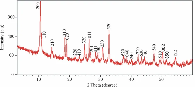
3.1. X-ray Diffraction
The structural properties of the prepared samples were studied by using the X-ray diffraction (XRD). The X-ray diffraction (XRD) pattern of the as-obtained powder after hydrothermal treatment is shown in Figure 1. All of dif-fraction peaks can be perfectly indexed to orthorhombic vanadium oxide crystalline phase V3O7·H2O·(H2V3O8) with lattice constants of a = 16.847 Å, b = 9.362 Å and c
= 3.634 Å (JCPDS 85-2401). No peaks of any other
phases or impurities were observed from the XRD pat-terns, indicating that V3O7·H2O crystalline phase with high purity could be obtained using the present synthetic process.
3.2. Scanning and Transmission Electron Microscopes
[image:2.595.128.468.550.703.2]The surface morphology and size of the as-prepared samples were studied by using the scanning and trans-mission electron microscopes (SEM and MET). The ob-servation by scanning electronic microscopy of the sam-ple (Figure 2) shows that the as-obtained V3O7·H2O is made of a homogenous phase with particles uniformly sized which display belt-like morphology, with a smooth
Figure 2. SEM micrographs (a and b) of the as-synthesized material.
surface and a rectangular cross-section, 2 - 10 min length. The transmission electron microscopy photos of the as-obtained V3O7·H2O shows that the nanobelts are typi-cally 90 nm wide and 40 nm thick (Figure 3). The high magnification HRTEM image clearly exhibits high
(a)
(b)
Figure 3. TEM photos (a and b) of V3O7·H2O nanobelts.
crystallinity (Figure 4).The lattice fringes correspond to a d spacing of 0.47 nm which is consistent with the dis-tance between two (020) crystal planes of the
ortho-rhombic V3O7·H2O crystal, according to JCPDS 85 -
2401 [25]. This result is good agreement with X-ray dif-fraction.
3.3. Infrared Spectroscopy
The structure information was further provided by FTIR spectroscopy. Figure 5 displays the infrared spectrum of
as-synthesized V3O7·H2O nanobelts. The bands at 1021
cm–1, 980 cm–1, and 557 cm–1 are attributed to the char-acteristic of V-O vibration bonds. Indeed, the bands at
1021 cm–1 and 980 cm–1 correspond to the symmetric
stretching of the s (V5+ = O) and s(V4+ = O) bonds, respectively, which indicates the similarity of the as-prepared phase with the structure of the layered or-thorhombic V2O5 [26]. The band at 534 cm–1 corresponds to the stretching vibrations of the s (V-O-V) bridging
bonds [27]. The bands located at 3407 cm–1 and 1634
[image:3.595.91.263.338.701.2]cm–1 come from the s (H-O-H) and s(H2O) vibration, which might indicate that a certain amount of water molecules is embedded between the layers [23]. The re-sults demonstrates that the
Figure 4. HRTEM image of V3O7·H2O nanobelts.
[image:3.595.335.518.562.696.2]Figure 5. FTIR spectrum of V3O7·H2O nanobelts.
Figure 6. Cyclic voltammogams of the as-synthesized V3O7·H2O nanobelts.
material consist of mixed valance state vanadium atoms and water.
3.4. Electrochemical Properties
As an intercalation compound, V3O7·H2O is promising cathode material in lithium-ion batteries [23]. It was re-ported that the electrochemical properties of the electrode materials are influenced by many factors such as instinc-tive structure, morphology, and preparation processes. Therefore, in this paper, we also investigated the elec- trochemical properties of lithium ion intercalation/dein- tercalation of V3O7·H2O nanobelts. The cyclic voltam-mogram (CV) curves of V3O7·H2O are shown in Figure
6. This clearly exhibits that there is a broad cathodic re-duction peak at–0.5 V which result from the lithium ion intercalation process and anodic oxidation peak at 1.2 V, which is attributed to lithium ion deintercalation process, which means that the crystalline structure is reversible. This is a typical phenomenon of VOx active materials and is often reported in the literature [28]. The lithium ion intercalation and deintercalation process can be described by the following process:
V3O7·H2O +x Li+ + x e-↔ LixV3O7·H2O
3.5. Brunauer-Emmett-Teller (BET)
Figure 7. N2 adsorption/desorption isotherms of V3O7·H2O nanobelts.
average pore size (dpor) of V3O7·H2O nanobelts were measured by physisorption of nitrogen according to BET. The obtained results are SBET = 13 m2·g–1, Vpor = 47.10–3 cm3·g–1 and dpor = 141 Ǻ. Furthermore, the analysis of the N2 adsorption and desorption isotherms of the sample leads to identification of the isotherm profiles, as type IV in the BDDT system [29] as shown in Figure 7. This profile is typical of mesoporous materials having pore diameter between 2.0 and 50.0 nm. A hysteresis loop is apparent identified as H3 type (IUPAC), which is char-acteristic of cylindrical pores [29]. H3-type hysteresis loops are usually observed with rigid bulk particles hav-ing a uniform sizes [30]. This result is confirmed by the specific surface area and the average pore size values. These materials are promising for chemical and en-ergy-related applications such as catalysts, and electro-chemical device.
Although the formation mechanism of V3O7·H2O nanobelts remains an open question, experimental data and close investigation of intermediate compounds make it possible to propose a model for the belt formation. To the best of our knowledge and experiments, the whole formation process of V3O7·H2O nanobelts under hydro-thermal conditions can be described as follows.
To explain the mechanism which controls the mor-phology of the material after hydrothermal treatment, a possible intermediate process was suggested on the basis of the layered structure of the precursors [31]. Whereas this structure is stabilized by the phenolphthalein mole-cules, it is sensible that the lamellate structure would break down after displacement of the organic com-pounds.
Through the hydrothermal treatment, it is plausible that the layered precursors would split into nanobelts, and the products obtained would show striated mor-phologies. However, in the hydrothermal synthesis, the lamellar intermediate product would split later in small stems because of the loss of organic molecules. Indeed, the phenolphthalein molecules make the immobilization of this lamellar structure difficult, resulting in the col-lapse of the final lamellar precursor, supporting the exfo-liation of the precursor and the formation of vanadium oxide belts. We think that the phenolphthalein played a double role of reducing and structuring agent in the for-mation of V3O7·H2O nanobelts.
4. Conclusions
In summary, a facile synthetic route to monodisperse crystalline vanadium oxide hydrate (V3O7·H2O) nano-belts has been developed. Indeed, the synthesized V3O7·H2O nanobelts were reported for the first time us-ing phenolphthalein as structure-directus-ing template. The lengths of nanobelts up to several hundreds of nanome-ters, the widths and thicknesses are 90 and 40 nm, re-spectively. The phenolphthalein molecules were mainly as reactants in the development of the formation of V3O7·H2O nanobelts. The formation mechanism of V3O7·H2O nanobelts was clearly explained. Finally, the as-obtained V3O7·H2O nanobelts are promising cathode materials in lithium-ion batteries
REFERENCES
Dehydrogena-chemical Properties of Vanadium Oxide Films on Their nano- and Microstructures,” Journal Physics Chemical B, Vol. 109, No. 35, 2005, pp. 16700-16704.
doi:10.1021/jp051686q
[5] Y. Wang, K. Takahashi, K. Lee and G. Cao, “Nanostruc-tured Vanadium Oxide Electrodes for Enhanced Lith-ium-Ion Intercalation,” Advanced Functional Materials, Vol. 16, No. 9, 2006, pp. 1133-1144.
doi:10.1002/adfm.200500662
[6] M. S. Whittingham, Y. N. Song, S. Lutta, P. Y. Zavalij and N. A. Chernova, “Some Transition Metal (Oxy) Phosphates and Vanadium Oxides for Lithium Batteries”, Journal of Materials Chemistry, Vol. 15, No. 33, April 2005, pp. 3362-3379. doi:10.1039/b501961c
[7] J. Liu, X. Wang, Q. Peng and Y. Li, “Vanadium Pentox-ide Nanobelts: Highly Selective and Stable Ethanol Sen-sor Materials,” Advanced Materials, Vol. 17, No. 6, March 2005, pp. 764-766.
[8] B. X. Li, Y. Xu, G. X. Rong, M. Jing and Y. Xie, “Vana-dium Pentoxide Nanobelts and Nanorolls: from Control-lable Synthesis to Investigation of Their Electrochemical Properties and Photocatalytic Activities,” Nanotechnol-ogy, Vol. 17, No. 10, April 2006, pp. 2560-2566. [9] M. A. Gimenes, L. P. R. Profeti, T. A. F. Lassali, C. F. O.
Graeff and H. P. Oliveira, “Synthesis Characterization, Electrochemical, and Spectroelectrochemical Studies of an N-Cetyl-Trimethylammonium Bromide/V2O5 Nano-composite,” Langmuir, Vol. 17, No. 6, March 2001, pp. 1975-1982.
[10] F. Huguenin, M. Ferreira, V. Zucolotto, F. C. Nart, R. M. Torresi and O. N. Oliveira Jr, “Molecular-Level Manipu-lation of V2O5/Polyaniline Layer-by-Layer Films to Con-trol Electrochromogenic and Electrochemical Properties,” Chemistry of Materials, Vol. 16, No. 11, April 2004, pp. 2293-2299.
[11] Y. Wang and G. Z. Cao, “Synthesis and Enhanced Inter-calation Properties of Nanostructured Vanadium Oxides” Chemistry of Materials, Vol. 18, No. 12, May 2006, pp. 2787-2804.
[12] Y. Wang, K. Takahashi, K. Lee and G. Z. Cao, “Nanos-tructured Vanadium Oxide Electrodes for Enhanced Lith-ium-Ion Intercalation,” Advanced Functional Materials, Vol. 16, No. 9, June 2006, pp. 1133-1144.
[13] H. Qiao, X. J. Zhu, Z. Zheng, L. Liu and L. Z. Zhang, “Synthesis of V3O7·H2O Nanobelts as Cathode Materials
“Vanadium Oxide Nanostructure: from Zero to Three- Dimensional,” Journal Physical-Condemns Matter, Vol. 18, No. 4, 2006, pp. R1-R14.
doi:10.1088/0953-8984/18/4/R01
[17] H. Y. Xu, H. Wang, Z. Q. Song, Y. W. Wang, H. Yan and M. Yoshimura, “Novel Chemical Method for Synthesis of LiV3O8 Nanorods as Cathode Materials for Lithium-Ion Batteries,” Electrochimica Acta, Vol. 49, No. 2, January 2004, pp. 349-353.
[18] F. Sediri and N. Gharbi, “From Crystalline V2O5 to Nanostructured Vanadium Oxides Using Aromatic Amines as Templates,” Journal of Physics and Chemistry of Sol-ids, Vol. 68, No. 10, October 2007, pp.1821-1829. [19] F. Théobald, “Etude Hydrothermale du Système
VO2-VO2,5-H2O,” Journal of the Less-Common Metals, Vol. 53, September 1977, pp. 55-71.
[20] Y. Oka, T. Yao and N. Yamamoto, “Structure Determina-tion of H2V3O8 by Powder X-ray DiffracDetermina-tion,” Journal of Solid State Chemistry, Vol. 89, No. 2, December 1990, pp. 372-377.
[21] T. Chirayil, P. Y. Zavalij and M. S. Whittingham, “Hy-drothermal Synthesis of Vanadium Oxides,” Chemistry of Materials, Vol. 10, No. 10, September 1998, pp. 2629-2640.
[22] G. C. Li, S. P. Pang, Z. B. Wang, H. R. Peng and Z. K. Zhang, “Synthesis of H2V3O8 Single-Crystal Nanobelts”, European Journal of Inorganic Chemistry, Vol. 11, No. 5, June 2005, pp. 2060-2063.
[23] S. F. Shi, M. H. Cao, X. Y. He and H. M. Xie, “Surfac-tant-Assisted Hydrothermal Growth of Single-Crystalline Ultrahigh-Aspect-Ratio Vanadium Oxide Nanobelts,” Crys-tal Growth & Design, Vol. 7, No. 9, July 2007, pp. 1893-1897.
[24] J. Livage, “Vanadium Pentoxide Gels,” Chemistry of Materials, Vol. 3, No. 4, July 1991, pp. 578-593.
[25] S. Gao, Z. Chen, M. Wei, K. Wie and H. Zhou, “Single crystal Nanobelts of V3O7. H2O: A Lithium Intercalation host with a Large Capacity,” Electrochimica Acta, Vol. 54, No. 3, January 2009, pp. 1115-1118.
[26] T. R. Gilson, O. F. Bizri and N. Cheetham, “Dalton Transaction,” Journal of the Chemical Society, Vol. 33, No. 3, 1973, pp. 291-294.
En-gineering B, Vol. 54, No. 3, June 1998, pp. 168-173. [28] C. Delmas, H. Cognac-Auradou, J. M. Cocciantelli, M.
Ménétrier and J. P. Doumerc, “The LixV2O5 System: An overview of the Structure Modifications Induced by the Lithium Intercalation,” Solid State Ionics, Vol. 69, No. 3-4, August 1994, pp. 257-264.
[29] K. S. W. Sing, D. H. Everett, R. A. W. Haul, L. Moscou, R. A. Pierotti, J. Rouquerol and T. Siemieniewska, “Re-porting Physisorption Data for Gas/Solid Systems with Special Reference to the Determination of Surface Area
and Porosity,” Pure and Applied Chemistry, Vol. 57, No. 4, 1985, pp. 603-619.
[30] M. Toba, F. Mizukami, S. Niwa, T. Sano, K. Maeda, A. Annila and V. Komppa, “The Effect of Preparation Methods on the Properties of Zirconia/Silicas,” Journal of Molecular Catalysis, Vol. 94, No. 1, 1994, pp. 85-96. [31] F. Sediri and N. Gharbi, “Controlled Hydrothermal
Syn-thesis of VO2(B) Nanobelts,” Materials Letters, Vol. 63,