doi:10.4236/pp.2011.23022 Published Online July 2011 (http://www.scirp.org/journal/pp)
Determination of Capsaicin Induced Increase in
Dermal Blood Flow Using Laser Doppler
Flowmetry Technique
Sunil Kumar Reddy Khambam, Madireddy Umamaheshwar Rao Naidu*, Pingali Usha Rani, Takallapalli Ramesh Kumar Rao
ICMR Advance Centre for Clinical Pharmacodynamics, Departments of Clinical Pharmacology & Therapeutics, Nizam’s Institute of Medical Sciences, Panjagutta, Hyderabad, India.
Email: *murnaidu@yahoo.com
Received April 28th, 2011; revised May 20th, 2011; accepted June 30th, 2011.
ABSTRACT
In the present study, we evaluated laser Doppler flowmetry technique using LDF100C (Biopac systems) by determining blood flow changes following acute application of capsaicin on 12 healthy human subjects. Capsaicin applied locally
(topical application) at a dose of 0.075% produced significant increase in mean dermal blood flow from 31.4 ± 3.1
Blood Perfusion Units (BPU) to 115. 7 ± 24.6 Blood Perfusion Units (BPU) after 30 minutes, also there was significant difference in dermal blood flow change between placebo (32.1 ± 2.7 BPU) and capsaicin (115.7 ± 24.6 BPU) applica- tion. Capsaicin application produced significant percentage change in dermal blood flow by 291.0 ± 85.3% from base- line, while the change was insignificant with placebo (13.2 ± 7.4%). Therefore, it is suggested that this technique which is technically sound, non-invasive and inexpensive can be adopted in various fields of research to determine blood flow changes and this technique can also be utilized to determine the antagonists of the mediators involved in capsaicin in- duced vasodilatation.
Keywords: Pharmacodynamics, Neurogenic Inflammation, Capsaicin
1.
Introduction
Laser Doppler Flowmetry (LDF) is an established and reliable method for measurement of blood perfusion in microvascular research. Laser Doppler signals from the tissue are recorded in BPU (Blood Perfusion Units) which is a relative units scale defined using a carefully controlled motility standard comprising a suspension of latex spheres undergoing Brownian motion. The LDF technique (Figure 1) offers substantial advantages over other methods in the measurement of microvascular blood perfusion. This technique provides promise and opportunity to adapt the methodology in various fields of research for example in cerebral monitoring (stroke, in- jury …), transplantation surgery (skin grafts, free flaps …), vital organ monitoring (organ viability), tumor vascular research (angiogenesis) and peripheral vascular research (diabetes). Studies have shown that it is both highly sen- sitive and responsive to local blood perfusion and is also versatile and easy to use for continuous monitoring [1,2].
Capsaicin, the pungent ingredient in a wide variety of
hot peppers, has been used extensively in human pain models to induce experimental pain [3-6]. Application of capsaicin to the skin activates the transient receptor po- tential vanilloid type 1 receptor (TRPV1), producing neu- rogenic inflammation and vasodilation [7,9-12]. Most evidence indicates that the release of calcitonin gene- related peptide (CGRP) is a major initiator of this re- sponse [13,8]. Other putative bioactive mediators are substance P (SP), neurokinin (NK) A, somatostatin, ni- tric oxide (NO), histamine and prostaglandins, but their role in capsaicin-induced neurogenic inflammation in the normal human skin is not well established [14-18].
The aim of the study was to determine microvascular changes following acute application of capsaicin using laser Doppler flowmetry Technique.
2.
Materials and Methods
Table 1. Showing demographic characteristics of study par-ticipants.
Parameter N (12)
Age (yrs) Weight (kgs) Height (cms) BMI (kg/m2)
27.8 ± 3.9 68.6 ± 7.7 170.7 ± 4.9
23.5 ± 2.0
[image:2.595.57.289.112.428.2]Values are Mean ±SD.
Figure 1. Shows laser Doppler flowmetry technique. Low power laser light is used to illuminate tissue using a fibre optic; The light is scattered by the static tissue structures and moving blood cells; The moving blood cell impart a Doppler Shift; An adjacent fibre detects light returned from the tissue; This light contains Doppler shifted and Un shifted light; The signal is processed to extract the signal related to the moving red blood cells.
1. All completed study per protocol. Exclusion criteria included: obesity (body mass index >30 kg/m2), under weight (body mass index <18.5 kg/m2), any medication with the potential to alter cardiovascular or thermaoregu- latory control or response, allergies to hot peppers, and various dermatological conditions or diseases.
The study protocol was approved by the institutional human studies committee, Nizam’s Institute of Medical Sciences, Hyderabad, India and complied with the Dec- laration of Helsinki on Biomedical research Involving Human Subjects. Before enrolment, all participants gave informed consent in writing.
Each participant had an initial visit to the experimental laboratory, for a physical examination and a medical history assessment. A 0.075% capsaicin cream (Asian Herbex LTD, Hyderabad, India) was applied on the volar
Figure 2. Shows area of flare 30 minutes post-capsaicin application.
aspect of forearm of one hand to detect any adverse reac- tions to capsaicin. In the absence of symptoms indicating hypersensitivity, including an unusually painful or hy- peremic response to capsaicin, the subjects were cleared to participate in the study.
A Laser Doppler Flowmetry amplifier (BIOPAC Sys- tems, Goleta, CA, USA) with an associated Laser Dop- pler Flowmetry probe (TSD 142) was used to perform the necessary measurements. Experiment was performed in environmental conditions where the temperature was maintained at ~23˚C ± 2˚C.
Application site (10 cm distal to elbow crease) were marked on the volar aspect of the forearm of both non- dominant and dominant hand with a soft pen. Prior to any applications, Laser Doppler Flow (LDF) recording were taken on volar aspect of both the forearms to serve as a baseline measurement.
After the initial measurement, acute capsaicin applica- tions (0.075%) were preceded on the non-dominant fore- arm and placebo (containing ointment base only) appli- cation to the dominant forearm. The blood flow response was measured at 30 minutes post-capsaicin/placebo ap- plication in the area within ring (Figure 2).
Statistical Analysis
Participant’s demographic data is presented as mean ± SD, and pharmacodynamic parameters are presented as mean, Standard Deviation (SD), Standard Error (SE) and 95% of Confidence Interval (CI).
were performed using the Graph pad PRISM software 4 (Graph pad software Inc. San Diego, California, USA).
3.
Results
Dermal blood flow was determined in 12 healthy male subjects’ with mean age, weight, height, and BMI (27.8 ± 3.9 yrs), (68.6 ± 7.7 kgs), (170.7 ± 4.9 cms), and (23.5 ± 2.0 kg/m2) respectively shown in Table 1.
Application of capsaicin didn’t produce any major adverse events; however, there was local flare and mild tingling sensation which disappeared after 4 hrs of ap- plication.
[image:3.595.311.540.86.326.2] [image:3.595.58.286.470.689.2]There was no difference in blood flow at baseline be- tween placebo and capsaicin forearm. Dermal blood flow significantly increased in all the 12 subjects’ thirty min- utes after 0.075% application of capsaicin ointment. Dermal blood flow represented as Blood Perfusion Units (BPU) obtained with placebo and capsaicin is given in
Table 2.
After 30 minutes of capsaicin application the mean dermal blood flow significantly increased from 31.4 ± 3.1 BPU to 115. 7 ± 24.6 BPU, this increase was signifi-cant (p < 0.001). Similarly, there was a signifisignifi-cant dif- ference in dermal blood flow change after 30 minutes between placebo (32.1 ± 2.7 BPU) and capsaicin (115.7 ± 24.6 BPU) application (p < 0.001).
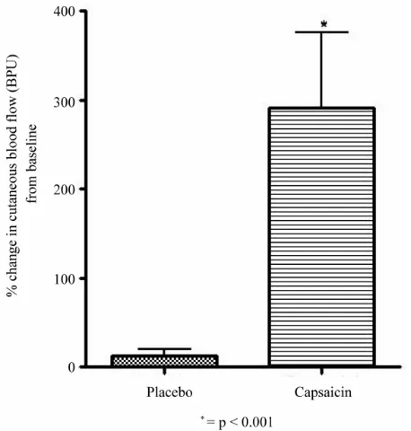
The percentage change from baseline in dermal blood flow in placebo arm and in capsaicin arm is shown in
Figure 3. Placebo application produced insignificant rise
Table 2. Showing cutaneous blood flow (in BPU) before and post placebo/capsaicin application.
Blood Perfusion Units (BPU)
Blood Perfusion Units (BPU)
Placebo Capsaicin Parameter
Baseline 30 minutes
post-application Baseline
30 minutes post-application
Mean 29.8 32.1 31.4ns 115.7*,$
SD 11.9 9.5 10.7 85.4
SE 3.4 2.7 3.1 24.6
Lower 95%
CI of mean 22.2 26.0 24.6
61.4
Upper 95%
CI of mean 37.4 38.1 38.2
169.9
Abbreviations: SD; Standard Deviation, SE; Standard Error, CI; Con-fidence Interval; ns = not significant compared to placebo; * = p < 0.001 compared to baseline; $ = p < 0.001 compared to placebo.
Figure 3. Showing % change in cutaneous blood flow (in BPU) from baseline with placebo and capsaicin.
in blood flow by 13.2 ± 7.4%, while there was a signifi- cant increase in dermal blood flow 30 minutes after the application of 0.075% capsaicin (291.0 ± 85.3%). This difference between placebo and capsaicin was highly significant (p < 0.001).
4.
Discussion
This is the first study to determine capsaicin-induced neurogenic inflammation using Laser Doppler flowmetry technique.
In the present study we have demonstrated the effect of local application of capsaicin on microcirculation of forearm skin in healthy male subjects using laser Dop- pler flowmetry technique. Earlier technique of Laser Doppler perfusion imaging has been described to have good reproducibility to detect microvascular changes on skin surface.
Exploration of microcirculation by laser Doppler technology has often been considered poorly reproduci- ble [19]. Introduction of laser Doppler perfusion imaging has considerably improved reproducibility of this tech-nique [20-22]. As the clear guideline for measurement of digital blood flow by laser doppler perfusion imaging are available now, it has been regularly used to assess cuta- neous blood flow of normal and irritated skin [20]. Ear- lier study has revealed a dose-dependent increase in digital blood flow after capsaicin application on forearm [23].
significantly increased dermal blood flow as compared to placebo. Van der Schueren et al., have demonstrated the increase in blood flow after application of 300 and 1000 micro grams of capsaicin while the application of 100 micro grams didn’t produce any increase in blood flow [23].
In our study the blood flow measurement was carried- out after 30 minutes application of capsaicin. It has been shown that 1000 micrograms of capsaicin produced the maximum response between 30 and 45 minutes after application and at 60 minutes time point there was de- crease in blood flow [23]. There was increase in blood flow (291 ± 85.3 %) with 0.075% capsaicin application compared to placebo in our study. Helme and McKernan have extensively investigated the wide variation in size and intensity of capsaicin induced flare response [7]. According to these authors, site of application and age were the factors responsible for large variation. Gazerani
et al. recently found that capsaicin induced sensory and vasomotor responses were also gender specific [24]. The present study was conducted only in male gender. Het- erogeneity in the density and function of capsaicin sensi- tive nociceptive nerve endings of the dermal microcircu- lation seem to be the most plausible explanation. There may be variation in the response due to difference be- tween the proximal and distal forearm, due to difference in skin thickness [23]. We have thus applied the capsai- cin 10 cms distal to elbow crease to avoid this site varia- tion.
Munce and Kenney reported decrease in local skin blood flow response in older participants than healthy males [25]. Age had a significant effect on local vasodi- lation with younger individuals having percentage of maximal cutaneous vascular conductance (% CVCmax) at
least 2 times greater than seen in older group. Previous observations of decrease flare size in older individuals after acute application of capsaicin suggest that there is reduction in reduced skin blood flow in these subjects [7]. We included only younger individuals, the average age of participants in our study was 27.8 ± 3.9 yrs. Acute capsaicin stimulates a specific population of sensory nerves in the skin that possess capsaicin receptors, elic- iting local release of vasoactive neurotransmitters from their nerve endings [25]. The further use of our model can have advantage in dose finding and proof of concept studies.
5.
Conclusions
The present study has shown an increase in the dermal blood flow induced by topical application capsaicin on the human forearm using laser Doppler flowmetry tech- nique. Hence, this pharmacodynamic model which is non- invasive, technically uncomplicated and sound, might
therefore facilitate the early evaluation of antagonists of mediators involved in neurogenic inflammation, includ- ing CGRP, TRPV1 and possibly, SP antagonists in hu- mans.
6.
Acknowledgements
The study was funded through the Indian Council of Medical Research (ICMR) fund, Government of India. The authors declare no financial conflict of interest con- nected to this study and its results.
We thank the Director, Nizam’s institute for provid- ing us the necessary infrastructure.
7.
References
[1] M. F. Swiontkowski, “Laser Doppler Flowmetry—De- velopment and Clinical Application,” The Iowa Ortho-
paedic Journal, Vol. 11, 1991, pp. 119-126.
[2] A. Sandner-Kiesling, G. Litscher, H. Voit-Augustin, R. L. James and G. Schwarz, “Laser Doppler Flowmetry in Combined Needle Acupuncture and Moxibustion: A Pilot Study in Healthy Adults,” Lasers in Medical Science, Vol. 16, No. 3, 2001, pp. 184-191.doi:10.1007/PL00011353 [3] J. Dirks, P. Fabricius, K. L. Petersen, M. C. Rowbotham
and J. B. Dahl, “The Effect of Systemic Lidocaine on Pain and Secondary Hyperalgesia Associated with Heat/ Capsaicin Sensitization Model in Healthy Volunteers,”
Anesthesia & Analgesia, Vol. 91, No. 4, 2000, pp.
967-972.doi:10.1097/00000539-200010000-00037
[4] K. L. Petersen and M. C. Rowbotham, “A New Human Experimental Pain Model: The Heat/Capsaicin Sensitiza-tion Model,” NeuroReport, Vol. 10, No. 7, 1999, pp. 1511- 1516.doi:10.1097/00001756-199905140-00022
[5] K. L. Petersen, B. Jones, V. Segredo, J. B. Dahl and M. C. Rowbotham, “Effect of Remifentanil on Pain and Sec- Ondary Hyperalgesia Associated with the Heat-Capsaicin Sensitization Model in Healthy Volunteers,” Anesthesiol- ogy, Vol. 94, No. 1, 2001, pp. 15-20.
doi:10.1097/00000542-200101000-00008
[6] H. Sumikura, O. K. Andersen, A. M. Drewes and L. Ar- endt-Nielsen, “Spatial and Temporal Profiles of Flare and Hyperalgesia after Intradermal Capsaicin,” Pain, Vol. 105, No. 1-2, 2003, pp. 285-291.
doi:10.1016/S0304-3959(03)00243-4
[7] R. D. Helme and S. McKernan, “Neurogenic Flare Re- Sponses Following Topical Application of capsaicin in Humans,” Neurology, Vol. 18, No. 4, 1985, pp. 505-509.
doi:10.1002/ana.410180414
[8] S. R. Hughes and S. D. Brain, “A Calcitonin Gene-Re- lated Peptide (CGRP) Antagonist (CGRP8-37) Inhibits Micro-Vascular Responses Induced by CGRP and Cap-saicin in Skin,” British Journal of Pharmacology, Vol. 104, No. 3, 1991, pp. 738- 742.
[9] P. Holzer, “Capsaicin: Cellular Targets, Mechanisms of Action, and Selectivity for Thin Sensory Neurons,” Phar-
macol Review, Vol. 43, No. 2, 1991, pp. 143-201.
Kotzmann and R. Payne, “Effects of Capsaicin on Cuta- Neous Vasodilator Responses in Humans,” Inflammation
Research, Vol. 37, No. 1-2, 1992, pp. 53-59.
doi:10.1007/BF01987890
[11] D. A. Simone and J. Ochoa, “Early and Late Effects of Prolonged Topical Capsaicin on Cutaneous Sensibility and Neurogenic Vasodilatation in Humans,” Pain, Vol. 47, No. 3, 1991, pp. 285-294.
doi:10.1016/0304-3959(91)90217-L
[12] D. P. Stephens, N. Charkoudian, J. M. Benevento, J. M. Johnson and J. L. Saumet, “The Influence of Topical Capsaicin on the Local Thermal Control of Skin Blood Flow in Humans,” American Journal of Physiology, Vol. 281, No. 3, 2001, pp. R894-R901.
[13] S. D. Brain and T. J. Williams, “Inflammatory Oedema Induced by Synergism between Calcitonin Gene-Related Peptide (CGRP) and Mediators of Increased Vascular Permeability,” British Journal of Pharmacology, Vol. 86, Vol. 4, 1985, pp. 855-860.
[14] M. K. Herbert, R. Tafler, R. F. Schmidt and K. H. Weis, “Cyclooxygenase inhibitors acetylsalicylic acid and In- Domethacin do not Affect Capsaicin-Induced Neurogenic Inflammation in Human Skin,” Agents Actions, Vol. 38, 1993, pp. C25-C27.doi:10.1007/BF01991126
[15] M. Huttunen, I. T. Harvima, L. Ackermann, R. J. Har- vima, A. Naukkarinen and M. Horsmanheimo, “Neu- Ropeptide- and Capsaicin-Induced Histamine Release in Skin Monitored with the Microdialysis Technique,” Acta
dermatovenereologica, Vol. 76, No. 3, 1996, pp. 205-209.
[16] L. J. Petersen, K. Winge, E. Brodin and P. S. Skov, “No release of Histamine and Substance P in Capsaicin-In- duced Neurogenic Inflammation in Intact Human Skin in vivo: A Microdialysis Study,” Allergy & Clinical
Immu-nology, Vol. 27, No. 8, 1997, 957-965.
doi:10.1111/j.1365-2222.1997.tb01239.x
[17] R. Tafler, M. K. Herbert, R. F. Schmidt and K. H. Weis, “Small Reduction of Capsaicin-Induced Neurogenic In- Flammation in Human Forearm Skin by the Glucocorti-coid Prednicarbate,” Agents Actions, Vol. 38, 1993, pp. C31- C34.doi:10.1007/BF01991128
[18] J. Wallengren, “Vasoactive Peptides in the Skin,”Journal
of Investigative Dermatology Symposium Proceedings,
Vol. 2, No. 1, 1997, pp. 49-55.
[19] A. M. Schabauer and T. W. Rooke, “Cutaneous Laser Doppler Flowmetry: Applications and Findings,” Mayo
Clinic Proceeding, Vol. 69, No. 6, 1994, pp. 564-574.
[20] A. Fullerton, M. Stucker, K. P. Wilhelm, K. Wardell, C. Anderson, T. Fischer, G. E. Nilsson and J. Serup, “Guide- lines for Visualization of Cutaneous Blood Flow by Laser Doppler Perfusion Imaging. A Report from the Stan-dardization Group of the European Society of Contact Dermatitis Based upon the HIRELADO European com- munity Project,” Contact Dermatitis, Vol. 46, No. 3, 2002a, pp. 129-140.
doi:10.1034/j.1600-0536.2002.460301.x
[21] S. Kubli, B. Waeber, A. Dalle-Ave and F. Feihl, “Repro- ducibility of Laser Doppler Imaging of Skin Blood Flow as a Tool to Assess Endothelial Function,” Journal of
Cardiovascular Pharmacology, Vol. 36, No. 5, 2000, pp.
640-648.doi:10.1097/00005344-200011000-00014
[22] A. Fullerton, B. Rode and J. Serup, “Studies of Cutane-ous Blood Flow of Normal Forearm Skin and Irritated Forearm Skin Based on High-Resolution Laser Doppler Perfusion Imaging (HR-LDPI),” Skin Research and
Technology, Vol. 8, No. 1, 2002b, pp. 32-40.
doi:10.1046/j.0909-752x.2001.10327.x
[23] B. J. Van der Schueren, J. N. de Hoon, F. H. Vanmolkot, A. Van Hecken, M. Depre, S. A. Kane, I. De Lepeleire and S. R. Sinclair, “Reproducibility of the Capsaicin-In- duced Dermal Blood Flow Response as Assessed by La-ser Doppler Perfusion Imaging,” British Journal of Clinic
Pharmacology, Vol. 64, No. 5, 2007, pp. 580-590.
doi:10.1111/j.1365-2125.2007.02939.x
[24] P. Gazerani, O. K. Andersen and L. Arendt-Nielsen, “A Human Experimental Capsaicin Model for Trigeminal Sensitization, Gender-specific Differences,” Pain, Vol. 118, No. 1-2, 2005, pp. 155-163.
doi:10.1016/j.pain.2005.08.009
[25] T. A. Munce and W. Larry Kenney, “Age-Specific Skin Blood Flow Responses to Acute Capsaicin” Journal of