6-Methylpyridine-2(1
H
)-thione
Guo-Jun Lian,aBo Chen,bTing-Ting Zhang,bLi-Zhen Zhuangband Hong-Ze Liangb*
a
School of Public Health of Wenzhou Medical College, Wenzhou 325035, People’s Republic of China, andbState Key Laboratory Base of Novel Functional Materials and Preparation Science, Faculty of Materials Science and Chemical Engineering, Ningbo University, Ningbo 315211, People’s Republic of China
Correspondence e-mail: [email protected]
Received 9 April 2010; accepted 19 April 2010
Key indicators: single-crystal X-ray study;T= 295 K; mean(C–C) = 0.003 A˚; Rfactor = 0.043;wRfactor = 0.148; data-to-parameter ratio = 20.2.
There are two unique molecules in the asymmetric unit of the title pyridinethione derivative, C6H7NS, each of which adopts
the thione rather than the mercaptan form. The rings in both molecules are essentially planar, with maximum deviations from the least-squares planes through all non-H atoms of 0.021 (2) and 0.017 (2) A˚ . In the crystal structure, the molecules form centrosymmetric cyclic dimers through inter-molecular N—H S hydrogen bonds. Additional C— H(methyl) S interactions generate a three-dimensional network.
Related literature
For the synthesis of 2-mercaptopyridines, see: Thirtle (1946). For background to the applications of organic sulfur-containing compounds, see: Cui et al. (2009); Saadat et al. (2004); Qianet al.(2007). For metal complexes of 2-mercapto pyridine N-oxide and 6-methyl substituted derivatives, see: Hamaguchiet al.(2007); Chunchuryukinet al.(2006); Cotton et al.(1978); Westet al.(1998); Fieldinget al.(1997); Berardini et al.(1997); Tylickiet al.(1995); Honget al.(1999); Cabezaet al.(2007).
Experimental
Crystal data
C6H7NS
Mr= 125.19
Monoclinic,P21=c
a= 7.4608 (15) A˚
b= 14.902 (3) A˚
c= 11.665 (2) A˚
= 94.85 (3)
V= 1292.3 (4) A˚3
Z= 8
MoKradiation
= 0.39 mm1
T= 295 K
0.330.330.20 mm
Data collection
Rigaku R-AXIS RAPID diffractometer
Absorption correction: multi-scan (ABSCOR; Higashi, 1995)
Tmin= 0.880,Tmax= 0.926
12472 measured reflections 2944 independent reflections 2088 reflections withI> 2(I)
Rint= 0.030
Refinement
R[F2> 2(F2)] = 0.043
wR(F2) = 0.148
S= 1.12 2944 reflections
146 parameters
H-atom parameters constrained
max= 0.36 e A˚ 3
min=0.26 e A˚3
Table 1
Hydrogen-bond geometry (A˚ ,).
D—H A D—H H A D A D—H A
N1—H1A S1i
0.86 2.50 3.3376 (19) 165 N2—H2A S2ii 0.86 2.50 3.340 (2) 166 C1—H1B S1i
0.96 2.79 3.678 (3) 154 C7—H7A S2ii
0.96 2.74 3.639 (3) 156
Symmetry codes: (i)xþ1;y;zþ2; (ii)xþ2;y;zþ1.
Data collection:RAPID-AUTO (Rigaku, 1998); cell refinement:
RAPID-AUTO; data reduction: CrystalStructure (Rigaku/MSC, 2004); program(s) used to solve structure: SHELXS97 (Sheldrick, 2008); program(s) used to refine structure:SHELXL97(Sheldrick, 2008); molecular graphics: SHELXTL (Sheldrick, 2008); software used to prepare material for publication:SHELXL97.
The authors thank the Critical Projects in Science and Technology Department of Zhejiang Province (No. 2007 C21113), the Cultivation Program of Young and Middle-aged Academic Leaders in Zhejiang Higher Education Institutions, the Natural Science Foundation of Ningbo City (No. 2009 A610047) and the K. C. Wong Magna Fund of Ningbo University for financial support.
Supplementary data and figures for this paper are available from the IUCr electronic archives (Reference: SJ2766).
References
Berardini, M., Lee, J., Freedman, D., Lee, J., Emge, T. J. & Brennan, J. G. (1997).Inorg. Chem.36, 5772–5776.
Cabeza, J. A., del Rı´o, I., Garcia-A´ lvarez, P. & Miguel, D. (2007). J. Organomet. Chem.692, 3583–3587.
Chunchuryukin, A. V., Chase, P. A., Mills, A. M., Lutz, M., Spek, A. L., van Klink, G. P. M. & van Koten, G. (2006).Inorg. Chem.45, 2045–2054. Cotton, F. A., Fanwick, P. E. & Fitch, J. W. III (1978).Inorg. Chem.17, 3254–
3257.
Cui, H., Turn, S. Q. & Reese, M. A. (2009).Catal. Today,139, 274–279. Fielding, C., Parsons, S. & Winpenny, R. E. P. (1997).Acta Cryst.C53, 174–176. Hamaguchi, T., Ujimoto, K. & Ando, I. (2007).Inorg. Chem.46, 10455–10457. Higashi, T. (1995).ABSCOR. Rigaku Corporation, Tokyo, Japan.
Hong, M., Su, W., Cao, R., Zhang, W. & Lu, J. (1999).Inorg. Chem.38, 600– 602.
Qian, X., Li, Z. & Yang, Q. (2007).Bioorg. Med. Chem.15, 6846–6851. Rigaku (1998).RAPID-AUTO. Rigaku Corporation, Tokyo, Japan. Rigaku/MSC (2004). CrystalStructure. Rigaku/MSC Inc., The Woodlands,
Texas, USA.
organic compounds
o1154
Lianet al. doi:10.1107/S1600536810014273 Acta Cryst.(2010). E66, o1154–o1155 Acta Crystallographica Section EStructure Reports
Online
Saadat, M., Bahaoddini, A. & Nazemi, S. (2004). Biochem. Biophys. Res. Commun.313, 568–569.
Sheldrick, G. M. (2008).Acta Cryst.A64, 112–122. Thirtle, J. R. (1946).J. Am. Chem. Soc.68, 342–343.
Tylicki, R. M., Wu, W., Fanwick, P. E. & Walton, R. A. (1995).Inorg. Chem.34, 988–991.
supporting information
sup-1 Acta Cryst. (2010). E66, o1154–o1155
supporting information
Acta Cryst. (2010). E66, o1154–o1155 [https://doi.org/10.1107/S1600536810014273]
6-Methylpyridine-2(1
H
)-thione
Guo-Jun Lian, Bo Chen, Ting-Ting Zhang, Li-Zhen Zhuang and Hong-Ze Liang
S1. Comment
Organic sulfur-containing compounds have been frequently encountered or used in chemical industry (Cui et al., 2009),
life science (Saadat et al., 2004) and pharmacy (Qian et al., 2007). Some of them have found their applications in crystal
engineering. 2-mercaptopyridine, its 6-methyl substituted and N-oxide derivatives have exhibited rich coordination
motifs. They can serve as monodentate (Hamaguchi et al., 2007; Chunchuryukin et al., 2006), bidentate (Cotton et al.,
1978; West et al., 1998; Fielding et al., 1997; Berardini et al., 1997), and bridging (Tylicki et al., 1995; Hong, et al.,
1999; Cabeza, et al., 2007) ligands to coordinate metals. Though syntheses and crystal structures of these metal
complexes have been reported, the crystal structure of 2-mercapto-6-methylpyridine itself has not been yet reported. Here
we report its crystal structure and packing pattern.
A perspective view of the title compound is shown in Fig. 1. The C—S bond lengths were 1.694 (3) and 1.700 (2) Å,
shorter than those in the above-mentioned metal complexes (typically 1.740 Å). This clearly indicates that the neutral title
compound in solid state exists as a pyridinethione, while in metal complexes it ligates to metal centers as a
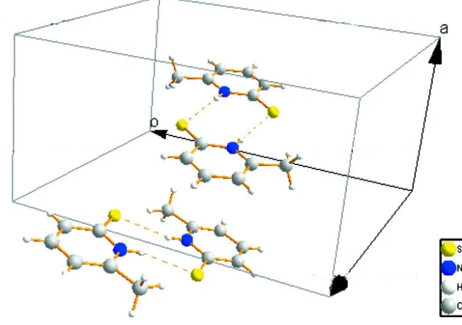
pyridinethiol-ate anion. As shown in Fig. 2, adjacent two molecules are linked by intermolecular N–H···S interactions, forming a cyclic
dimer.
S2. Experimental
6-methylpyridine-2(1H)-thione was synthesized by a literature method (Thirtle 1946). X-ray quality single crystals were
grown from ethyl acetate and petroleum ether (1/10, v/v).
S3. Refinement
H atoms bonded to C atoms were placed in geometrically calculated position and were refined using a riding model, the
C–H bond lengths are 0.93 or 0.96 [Uiso(H) = 1.2 Ueq(C)] or [Uiso(H) = 1.5 Ueq(C)] for the methyl group, H atoms
bonded to N atoms were placed in geometrically calculated positions and were also refined as riding [Uiso(H) = 1.2
Figure 1
A perspective view of the title compound. Displacement ellipsoids are drawn at the 45% probability level.
Figure 2
Part of the packing of the title compound showing the formation of centrosymmetric dimers.
6-Methylpyridine-2(1H)-thione
Crystal data
C6H7NS Mr = 125.19 Monoclinic, P21/c
Hall symbol: -P 2ybc a = 7.4608 (15) Å b = 14.902 (3) Å c = 11.665 (2) Å
β = 94.85 (3)° V = 1292.3 (4) Å3 Z = 8
F(000) = 528 Dx = 1.287 Mg m−3
[image:4.610.141.472.318.551.2]supporting information
sup-3 Acta Cryst. (2010). E66, o1154–o1155
θ = 3.1–27.4° µ = 0.39 mm−1 T = 295 K
Block, yellow
0.33 × 0.33 × 0.20 mm
Data collection
Rigaku R-AXIS RAPID diffractometer
Radiation source: fine-focus sealed tube Graphite monochromator
Detector resolution: 0 pixels mm-1 ω scans
Absorption correction: multi-scan (ABSCOR; Higashi, 1995) Tmin = 0.880, Tmax = 0.926
12472 measured reflections 2944 independent reflections 2088 reflections with I > 2σ(I) Rint = 0.030
θmax = 27.4°, θmin = 3.1° h = −9→9
k = −19→19 l = −15→15
Refinement
Refinement on F2
Least-squares matrix: full R[F2 > 2σ(F2)] = 0.043 wR(F2) = 0.148 S = 1.12 2944 reflections 146 parameters 0 restraints
Primary atom site location: structure-invariant direct methods
Secondary atom site location: difference Fourier map
Hydrogen site location: inferred from neighbouring sites
H-atom parameters constrained w = 1/[σ2(F
o2) + (0.0738P)2 + 0.3029P]
where P = (Fo2 + 2Fc2)/3
(Δ/σ)max < 0.001
Δρmax = 0.36 e Å−3
Δρmin = −0.26 e Å−3
Special details
Geometry. All esds (except the esd in the dihedral angle between two l.s. planes) are estimated using the full covariance matrix. The cell esds are taken into account individually in the estimation of esds in distances, angles and torsion angles; correlations between esds in cell parameters are only used when they are defined by crystal symmetry. An approximate (isotropic) treatment of cell esds is used for estimating esds involving l.s. planes.
Refinement. Refinement of F2 against ALL reflections. The weighted R-factor wR and goodness of fit S are based on F2,
conventional R-factors R are based on F, with F set to zero for negative F2. The threshold expression of F2 > σ(F2) is used
only for calculating R-factors(gt) etc. and is not relevant to the choice of reflections for refinement. R-factors based on F2
are statistically about twice as large as those based on F, and R- factors based on ALL data will be even larger.
Fractional atomic coordinates and isotropic or equivalent isotropic displacement parameters (Å2)
x y z Uiso*/Ueq
S1 0.52754 (9) 0.10281 (4) 0.87258 (5) 0.0561 (2)
S2 0.90012 (12) 0.13575 (4) 0.50014 (6) 0.0743 (3)
N1 0.5873 (2) −0.07318 (12) 0.85963 (15) 0.0470 (4)
H1A 0.5725 −0.0723 0.9319 0.056*
N2 0.9262 (2) 0.02468 (12) 0.32400 (15) 0.0488 (4)
H2A 0.9555 −0.0139 0.3771 0.059*
C1 0.6385 (5) −0.23318 (17) 0.8905 (2) 0.0746 (8)
H1B 0.6160 −0.2149 0.9669 0.112*
H1C 0.7568 −0.2587 0.8916 0.112*
H1D 0.5509 −0.2772 0.8633 0.112*
C2 0.6257 (3) −0.15375 (16) 0.8125 (2) 0.0541 (6)
C3 0.6503 (4) −0.15630 (18) 0.6979 (2) 0.0626 (6)
C4 0.6350 (4) −0.0782 (2) 0.6339 (2) 0.0658 (7)
H4A 0.6513 −0.0800 0.5557 0.079*
C5 0.5961 (3) 0.00181 (18) 0.68372 (19) 0.0593 (6)
H5A 0.5868 0.0536 0.6392 0.071*
C6 0.5702 (3) 0.00646 (15) 0.80167 (18) 0.0469 (5)
C7 0.9694 (4) −0.10058 (16) 0.1955 (2) 0.0609 (6)
H7A 0.9963 −0.1291 0.2687 0.091*
H7B 1.0723 −0.1042 0.1516 0.091*
H7C 0.8693 −0.1303 0.1546 0.091*
C8 0.9229 (3) −0.00394 (16) 0.21353 (19) 0.0499 (5)
C9 0.8776 (3) 0.05620 (18) 0.1275 (2) 0.0590 (6)
H9A 0.8744 0.0386 0.0508 0.071*
C10 0.8365 (4) 0.14393 (18) 0.1558 (2) 0.0634 (7)
H10A 0.8072 0.1853 0.0975 0.076*
C11 0.8384 (3) 0.17037 (16) 0.2678 (2) 0.0581 (6)
H11A 0.8074 0.2290 0.2849 0.070*
C12 0.8870 (3) 0.10976 (15) 0.3583 (2) 0.0517 (5)
Atomic displacement parameters (Å2)
U11 U22 U33 U12 U13 U23
S1 0.0712 (4) 0.0439 (3) 0.0542 (4) 0.0043 (3) 0.0104 (3) 0.0009 (2)
S2 0.1163 (7) 0.0487 (4) 0.0574 (4) 0.0151 (4) 0.0044 (4) −0.0032 (3)
N1 0.0524 (11) 0.0453 (9) 0.0438 (9) 0.0011 (8) 0.0076 (7) −0.0034 (8)
N2 0.0570 (11) 0.0408 (9) 0.0485 (10) 0.0023 (8) 0.0037 (8) 0.0057 (8)
C1 0.108 (2) 0.0494 (14) 0.0673 (16) 0.0165 (14) 0.0117 (15) −0.0044 (12)
C2 0.0553 (14) 0.0508 (13) 0.0567 (13) 0.0033 (10) 0.0078 (10) −0.0093 (10)
C3 0.0703 (17) 0.0623 (15) 0.0561 (13) 0.0028 (12) 0.0102 (11) −0.0160 (12)
C4 0.0716 (17) 0.0807 (18) 0.0463 (12) −0.0021 (14) 0.0114 (11) −0.0101 (12)
C5 0.0671 (16) 0.0642 (15) 0.0468 (12) −0.0044 (12) 0.0059 (10) 0.0038 (11)
C6 0.0424 (12) 0.0508 (12) 0.0475 (11) −0.0029 (9) 0.0044 (8) −0.0009 (9)
C7 0.0648 (16) 0.0570 (14) 0.0614 (14) 0.0012 (12) 0.0087 (11) −0.0063 (11)
C8 0.0442 (12) 0.0530 (12) 0.0531 (12) −0.0045 (9) 0.0073 (9) −0.0001 (10)
C9 0.0590 (15) 0.0682 (16) 0.0502 (12) −0.0028 (12) 0.0057 (10) 0.0077 (11)
C10 0.0646 (16) 0.0623 (15) 0.0619 (15) −0.0042 (12) −0.0030 (11) 0.0229 (12)
C11 0.0595 (15) 0.0423 (11) 0.0716 (15) −0.0034 (10) −0.0006 (11) 0.0120 (11)
C12 0.0534 (13) 0.0418 (11) 0.0601 (13) −0.0022 (10) 0.0053 (10) 0.0047 (10)
Geometric parameters (Å, º)
S1—C6 1.700 (2) C4—C5 1.368 (4)
S2—C12 1.694 (3) C4—H4A 0.9300
N1—C2 1.361 (3) C5—C6 1.407 (3)
N1—C6 1.367 (3) C5—H5A 0.9300
N1—H1A 0.8600 C7—C8 1.500 (3)
N2—C8 1.356 (3) C7—H7A 0.9600
N2—C12 1.369 (3) C7—H7B 0.9600
supporting information
sup-5 Acta Cryst. (2010). E66, o1154–o1155
C1—C2 1.491 (4) C8—C9 1.367 (3)
C1—H1B 0.9600 C9—C10 1.389 (4)
C1—H1C 0.9600 C9—H9A 0.9300
C1—H1D 0.9600 C10—C11 1.363 (4)
C2—C3 1.366 (3) C10—H10A 0.9300
C3—C4 1.383 (4) C11—C12 1.413 (3)
C3—H3A 0.9300 C11—H11A 0.9300
C2—N1—C6 125.47 (18) N1—C6—C5 115.2 (2)
C2—N1—H1A 117.3 N1—C6—S1 120.41 (15)
C6—N1—H1A 117.3 C5—C6—S1 124.34 (19)
C8—N2—C12 125.61 (19) C8—C7—H7A 109.5
C8—N2—H2A 117.2 C8—C7—H7B 109.5
C12—N2—H2A 117.2 H7A—C7—H7B 109.5
C2—C1—H1B 109.5 C8—C7—H7C 109.5
C2—C1—H1C 109.5 H7A—C7—H7C 109.5
H1B—C1—H1C 109.5 H7B—C7—H7C 109.5
C2—C1—H1D 109.5 N2—C8—C9 118.4 (2)
H1B—C1—H1D 109.5 N2—C8—C7 116.7 (2)
H1C—C1—H1D 109.5 C9—C8—C7 124.9 (2)
N1—C2—C3 118.1 (2) C8—C9—C10 119.2 (2)
N1—C2—C1 117.3 (2) C8—C9—H9A 120.4
C3—C2—C1 124.6 (2) C10—C9—H9A 120.4
C2—C3—C4 119.6 (2) C11—C10—C9 121.0 (2)
C2—C3—H3A 120.2 C11—C10—H10A 119.5
C4—C3—H3A 120.2 C9—C10—H10A 119.5
C5—C4—C3 121.0 (2) C10—C11—C12 120.8 (2)
C5—C4—H4A 119.5 C10—C11—H11A 119.6
C3—C4—H4A 119.5 C12—C11—H11A 119.6
C4—C5—C6 120.7 (2) N2—C12—C11 114.9 (2)
C4—C5—H5A 119.7 N2—C12—S2 120.08 (17)
C6—C5—H5A 119.7 C11—C12—S2 125.00 (19)
Hydrogen-bond geometry (Å, º)
D—H···A D—H H···A D···A D—H···A
N1—H1A···S1i 0.86 2.50 3.3376 (19) 165
N2—H2A···S2ii 0.86 2.50 3.340 (2) 166
C1—H1B···S1i 0.96 2.79 3.678 (3) 154
C7—H7A···S2ii 0.96 2.74 3.639 (3) 156