Selective reduction of APP-BACE1 activity improves memory via
NMDA-NR2B receptor-mediated mechanisms in aged PDAPP mice
Charles E. Evans
a,b, Rhian S. Thomas
b,c, Thomas J. Freeman
a,b, Martha Hvoslef-Eide
d,
Mark A. Good
a,*, Emma J. Kidd
baSchool of Psychology Cardiff University, Cardiff, UK
bSchool of Pharmacy & Pharmaceutical Sciences, Cardiff University, Cardiff, UK cDepartment of Applied Sciences, University of the West of England, Bristol, UK dDepartment of Biosciences, University of Oslo, Olso, Norway
a r t i c l e i n f o
Article history:
Received 10 November 2017
Received in revised form 26 October 2018 Accepted 12 November 2018
Available online 23 November 2018 Keywords:
Amyloid precursor protein BACE1
bCTF antibody Amyloid Hippocampus NMDA Memory
a b s t r a c t
b-Amyloid (Ab) accumulation is an early event of Alzheimer’s disease (AD) pathogenesis. Inhibition of Ab production byb-secretase (BACE) has been proposed as a potential therapeutic strategy for AD. However, BACE inhibitors lack specificity and have had limited clinical benefit. To better study the consequences of reducing BACE metabolism, specifically of APP, we used an antibody, 2B3, that binds to APP at the BACE cleavage site, inhibiting Abproduction. 2B3 was administered either directly into the lateral ventricles or by intraperitoneal injection to (platelet-derived growth factor promoter hAPP717V (PDAPP) mice and WT mice. 2B3 reduced soluble Ab40 and bCTF (b-amyloid derived C-terminal fragment) and improved memory for object-in-place associations and working memory in a foraging task in PDAPP mice. 2B3 also normalized the phosphorylation of the N-methyl-D-aspartate receptor NR2B subunit and subsequent extracellular signaleregulated kinase signaling. The importance of this NR2B pathway for OiP memory was confirmed by administering the NR2B antagonist, Ro25-6981, to 18-month-old WT. In contrast, 2B3 impaired associative recognition memory in young WT mice. These data provide novel insights into the mechanism by which selective modulation of APP metabolism by BACE influences synaptic and cognitive processes in both normal mice and aged APP transgenic mice.
Ó2018 The Author(s). Published by Elsevier Inc. This is an open access article under the CC BY license
(http://creativecommons.org/licenses/by/4.0/).
1. Introduction
The excess accumulation of
b
-amyloid (Ab
) in the brain with ageis considered an important factor in the cascade of cellular, neural network, and cognitive changes that characterize the early stages of
Alzheimer’s disease (AD) (Hardy and Selkoe, 2002; Selkoe, 2001).
A
b
is produced by the proteolytic cleavage of amyloid precursorprotein (APP) by the
b
-secretase cleavage enzyme (BACE1). BACE1 islocated in the presynaptic terminals of neurons and is thought to be
critical for the production of A
b
and subsequent disruption ofsynaptic connectivity that characterizes early-stage AD (Sadleir
et al., 2016).
One approach to altering APP metabolism is through the use of
BACE1 inhibitors (Yan and Vassar, 2014). However, this approach has
been problematic because BACE1 has multiple substrates other than
APP (Hemming et al., 2009). Although BACE1 modulation in people
with clinically diagnosed dementia appears to lack therapeutic
ef-ficacy, the targeting of this pathway in the elderly with cognitive
decline or those at high risk of dementia remains an area of current interest. Therefore, understanding the effects of selectively modi-fying BACE1-APP processing on synaptic function and cognition is vital to understanding the mechanism and the conditions under which such an intervention may have therapeutic value.
In the present study, we used a monoclonal antibody, 2B3, directed against the BACE1 cleavage site of APP to reduce BACE1
cleavage without influencing other BACE1 substrates (Thomas et al.,
2013, 2011b). We hypothesized that in vivo administration of 2B3 either directly into the brain or systemically into an aged mouse
model of excess A
b
accumulation would (1) reduce Ab
production,(2) improve aberrant synaptic processes in the hippocampus, and (3) improve visuospatial associative recognition memory. In
experiment 1, wefirst established the age of onset of a selective
associative (object-in-place [OiP]) recognition memory deficit in
transgenic platelet-derived growth factor promoter hAPP717V (PDAPP) mice; a task that relies on a brain network that includes the
hippocampus (Barker and Warburton, 2011). In experiment 2a, we
*Corresponding author at: School of Psychology, Cardiff University Park Place, Cardiff CF10 3AT, UK. Tel.: (þ44) 02920 875867; fax:þ44 2920 874858.
E-mail addresses:[email protected],[email protected](M.A. Good).
Contents lists available atScienceDirect
Neurobiology of Aging
j o u rn a l h o m e p a g e : w w w . e l s e v ie r . c o m / l o c a t e / n e u a g i n g
used the mice from experiment 1 to assess the effects of intra-cerebroventricular (ICV) administration of the antibody, 2B3, on associative memory dysfunction in aged PDAPP mice. In experi-ment 2b, we addressed the question of whether disruption of
endogenous APP processing by 2B3 in normal mice influenced
associative recognition memory. In experiment 3, we investigated the effects of chronic intraperitoneal (IP) administration of 2B3, initiated before the onset of behavioral impairment, on the subse-quent development of cognitive dysfunction in aged PDAPP mice. Finally, experiment 4 used a within-subject Latin-square design to test the hypothesis that disruption of glutamate signaling through N-methyl-D-aspartate (NMDA) NR2B receptors, evident in PDAPP mice from experiments 2 and 3, was required for normal associative
recognition memory in WT mice. Our results show for thefirst time
that selective reduction in APP metabolism by BACE1 using steric hindrance both improved and protected mice from memory dysfunction and altered synaptic NMDA-NR2B expression in PDAPP mice.
2. Materials and methods
2.1. Animals
Male PDAPP mice (Games et al., 1995), and their WT littermates,
were bred and maintained on a C57Bl/6 genetic background as
previously described (Evans et al., 2018). Mice were housed either
individually (if they showed signs of aggressive behavior) or in groups. One WT and one transgenic mouse were housed separately. All animals were housed using standard environmental and cage conditions, including nesting cardboard tubes and clean bedding. Behavioral testing was carried out during the light hours of the cycle.
Animals were maintained according to the UK Home Office under
the Animal Scientific Procedures Act (1986) and EU regulations.
Before assignment to groups, the mice were genotyped
ac-cording to protocols described previously (Evans et al., 2018).
Briefly, an ear biopsy sample was collected from each mouse at
6-8 weeks of age as part of animal husbandry identification
proced-ures, which was then digested and DNA extracted using DNeasy Blood and Tissue kits (Qiagen). A polymerase chain reaction was
used to amplify the human APP V717F transgene DNA.
PDAPP-specific primers forward: 5’-ATCTGGCCCTGGGGAAAAAAG-3’ and
reverse: 5’-GATGTCCTTCCTCCTCTGTTC-3’(Eurofins,
Wolverhamp-ton, UK) amplified thehAPP V717Fmutation. Control primers for
MusA-Actin forward: 5’-CACCACACCTTCTACAATGAGCTG-3’ and
reverse: 5’-TCATCAGGTAGTCAGTGAGGTCGC-3’(Eurofins) targeted
MusA-Actin.
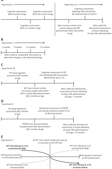
Experiment 1 established the age of onset of cognitive impair-ment, and experiment 2 assessed the effects of ICV infusion of 2B3 on cognition in the same animals. ICV administration was considered the most appropriate means to ensure the delivery of the antibody in the brain and thus provide a test of its putative actions in vivo as evidenced from our previous cell culture experiments. Experiments
1 and 2a consisted of 2 replications: Thefirst replication comprised
PDAPP (n¼14) and WT littermates (n¼15), which underwent a
battery of object memory tests at 6-8, 10-12, and 14-16 months of
age. Subsequently, these mice received ICV 2B3 (Thomas et al.,
2011b, 2013) or vehicle at 17-18 months of age using osmotic min-ipumps (experiment 2a). A second replication of 17- to
18-month-old WT (n¼8) and PDAPP (n¼8) mice received the same schedule of
prior training (age-of-onset testing; data not shown) and 2B3
administration. Together, these subgroups yielded a final total
number of 11 WT untreated,10 WT vehicle,11 vehicle PDAPP vehicle, and 10 2B3 PDAPP mice (1 WT and 1 PDAPP mouse died during osmotic minipump implantation). A separate third cohort of PDAPP
mice was used only to confirm and quantify aging-related changes in
human A
b
levels at 3 (n¼5), 7 (n¼7), 11 (n¼7), and 15 (n¼7)months of age; these mice did not undergo behavioral testing. For experiment 2b, a separate cohort of 5- to 6-month-old WT mice
were administered either 2B3 or a control mouse monoclonal IgG1
k
antibody (Millipore; MAB201) by osmotic minipump to investigate the effects on cognition of disrupting endogenous mouse APP pro-cessing with 2B3. Experiment 2b consisted of 8 WT mice that received the control IgG antibody and 8 mice that received ICV 2B3 via osmotic minipumps. As 1 mouse died postoperatively before the start of behavioral testing, it was removed from subsequent
ana-lyses. Thefinaln’s were, therefore, 8 WT control and 7 WT 2B3.
In experiment 3, PDAPP (n ¼19) and WT littermate controls
(n¼20) were used to assess the protective effects of long-term
administration of 2B3 that was initiated before the onset of cognitive impairment. As long-term administration was not feasible using an ICV minipump delivery method, 2B3 was administered to
PDAPP mice (n¼9) by once weekly IP injection at 20 mg/kg for a
period of 15 weeks from 11 months of age. Vehicle was
adminis-tered at an equal volume in control WT (n¼10) and PDAPP mice
(n¼10). An additional WT untreated group (n¼10) was included
to assess the potential impact of IP treatment on performance. The initial behavioral tests were carried out at 11 months of age, before 2B3 administration. All treatment groups were then subsequently matched in terms of their object contact times and discrimination ratios (data not shown). After a further 15 weeks of IP injections, memory function was tested at 15 months of age. These mice were tested on object memory tasks and a spatial working memory
foraging task (Evans et al., 2018) to establish the generality of the
behavioral effects of 2B3 administration across different perfor-mance measures. A breakdown of the experimental designs is
illustrated inFigure 1.
Experiment 4 evaluated the hypothesis that NMDA-NR2B re-ceptor function was necessary for associative recognition memory in WT mice. A naïve cohort of 18-month-old WT littermate controls
(n¼10) was administered the NR2B receptor antagonist Ro25-6981
or vehicle 30 minutes before behavioral testing. The experiment was run as a within-subject Latin-square design. A Latin-square is a commonly used experimental design to assess the effects of drug(s) and vehicle in the same animals on performance in a counter-balanced fashion.
Mice were given a 48-hour rest interval between each injection as a washout period before the next phase of testing. Novel objects were used in each phase of drug testing, and their order was counterbalanced across animals.
2.2. Behavior
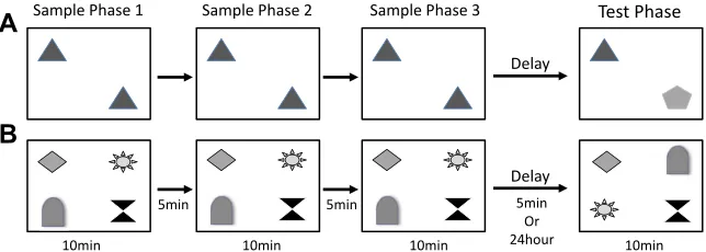
In experiments 1 and 2, mice received 2 different object-based memory tasks; a novel object recognition task and a visuospatial
OiP task (Barker and Warburton, 2011; Good and Hale, 2007; Hale
and Good, 2005) (Fig. 2A and B). Briefly, before testing object
memory, animals were habituated to the arena (60 cm 60 cm
square40 cm high). Mice were allowed to explore the arena freely
for 10 minutes on day 1. Mice were then habituated for 2 consec-utive days to the arena containing 2 identical objects for 10 minutes each day. Each mouse received 2 rounds of testing on each task, 1 day with a 5-minute and 1 day with a 24-hour delay period (in a counterbalanced order). The objects were a collection of every day
items and ornaments as described previously (Hale & Good, 2005).
2.2.1. Novel object recognition
Cognive assessment @6-8 months of age
Cognive assessment @10-12 months of age
Cognive assessment @14-16 months of age
Mice receive osmoc mini-pump surgery and 2B3 administraon @17-18 months
of age
Cognive assessment following 2B3 intervenon @ treatment day 12 and 14
Mice culled for Biochemical assessment
of ssue following 14-days 2B3 administraon
Experiment 1 Experiment 2a
A
3 months 7 months 11 months 15 months
Mice culled at comparable me-points to determine changes in β-amyloid pathology
B
Experiment 1
Pre-drug cognive assessment @5 months
of age
WT mice receive osmoc mini-pump surgery with either 2B3 or control IgG administraon
@5.5 months of age
Cognive assessment of WT mice following 2B3 intervenon
@treatment day 11-14
Mice culled for biochemical assessment of ssue following
14-days 2B3 administraon End age – 6 months Experiment 2b
C
Pre-drug behavioral assessment @11 months
of age
PDAPP mice received weekly intraperitoneal 2B3 administraon
@12 months of age
Behavioral assessment of PDAPP mice during treatment weeks 13-15
of 2B3 intervenon
Mice culled for biochemical assessment of ssue following
15-week 2B3 administraon End age – 15 months Experiment 3
D
WT mice (Group A; n=5) received Ro25-6981 Experiment 4
WT mice (Group B; n=5) received vehicle
All WT mice switch treatments and are re-assessed on the OiP task
WT mice (Group A; n=5) received vehicle WT mice (Group B; n=5)
received Ro25-6981
Both groups assessed on OiP performance
Both groups re-assessed on OiP performance
[image:3.594.108.468.72.613.2]E
separated by a 5-minute retention interval, during which the mouse was returned to its home cage. Following the third sample phase, mice received either a 5-minute or 24 hour delay period, the order of which was counterbalanced within groups. During the test phase, 1 familiar and 1 novel object were then placed into the arena
in identical spatial locations as the sample objects (Fig. 2A). The
mouse was then returned to the arena for 10 minutes to explore the object array.
2.2.2. Associative OiP memory
Four different objects were placed in the center of the arena in a square formation. Each object was approximately 15 cm from the walls and 25 cm apart from each other. Mice were placed in the center of the arena and exposed to the 4 different objects for 3 sample phases and a test phase as described for the object novelty test. In the test phase, conducted 5 minutes after the sample phase, the spatial location of 2 objects that were positioned diagonally
opposite each other was switched (Fig. 2B). The specific pair of
objects that underwent the switch was counterbalanced within and between groups.
2.2.3. Behavioural measures
For both recognition tasks, the animals’ exploratory behavior
was assessed during both the sample and test phases. Time spent exploring the objects was recorded in each phase. Object
explora-tion was defined according to the methods described previously by
Ennaceur and Delacour (1988). In brief, object contact was defined as when an animal was within a 2 cm radius of the object and
directly facing, sniffing, gnawing, but not climbing or sitting on, the
objects. A discrimination ratio (DR) was used to index the animals’
discriminative performance in the test phase that was independent of individual differences in object contact times; this was calculated as the time spent exploring the novel object (or objects in novel locations)/the time spent exploring all objects. All objects were cleaned before each phase of testing to reduce the use of odor cues introduced from handling the objects.
2.2.4. Foraging behavior
To establish the generality of the cognitive changes promoted by 2B3 administration, mice that received longitudinal IP 2B3 in-jections were also assessed on a foraging-based spatial working memory task sensitive to age-dependent changes in PDAPP mice (Evans et al., 2018). Briefly, throughout the training and test phase, mice were water-deprived to approximately 90% of their pre-training weight. Water was given for 4 hours immediately after training or testing each day. Mice were trained to forage from white ceramic pots (6.5 cm diameter, 3.5 cm depth; Lakeland, UK), which were mounted on a wooden cube base. Pots were secured to the
floor of the cage/arena with blue-tack. In the initial training stages
(day 1-3), pots containing 10 mL of water were placed in the home cage of mice for 1 hour to encourage mice to consume liquid re-wards from within the pot. The following 3 days (day 4-6), 2 pots
were baited with a 30
m
L liquid reward (1:3 sweetened condensedmilk [Nestle] solution, prepared in water; H20) in an open arena
with 1-cm-deep sawdust covering the arena floor. Mice were
placed in the center of the arena and given 3 minutes to forage from each pot. Mice were removed once the liquid reward was consumed from each pot or the 3-minute limit was reached. Mice were given 1 training session/day. On each day, the location of the pots was moved to a new location to prevent the development of any sys-tematic search bias in the test phase.
Mice were then tested over the next 4 consecutive days with 1 session per day. During these sessions, the arena was set up with 6 pots arranged in a circular pattern, each 20 cm apart. The mouse was placed in the center of the arena and allowed to forage pots until they had consumed all 6 rewards or until a 10-minute period
had elapsed from when thefirst pot was foraged. Following the
trial, mice were returned to their home cage. The pots were then wiped clean with 70% ethanol wipes, and the milk solution replenished before the next mouse was tested. All test sessions were recorded onto a DVD player using an overhead camera.
Foraging behavior was defined as a mouse jumping onto the rim
of a pot and directing its nose in toward the bottom to consume a
reward. An error was defined as a mouse returning to forage in a pot
that had previously been foraged. A full description of errors can be
found inEvans (2018).
2.3. Antibody production and administration
Full details of the immunization protocol, hybridoma
develop-ment, antibody characterization, production, and purification are
provided elsewhere (Thomas et al., 2006, 2011b, 2013).
In Experiments 2a and 2b, purified and sterilized 2B3 (1.2 mg/
mL), control monoclonal IgG1
k
antibody (experiment 2b) andvehicle (experiment 2a) were administered
intra-cerebroventricularly via a 28G cannula surgically implanted into the left lateral ventricle (Alzet; 0004760). The minipumps were attached to the infusion cannula via a catheter and implanted
subcutaneously between the scapulae. Minipumps werefilled with
200
m
L of antibody or vehicle (Model number 1002; Alzet,Califor-nia, USA;flow rate 0.25
m
L/hour for a period of 14 days). Inexper-iment 2a, the vehicle was sterile PBS and was administered to both
PDAPP mice (n¼11) and WT littermate control groups (n¼10). In
experiment 2b, the WT control group received ICV infusion of a
monoclonal IgG1
k
.Sample Phase 1 Sample Phase 2 Sample Phase 3
Test Phase
Delay
Delay
A
B
10min 10min 10min 10min
5min 5min 5min
[image:4.594.143.465.65.180.2]Or 24hour
In experiment 3, mice received a weekly IP injection of 2B3 for 15 weeks. 2B3 was produced by ascites (ProMab, California, USA),
before being purified and sterilized as described previously. PDAPP
mice were administered 2B3 (IP) at a dose of 20 mg/kg at an average concentration of 3 mg/mL. Sterile PBS was administered at the same volume to vehicle PDAPP and WT mice control groups. After the
completion of behavioral testing and 3 days after thefinal IP
in-jection of 2B3, brain tissue was collected from all mice for biochemical analyses.
2.4. Surgical procedure
During stereotaxic surgery, the mouse was anesthetized using
an isoflurane/O2mix, and a small hole was drilled through the skull
of the animal 0.5 mm posterior and 1.2 mm lateral to Bregma. A 28G cannula was then inserted 3.0 mm ventral to the skull surface and
fixed in place by dental acrylic. The minipump was carefully
inserted into a subcutaneous pocket between the scapulae, the skin incision sutured, and the mouse allowed to recover in an incubator until independently mobile. Following standard postoperative care procedures, the mice were then housed individually for the dura-tion of the study.
Postoperative behavioral testing on the OiP associative recog-nition task occurred 9 days after surgery and used the procedure described in experiments 1 and 2. On day 14, following behavioral testing, the animals were culled and brain tissue collected for analyses.
2.5. Ro25-6981 administration
Ro25-6981 (Tocris, Abingdon, UK) was dissolved in sterile saline
at a final concentration of 5 mg/mL. Mice were administered a
single 10 mg/kg dose of Ro25-6981 or vehicle IP, 30 minutes before the start of the associative recognition OiP task. This dose was selected based on previously published behavioral work (e.g., Mikics et al., 2017).
2.6. Pathology: protein extraction and immunoblots
The hippocampus and cortex were dissected, snap-frozen in
liquid nitrogen, and stored at 80 C. Soluble proteins were
extracted from brain samples as previously described (Thomas
et al., 2011a). For the ICV study, all blots quantifying changes in soluble protein levels for the ICV study assessed 10 WT vehicle, 11 PDAPP vehicle, and 10 PDAPP mice administered 2B3 intra-cerebroventricularly. For the IP study analysis, 9 WT vehicle, 10 PDAPP vehicle, and 9 PDAPP 2B3-administered mice were assessed. Synaptosome extractions were performed using Syn-PER synaptic protein extraction reagent (Thermo Fisher, UK). Western blotting was performed using standard methods as described previously (Thomas et al., 2011a) using samples from all animals undergoing
treatment and behavioral testing. Briefly, after protein quantifi
ca-tion, 20
m
g of protein/sample was resolved on either a 10% or 7.5%polyacrylamide gel and detected with the relevant antibody (Table 1). The right hippocampus of all 10 WT vehicle, 11 PDAPP
vehicle, and 10 PDAPP mice administered 2B3
intra-cerebroventricularly were used to assess synaptic protein changes.
2.7. Sandwich ELISA for detection of APP metabolites
ELISAs for the quantification of A
b
were carried out as previouslydescribed (Thomas et al., 2011a,b), or as recommended by the
manufacturer: APP (R&D Systems, Abingdon, UK),
b
-amyloidderived C-terminal fragment (
b
CTF; IBL, Hamburg, Germany), andA
b
40 and Ab
42 (human and mouse; Invitrogen, California, USA).Data are presented as ng or pg/mg total protein concentration. With exception of experiment 2b, which assessed endogenous mouse
A
b
40 and Ab
42, all further ELISAs were sensitive to human proteinonly. Therefore, the PDAPP mice that expressed the human APPV717F were used. The ICV study used 11 PDAPP vehicle mice and 10 PDAPP 2B3 mice (however, 1 mouse was removed as an outlier as explained below). The IP study used 10 PDAPP vehicle mice and 9 PDAPP 2B3 mice to quantify protein levels by ELISA.
2.8. Statistical analyses
All statistical analyses were performed using IBM SPSS statistics. The behavioral data conformed to the assumptions of analysis of variance (ANOVA) and were analyzed using a mixed measures
design. Significant interactions were assessed using tests for simple
main effects. Western blot data were analyzed using one-way
ANOVA followed by Tukey’s post hoc analysis. WT data were
collapsed across treatment (untreated and vehicle) for the analysis of the cognitive and behavioral effects of 2B3 administration as no
group differences were observed between WT groups p’s >0.5
(data not shown). ELISA data were analyzed using either Student’s
t-test or a Kruskal-Wallis test with Dunn’s test along with a
Bon-ferroni correction for multiple comparisons. All data were subject to
Levene’s and Shapiro-Wilks’tests for data normality before
anal-ysis. Appropriate transformations were carried out when necessary.
Data generated from ELISA assays were quantified by comparing
data to standard curves from each plate using GraphPad Prism 4 and normalized to total protein concentration. One PDAPP 2B3-treated mouse was found to be an extreme outlier across the ELISA analyses of the 2B3 ex vivo tissue, as determined by SPSS Tukey box plots and more conservative methods for labeling
out-liers, as described previously (Carling, 1998; Hoaglin and Iglewicz,
1987). No transformation was able to normalize this outlier for
either parametric or nonparametric analysis. Therefore, the data from this 1 mouse were removed from all ELISA analyses but no other analysis. The result of this exclusion was that all remaining ELISA data sets were normally distributed, and its exclusion did not
change the pattern of results. Western blots were quantified using
Image J software (www.imagej.nih.gov/).
3. Results
Experiment 1 used object novelty and OiP tasks to determine the
nature and age of onset of memory deficits in PDAPP mice. In the
[image:5.594.301.551.82.220.2]second stage of the study, experiment 2a, the same animals were administered the APP antibody 2B3, at 17-18 months of age, via osmotic minipumps for a period of 14 days. Having established that
Table 1
Primary antibodies used in Western blotting
Primary antibody Species Dilution Source APP (22C11) Mouse 1:1000 Millipore
BACE1 Rabbit 1:1000 Cell Signalling Technology NMDA-NR1 Mouse 1:1000 BD Biosciences
NMDA-NR2B Rabbit 1:500 Millipore NMDA-NR2B
pY1472
Rabbit 1:750 Millipore
PSD95 Rabbit 1:1000 Abcam
PS1 Rabbit 1:500 Santa Cruz
Fyn Mouse 1:750 BD Biosciences
STEP Mouse 1:750 Novus
ERK Rabbit 1:2000 Cell Signalling Technology Phospho-ERK Rabbit 1:1000 Cell Signalling Technology GAPDH Pre-conjugated 1:50,000 Sigma
b-Actin Pre-conjugated 1:20,000 Sigma
2B3 reversed pre-existing associative recognition memory impair-ment, experiment 3 determined whether longitudinal peripheral administration of 2B3 prevented the onset of cognitive decline in PDAPP mice.
3.1. Experiment 1
During development, WT mice showed a greater overall object contact time during sample phases across both tasks and at all ages,
than PDAPP mice,p’s<0.01 (Supplementary Table 1). However,
analysis showed that both WT and PDAPP mice habituated nor-mally across the 3 sample phases, as indicated by a decline in object
contact times (Supplementary Table 1). In the test phase, WT mice
showed a greater contact time with objects than PDAPP mice,p<
0.001 across all ages (Supplementary Table 2). However, both WT
and PDAPP mice showed a preference to explore objects in novel locations more than familiar locations both at 6-8 and
10-12 months of age,pvalues<0.01. However, at 14-16 months of age,
only WT but not PDAPP mice showed this preference, (p<0.001
andp>0.05, respectively; data collapsed across both delays). There
were no significant interactions involving genotype and delay for
either task (seeSupplementary Analysis). In contrast, both WT and
PDAPP mice showed a preference to explore novel objects across all
ages and delays,pvalues<0.01, and showed no genotype
[image:6.594.85.517.406.667.2]differ-ence, even at 14-16 months of age (p > 0.05; Supplementary
Table 2).
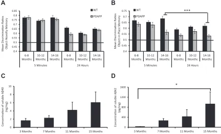
An analysis of DRs similarly showed an identical pattern with PDAPP mice performance, as well as WT mice, on the novelty test at
all ages as indicated by no main effect of genotype,F(1, 27)¼2.5,p
>0.1, and no genotypeage interaction,F(1, 27)¼2.8,p>0.1. In
contrast, PDAPP mice were significantly impaired relative to control
mice only at 14-16 months of age for the OiP task (seeFig. 3A and
3B). Analysis of the OiP task revealed a significant agegenotype
interaction,F(2, 54)¼9.2,p<0.05. Subsequent tests of simple main
effects revealed that PDAPP mice showed a significant main effect
of age with a reduction in OiP memory performance across age
ranges, F(2, 26)¼9.8, p< 0.001. Further analysis revealed that
PDAPP mice showed a significant memory deficit compared with
WT mice only at 14-16 months of age,F(1, 27)¼49.9,p<0.001.
There was no significant interaction involving delay,F <1. The
analysis of DR values confirmed that PDAPP mice showed an
age-dependent deficit in associative recognition memory, without
affecting object novelty/familiarity discriminations.
An analysis of age-related changes in hippocampal A
b
levels in aseparate group of PDAPP mice confirmed a numerical 3.6-fold
in-crease in soluble A
b
40 levels (Fig. 3C; nonsignificant followinganalysis by the Kruskal-Wallis test, X2(3)¼2.7,p>0.1) and a
sig-nificant, X2(3)¼10.5,p<0.05, 32-fold increase in soluble A
b
42levels by 15 months of age (Fig. 3D). Dunn’s test for multiple
comparisons showed a significant increase in the levels of soluble
A
b
42 when comparing mice at 3 months and 15 months of age,p<0.05. Thus, PDAPP mice showed an age-related increase in amyloid
pathology with a marked increase in A
b
production at the same ageas behavioral deficits emerged in a separate cohort of mice.
3.2. Experiment 2
3.2.1. Experiment 2a
After 2B3 or vehicle administration, PDAPP mice continued to
explore all 4 objects less than WT mice,pvalues<0.01. However, all
0.4 0.45 0.5 0.55 0.6 0.65 0.7 0.75 0.8 0.85
6-8 Months
10-12 Months
14-16 Months
6-8 Months
10-12 Months
14-16 Months
5 Minutes 24 Hours
Mean Discrimina
on Ra
os
Object
-Novelty Memory
WT
PDAPP
0.4 0.45 0.5 0.55 0.6 0.65 0.7 0.75
6-8 Months
10-12 Months
14-16 Months
6-8 Months
10-12 Months
14-16 Months
5 Minutes 24 Hours
Mean Discrimina
on Ra
os
Object
-in-Place Memory
WT
PDAPP
0 2 4 6 8 10
3 Months 7 Months 11 Months 15 Months
Concentra
on of
soluble
A
β
40
(ng/mg)
0 400 800 1200 1600
3 Months 7 Months 11 Months 15 Months
Concentra
on of
soluble
A
β
42
(pg/mg)
*
A
B
C
D
***
mice continued to show reduced contact times across sample trials,
p’s<0.001 (Supplementary Table 3A). During the test phase, overall
mice showed a preference to explore objects in novel locations over
familiar,p<0.001 (Supplementary Table 4A). However, WT mice
had higher contact times with objects both in familiar and novel
locations than vehicle PDAPP mice,pvalues<0.01, and PDAPP
2B3-treated mice,pvalues<0.05. No significant differences in contact
times were observed between PDAPP treatment groups,p>0.1.
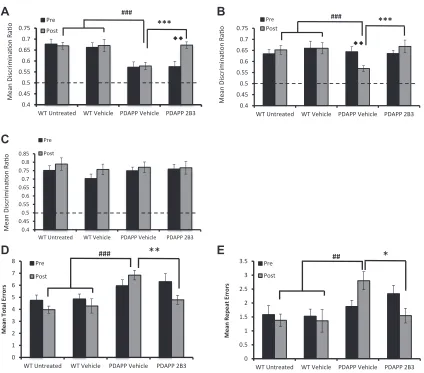
An analysis of the DRs, however, showed a significant
treatmenttime point interaction,F(2, 39)¼3.27,p<0.05. Tests
of simple main effects revealed that PDAPP mice administered ICV
2B3 showed a significant improvement in performance compared
to both their pre-administration,p<0.01, and to vehicle PDAPP
mice performance, p < 0.001. In contrast, vehicle PDAPP mice
remained significantly impaired relative to WT controls,p<0.001,
and 2B3 PDAPP mice were not significantly different to WT mice,p
> 0.5 (see Fig. 4A). Thus, ICV infusion of 2B3 in PDAPP mice
reversed an age-dependent deficit in associative recognition
memory.
3.2.2. Experiment 2b
To investigate further how inhibition of APP metabolism at the BACE1 cleavage site affected healthy WT control animals, a sepa-rate group of mice were directly administered ICV 2B3. Similar to PDAPP mice, 2B3 administration in WT mice showed no overall changes in object contact times across sample phases for object
novelty and OiP tasks relative to WT IgG1
k
controls,p’s>0.1 norduring the test phases,p’s>0.1 (Supplementary Table 3Cand4C,
respectively). Interestingly, while both 2B3 and control WT IgG1
k
mice explored novel objects in preference to familiar objects, a numerical reduction in contact times with objects in novel
loca-tions was observed in 2B3 mice (Supplementary Table 4C);
0.4 0.45 0.5 0.55 0.6 0.65 0.7 0.75
WT Untreated WT Vehicle PDAPP Vehicle PDAPP 2B3
Mean Discrimina
on Ra
o Pre
Post
***
###
A
0.4 0.45 0.5 0.55 0.6 0.65 0.7 0.75
WT Untreated WT Vehicle PDAPP Vehicle PDAPP 2B3
Mean Discrimina
on Ra
o
Pre
Post
***
0.4 0.45 0.5 0.55 0.6 0.65 0.7 0.75 0.8 0.85
WT Untreated WT Vehicle PDAPP Vehicle PDAPP 2B3
Mean Discrimina
on Ra
o
Pre
Post
B
C
###
0 0.5 1 1.5 2 2.5 3 3.5
WT Untreated WT Vehicle PDAPP Vehicle PDAPP 2B3
Mean Repeat Er
rors
Pre
Post
0 1 2 3 4 5 6 7 8
WT Untreated WT Vehicle PDAPP Vehicle PDAPP 2B3
Mean Total Errors
Pre
Post
D E
###**
[image:7.594.80.504.250.623.2]##
*
however, this failed to reach statistical significance. When the data
from both tasks were converted to DRs, there was no significant
difference between 2B3 and control IgG1
k
mice on the novelty test(t<1) but a significant deficit in 2B3 mice on the OiP task [t(13)¼
2.66, p < 0.05; Supplementary Figure 1B]. Furthermore, both
groups performed the novel object recognition memory task
above chance (0.5), p’s < 0.01 (Supplementary Figure 1A). In
contrast, 2B3 mice did not perform above chance level (0.5) on the OiP
task,p>0.5, unlike WT IgG1
k
control mice,p<0.01 (SupplementaryFigure 1B). These results indicate that 2B3 (but not a control IgG1
k
) administered to normal WT mice leads to a selective disruption of associative recognition memory.3.3. Experiment 3
Analysis of contact times with objects across sample trials for both recognition memory tasks showed that all groups explored the
objects at similar levels, p>0.05 (Supplementary Table 3B). All
mice also showed habituation of exploratory activity to objects, as indicated by a decrease in contact times when comparing sample
trial 1 to sample trial 3,p’s<0.001. Following contact time analysis
within the test phase of the object novelty and OiP tasks, mice
showed no significant differences in overall contact times with all
objectsp’s>0.05 (Supplementary Table 4B). In the OiP task, despite
treatment groups showing numerical differences in contact times
A
47kDa
WT Vehicle
PDAPP Vehicle
PDAPP 2B3
BACE1 70kDa
β-Ac n 42kDa
PS1
APP 110kDa
0 2 4 6 8 10
APP BACE1 PS1
Pr
ot
ein Density
(f
old of WT)
WT Vehicle PDAPP Vehicle PDAPP 2B3 ***
B
47kDa
WT Vehicle
PDAPP Vehicle
PDAPP 2B3
BACE1 70kDa
β-Ac n 42kDa
PS1
0 0.2 0.4 0.6 0.8 1 1.2 1.4 1.6 1.8
PS1 BACE1
Pr
ot
ein Density (fold WT)
WT Vehicle
PDAPP Vehicle
PDAPP 2B3
0 50 100 150 200 250 300 350 400 450
PDAPP Vehicle PDAPP 2B3
APP Concen
tr
a
on
(
ng
/mg)
0 10 20 30 40 50 60
PDAPP Vehicle PDAPP 2B3
β
CTF Concen
tr
a
on
(pg
/mg)
*
0 40 80 120 160 200
PDAPP Vehicle PDAPP 2B3
Soluble A
β
40 Concen
tr
a
on
(pg
/mg)
*
C
D
E
0 20 40 60 80 100 120 140 160
PDAPP Vehicle PDAPP 2B3
APP Concentra
on (ng/ml)
[image:8.594.114.497.67.506.2]F
between objects in novel and familiar locations, all mice continued to show a general preference to explore objects in novel locations
over familiar,p<0.001. The same pattern was also observed in the
object novelty task.
However, analysis of the OiP test DRs revealed a significant
time treatment group interaction, F(2, 34) ¼ 3.41, p < 0.05.
Testing for simple main effects further revealed that, although vehicle PDAPP mice showed a decline in OiP performance across
pre- and post-treatment time points,p<0.01, IP administration of
2B3 prevented this decline,p>0.05. Moreover, both WT and 2B3
PDAPP mice showed comparable performance (p>0.05) and both
groups performed better than vehicle PDAPP mice,pvalues<0.001
(Fig. 4B). Analysis of the object novelty task DRs showed no effect of
treatment group,F(2, 34)¼0.16,p>0.1, or treatment grouptime
interaction, F(2, 34)¼0.27,p>0.1, (Fig. 4C). To summarize, the
results showed that peripheral administration of 2B3 before the
onset of cognitive decline prevented the age-dependent deficit in
associative recognition, without influencing object novelty
detec-tion, in PDAPP mice.
To establish the generality of the improvement in associative OiP memory with 2B3, PDAPP mice were also tested on a spatial
working memory foraging task (Evans et al., 2018). Time to
com-plete the foraging task after vehicle or 2B3 administration was
similar between all groups, p > 0.05 (Supplementary Table 5),
which indicated that neither transgene expression nor 2B3
administration influenced the motivation to complete the task.
However, foraging accuracy was improved in 2B3 PDAPP relative to
vehicle PDAPP mice (Fig. 4D and E). More specifically, analysis of the
mean total number of errors (a measure of overall foraging
accu-racy) revealed a significant treatment timegroup interaction,F(2,
34)¼3.68,p<0.05. Tests of simple main effects showed that 2B3
PDAPP mice made less errors than their pre-drug assessment at
11 months of age,p<0.05 and less errors than vehicle PDAPP mice,
p<0.01 (Fig. 4D). In contrast, vehicle PDAPP mice made more errors
than WT vehicle controls,p<0.001 (Fig. 4D). WT mice and 2B3
PDAPP mice performed at a comparable level,p>0.05. A similar
pattern of results was observed when analyzing repeat errors, a
measure of working memory (Fig. 4E). An ANOVA revealed a
sig-nificant treatment timegroup interaction,F(2, 34)¼3.85,p<
0.05. Simple main effects analysis revealed that 2B3 PDAPP mice
made fewer repeat errors than vehicle PDAPP mice,p<0.05, and
that there was no significant change in the number of repeat errors
compared to pre-drug performance,p>0.05. In contrast, vehicle
PDAPP mice showed an age-dependent increase in the total number
of repeat errors compared to pre-drug assessment,p<0.05. Vehicle
PDAPP mice also made more repeat errors than WT mice,p<0.01.
This impairment was not present in 2B3 PDAPP mice,p>0.05.
3.4. Biochemical analyses: experiments 2a, 2b, and 3
In experiment 2a,Western blot analysis showed that ICV 2B3
administration caused no change in total levels of BACE1,F(2, 28)¼
0 200 400 600 800 1000
PDAPP Vehicle PDAPP 2B3
Soluble A
β
42 Concen
tr
a
on
(pg
/mg)
G
0 50 100 150 200 250 300 350 400 450
PDAPP Vehicle PDAPP 2B3
Soluble A
β
40 c
oncen
tr
a
on
(pg
/mg)
0 5 10 15 20 25 30 35 40 45
PDAPP Vehicle PDAPP 2B3
Insoluble A
β
40 c
o
ncen
tr
a
on
(pg
/mg)
0 1000 2000 3000 4000 5000 6000 7000
PDAPP Vehicle PDAPP 2B3
Soluble A
β
42 c
oncen
tr
a
on
(pg
/mg)
H
J
I
0 100 200 300 400 500 600 700 800
PDAPP Vehicle PDAPP 2B3
Insoluble A
β
42 c
oncen
tr
a
on
(pg
/mg)
[image:9.594.99.486.62.440.2]K
1.95,p>0.1, or presenilin 1 (PS1),F(2, 28)¼1.40,p>0.1, in the
hippocampus of PDAPP mice (Fig. 5A). These data were replicated in
the hippocampus of IP 2B3 mice for both BACE1,F(2, 25)¼2.30,p>
0.1, and PS1,F<1 (Fig. 5B), and WT ICV 2B3 mice for BACE1, t(13)¼
0.5, p > 0.5 (Supplementary Figure 1D). Western blot analysis
showed an overall change in APP levels,F(2, 28)¼8.49,p<0.01 in
ICV 2B3 PDAPP mice (Fig. 5A). Post hoc Tukey tests revealed this
difference was evident only when comparing WT mice to PDAPP
vehicle,p<0.01, and PDAPP 2B3 mice,p<0.05. No difference in
APP levels was reported between the PDAPP groups,p>0.1. ELISA
analysis further confirmed that 2B3 caused no change in APP levels
in PDAPP mice after either ICV, t(18)¼1.32,p>0.1, or IP
admin-istration, t(17) ¼ 1.45, p > 0.1 (Fig. 5C and D, respectively). In
experiment 2b, there was similarly no change in endogenous APP
levels in WT mice administered 2B3, relative to IgG1
k
controlst(13)¼0.81,p>0.5 (Supplementary Figure 1D).
In experiment 2a, ICV 2B3 administration caused a significant
16% reduction in levels of
b
CTF, t(18)¼2.22,p<0.05, a significant56% reduction in levels of soluble A
b
40, t(18)¼2.28,p<0.05, and a(nonsignificant) 66% reduction in levels of soluble A
b
42, t(18) ¼1.01,p>0.1, in the hippocampus (Fig. 5E, F, and G, respectively; see
also in vitro data fromThomas et al., 2011b, 2013). In experiment
2b, there was a significant 8.1% reduction in endogenous soluble
A
b
40, t(13)¼2.45,p<0.05, in the hippocampus of WT ICV 2B3mice relative to IgG1
k
control mice (Supplementary Figure 1E).Endogenous soluble A
b
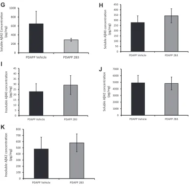
42 was undetectable by ELISA and,there-fore, could not be compared (data not shown). In experiment 3, analysis of hippocampal tissue from IP 2B3 PDAPP mice revealed no
significant change in soluble or insoluble A
b
40 or Ab
42 orb
CTFlevels (data not shown) compared with vehicle PDAPP mice, t’s<1
(Fig. 5H-K).
In experiment 2a, there were no significant differences in the
total expression levels of the synaptic marker PSD95 or NMDA re-ceptor subunits, NR1, or NR2B in hippocampal synaptosomes of ICV
[maximum NR2B,F(2, 28)¼2.72,p>0.05]. A similar pattern was
evident in experiment 3 after IP 2B3 administration (maximum
NR1,F(2, 26) ¼ 0.12,p >0.1, respectively; Fig 6A, B and E). In
experiment 2a, phosphorylation of the NR2B Y1472 residue appeared to be reduced in vehicle PDAPP mice, although this was
not significant relative to WT mice or ICV 2B3 PDAPP mice, F(2,
28)¼2.36,p>0.05 (Fig. 6A and B). However, after IP administration
of 2B3 (experiment 3), phosphorylation of the NR2B Y1472 residue
was significantly altered in IP 2B3 PDAPP mice,F(2, 25)¼3.86,p<
0.05, PDAPP vehicle mice showed reduced pY1472 compared with
WT vehicle mice,p<0.05 (Fig. 6E). When NR2B Y1472
phosphor-ylation was presented as a ratio of total NR2B, there was a signifi
-cant difference between groups in both the ICV (experiment 2a),
F(2, 28)¼16.11,p<0.001 and IP (experiment 3) studies,F(2, 25)¼
5.30,p<0.01. Post hoc Tukey’s analyses revealed a reduction in
vehicle PDAPP mice compared with WT mice in both ICV and IP
groups (p<0.001,p<0.05;Fig. 6B and E). Furthermore, the ratios
were also reduced in vehicle PDAPP mice compared with 2B3
PDAPP mice, (p<0.001,p<0.05, respectively). Neither ICV nor IP
2B3 PDAPP mice differed relative to their respective WT control
mice,pvalues>0.1.
Given the changes in NR2B phosphorylation, we investigated SRC kinase Fyn and striatal-tyrosine-enriched phosphatase (STEP) in hippocampal synaptosomes of ICV 2B3 PDAPP mice. These
ki-nases are key regulators of NR2B phosphorylation (Ittner et al.,
2010; Zhang et al., 2010). No change in total levels of Fyn be-tween any of the groups was evident in hippocampal
synapto-somes,F<1 (Fig. 6C). Conversely, both isoforms of STEP (46 and 61)
were numerically increased in vehicle PDAPP mice relative to WT
and 2B3 PDAPP mice (Fig. 6C). However, despite a numerical
in-crease in STEP61, there were no significant differences between
groups,F(2, 28)¼2.48,p>0.05. In contrast, STEP46 was altered
significantly,F(2, 28)¼4.39,p<0.05. Post hoc Tukey’s analysis
revealed STEP46 was increased in vehicle PDAPP mice compared
with WT mice,p<0.05 (there was a numerical difference between
PDAPP vehicle and 2B3 PDAPP mice, but it was not significant;p¼
0.074, 2B3;Fig. 6C).
Extracellular signaleregulated kinase (ERK) is downstream of
NMDA signaling and plays a role in regulating memory processes (Caccamo et al., 2010). There was a significant change in total ERK
levels [F(2, 28)¼15.29,p<0.001] in synaptosomes of ICV vehicle
PDAPP compared with WT (p<0.01) and ICV 2B3 PDAPP mice (p<
0.001;Fig. 6D). There were no changes between ICV WT and 2B3
PDAPP mice in total levels of ERK,p>0.1. Although phosphorylated
ERK appeared to be numerically reduced in ICV vehicle PDAPP mice,
there were no significant differences between groups, F< 1. In
contrast, when pERK was expressed as a ratio of total ERK, to better
determine ERK activity, ICV vehicle PDAPP mice showed a signifi
-cant difference,F(2, 28)¼7.03,p<0.01, with a significant reduction
compared with both WT (p < 0.05) and ICV 2B3 PDAPP mice
(p<0.01). There was no significant difference between WT and ICV
2B3 PDAPP mice,p>0.05 (Fig. 6D).
3.5. Experiment 4
The aforementioned analyses suggested that the NR2B receptor played an important role in associative recognition memory, although there are relatively little published data to support this conclusion. We therefore tested a separate group of 18-month-old WT mice on the OiP task after the administration of the NR2B re-ceptor antagonist, Ro25-6981. WT mice continued to show habit-uation to objects across 3 sample phases after Ro25-6981 or vehicle
administration (Supplementary Table 6). Ro25-6981 did not change
overall object contact times of WT mice in either the sample or test
phases when compared with vehicle contact times, p’s > 0.05
(Supplementary Tables 6and7). Moreover, despite numerical
dif-ferences, both vehicle (p<0.001) and Ro-25-6981 (p<0.01) mice
explored objects in novel locations more than those in familiar
lo-cations (Supplementary Table 7). However, an analysis of the test
DR scores showed that Ro25-6981 impaired performance relative
to vehicle administration, t(9)¼2.47,p<0.05 (Fig. 6F). These data
confirm that, under normal physiological conditions, the NR2B
re-ceptor plays a key role in associative recognition memory processes in normal aged WT mice.
4. Discussion
Male PDAPP mice showed a selective age-dependent impair-ment in associative recognition memory, while sparing object novelty detection. Similar to previous studies, the age-dependent decline in associative recognition memory coincided with a rise
in hippocampal A
b
levels (Barker and Warburton, 2013; Good andHale, 2007; Hale and Good, 2005; Selkoe, 2001). ICV administra-tion of the anti-APP antibody, 2B3, directed against the BACE1
cleavage site, reduced APP metabolism and lowered A
b
andb
CTFlevels in the hippocampus. The reduction in A
b
was accompaniedby restoration of associative recognition memory in aged PDAPP mice. Furthermore, longitudinal peripheral administration of 2B3 prevented the onset of associative recognition memory impairment as well as a decline in spatial working memory in PDAPP mice at 15 months of age. A critically important aspect of our results is that
both ICV and IP administration of 2B3 normalized a deficit in NMDA
receptor phosphorylation. Previous work in normal animals has shown that hippocampal NMDA receptors are required for asso-ciative recognition memory but not object novelty/familiarity
therefore, consistent with the view that the age-related
accumu-lation of A
b
changes hippocampal synaptic events that underpinsmemory (Guntupalli et al., 2016).
Previous work with PDAPP mice has reported an age-related
deficit in object novelty detection (Dodart et al., 1999). However,
this result has not been replicated across laboratories (Chen et al.,
2000). This discrepancy may be related to the test procedure. In
contrast to our own study and that conducted by Chen et al.,Dodart
et al. (1999)exposed mice to a single object in the sample phase and presented a familiar and novel object in the test phase. This test procedure confounded object novelty with object-location novelty.
Thus, the deficit in PDAPP mice reported by Dodart et al. may have
reflected impaired processing of location information. Importantly,
with the exception of the present experiments, no study has 0
0.2 0.4 0.6 0.8 1 1.2 1.4
NR2B pY1472 pY1472:NR2B Protein Density (fold
of
WT)
WT Vehicle PDAPP Vehicle PDAPP 2B3
0 1 2 3 4
Fyn STEP 61 STEP 46 Protein Density (fold
of
WT)
0 0.5 1 1.5
ERK p-ERK pERK:T-ERK Protein Density (fold
of
WT)
WT Vehicle
PDAPP Vehicle
PDAPP 2B3
NR2B 180 kDa
pY1472 180 kDa
NR1 110 kDa
PSD95 95 kDa
β-ac n 42 kDa
STEP 61 60 kDa
STEP 46 46 kDa
Fyn 59 kDa
Total ERK 44/42 kDa
Phospho-ERK 44/42 kDa
A
B
C
D
***
*
** *
E
F
0.4 0.45 0.5 0.55 0.6 0.65 0.7
WT Vehicle WT Ro25-6981
Mean Discrimina
Ɵ
on Ra
Ɵ
o
pY1472 NR2B
β-ac n
WT Vehicle PDAPP Vehicle PDAPP 2B3 180kDa
180kDa
42kDa
**
0 0.2 0.4 0.6 0.8 1 1.2
NR2B pY1472 pY1472:NR2B
Protein Density (fold of
WT)
WT Vehicle Tg Vehicle Tg 2B3
[image:11.594.102.479.68.502.2]*
*
compared the impact of aging on object novelty and associative OiP memory in the same PDAPP mice longitudinally, and our evidence indicates an age-related sensitivity to associative object-location memory or mismatch detection in PDAPP mice.
Before discussing the effects of 2B3 on APP processing and cognition, it is worth highlighting 2 drawbacks of the study. One drawback is that only male mice were tested. This strategy was undertaken to minimize extraneous sources of variability in both the behavioral and biological measures and thus maximize the detection of cognitive and synaptic changes induced by 2B3. Nevertheless, given the positive results of this study in male mice
and the fact that over 60% of patients with AD are female (“2015
Alzheimer’s disease facts andfigures,”2015), it would be important clearly to test the assumption that aged female PDAPP mice would
also demonstrate cognitive and pathology benefits from reducing
APP cleavage by BACE1.
A second drawback of the study is that the half-life of 2B3 has yet to be established in vivo. Although a single weekly injection of
2B3 was sufficient to alter NR2B expression and cognition in PDAPP
mice, there was no evidence of changes in brain amyloid levels, unlike the ICV administration study. However, this lack of
sensi-tivity to any changes probably reflects the much lower levels of 2B3
reaching the brain after peripheral IP administration, (because of the blood-brain barrier) compared with direct ICV administration. Further research is required to assess the temporal dynamics of the interactions of 2B3 with APP processing in vivo. Nevertheless, our data present clear evidence that, even under a restricted set of conditions, 2B3 has an impact on APP processing and memory.
Excess A
b
production leads to changes in the dynamic propertiesof hippocampal synaptic plasticity; promoting the induction of LTD
and impairing the induction of LTP (Hsieh et al., 2006). This change
in plasticity dynamics occurs through several mechanisms involving NMDA receptors and intracellular calcium signaling. Indeed, extra-synaptic NMDA-NR2B receptors have emerged as a key factor in mediating the effects of amyloid on synaptic
depres-sion and toxicity (Kessels et al., 2013; Wang et al., 2013); for
example, low nanomolar levels of soluble A
b
oligomers enhanceNR2B-mediated NMDA receptor currents and extra-synaptic
re-sponses (Li et al., 2011).
Excess A
b
production can also result in activation of Fyn kinase,which leads to phosphorylation of NR2B at Y1472 and stabilizes
expression of the receptor at the membrane surface. The effect of A
b
on Fyn kinase is counteracted by elevation of STEP (Ittner et al.,
2010; Zhang et al., 2010); for example, there is an age-related
in-crease in STEP levels in Tg2576 mice (Zhang et al., 2010). STEP, a
tyrosine phosphatase, dephosphorylates NR2B at the Y1472 site, as well as Fyn kinase and AMPA receptors. STEP therefore controls NMDA receptor activity by directly dephosphorylating NR2B and deactivating Fyn kinase. The enhanced expression of NR2B seen
with A
b
is thought to contribute ultimately to the loss of synapticconnections (Boehm, 2013). In the present study, 2B3 (both ICV and
IP) reversed the phosphorylation of NMDA-NR2B pY1472 in hip-pocampal synaptosomes, without affecting PSD95, NR1, or NR2B expression. We further showed an increase in total levels of STEP in vehicle PDAPP mice, which was numerically reduced after 2B3 administration. Activation of ERK, which is downstream of NMDA receptor activity, plays a role in regulating memory processes (Krapivinsky et al., 2003). Furthermore, dysregulation of NR2B/ERK signaling has been reported in 3xTg mice in association with
elevated levels of amyloid (Caccamo et al., 2010). In the present
study, ERK activity was reduced when expressed as a ratio of total ERK in hippocampal synaptosomes from PDAPP mice. The reduc-tion in ERK was, however, reversed after 2B3 administrareduc-tion. Taken together, this evidence suggests that the 2B3-mediated changes in NR2B synaptic signaling processes contributed to the improvement
in associative recognition memory in aged 2B3 PDAPP mice. In
support of this assertion, we showed for thefirst time that selective
antagonism of NR2B receptors impaired associative OiP recognition memory in WT mice.
It is important to acknowledge that other anti-APP-BACE1 and
immunization approaches have been investigated. Thus,Arbel et al.,
(2005) reported the activity of the antibody BBS1 also directed against the BACE1 cleavage site of APP. In vivo administration of BBS1 by osmotic minipumps to 3xTg mice for 28 days resulted in a
significant improvement in object-novelty memory at 17-18 month
of age (Rabinovich-Nikitin et al., 2012). These improvements in
behavior were complemented by a significant reduction in plaque
size, A
b
load, and a 24% reduction in soluble Ab
42 (Rabinovich-Nikitin et al., 2012). Peripheral administration of BBS1 also
resul-ted in improved object-novelty memory and reduced inflammatory
markers in Tg2576 mice (Rakover et al., 2007) as well as reduced A
b
plaques and intracellular A
b
load in mice with the hAPP V717LLondon mutation (Arbel-Ornath et al., 2009). Despite the changes in
A
b
production, BBS1 did not improve spatial reference memory, asassessed in the water maze. Our currentfindings confirm that steric
hindrance of APP processing by BACE1 can have beneficial effects
on amyloid levels and associative spatial recognition memory processes that rely upon the hippocampus. Unlike the present study, however, the effect of BBS1 on synaptic processes sensitive to
amyloid accumulation was not examined. A related study byChang
et al., (2007)immunized mice against memapsin-2 (BACE1) with the hypothesis that anti-memapsin-2 antibodies would bind to memapsin-2 located on the surface of neurons and, when
endo-cytosed, would interfere with the cleavage of APP and thus lower A
b
production. Immunization of Tg2576 with anti-M2 saw a reduction in amyloid load and improvement in performance in a water maze reference memory task. Unlike the present study, however, the extent of the cognitive change was not indexed against WT control mice but only vehicle-treated Tg2576 mice. However, taken
together, our results, and those of Chang (ibid), confirm that
selectively targeting BACE cleavage of APP can have positive effects on amyloid production and memory function. Importantly,
how-ever, our study is thefirst to show that an anti-APP antibody
tar-geting the BACE1 cleavage site improved associative recognition memory and spatial working memory and reduced phosphoryla-tion of NMDA-NR2B receptors. In addiphosphoryla-tion, it is worth noting that
2B3 caused a significant reduction in levels of
b
CTF, an effect thathas not previously been reported with BBS1 or anti-M2. Increased
levels of
b
CTF have been observed in patients with AD, and recentresearch has revealed an A
b
-independent mechanism causingdysregulation of endocytosis (Kim et al., 2015; Pimplikar et al.,
2010). Consistent with this evidence, genetic reduction in BACE1
activity in a mouse model of Down syndrome improved endosomal
volume and improved cholinergic markers without lowering A
b
levels (Jiang et al., 2016). It is possible, therefore, that the reduction
in
b
CTF levels by 2B3 may have had a beneficial impact on endocyticpathways, and this possibility requires further investigation.
In terms of the clinical relevance of our findings, evidence is
beginning to emerge that some antibody-based therapies have a positive impact on amyloid pathology and may be
disease-modifying in amyloid-positive patients (e.g., BAM2401, https://
www.eisai.com; aducanumab, Sevigny et al., 2016). Although
thesefindings are preliminary, the idea that such immunotherapies
may be useful in the context of treating individuals who are amy-loid positive in old age is gaining traction. However, our evidence that 2B3 disrupted associative (but not object novelty) recognition
memory in normal young mice confirms that metabolism of APP by
BACE1 is an important physiological process that contributes to
memory in healthy controls (see alsoBlume et al., 2018; Ou-Yang
Therefore, targeting this pathway in healthy presymptomatic aged participants should be combined with close monitoring for dele-terious changes in memory function.
As highlighted byPiton et al. (2018), modulation of BACE activity
remains a viable strategy in trials on prodromal and early AD. Our data add to a growing body of preclinical evidence that selectively modifying APP processing at an early stage of amyloid
accumula-tion in the aging amyloid-positive brain has a beneficial effect on
cognition. It also confirms that APP processing by BACE1 has a role
in normal memory function and that disruption to this equilibrium is detrimental. An antibody therapy that targets APP processing by BACE1 may therefore only have clinical relevance in the context of
amyloid-related cognitive changes in the elderly (seeFarrell et al.,
2017; Hedden et al., 2012).
Conclusion
In conclusion, the present study has shown that selectively
influencing APP metabolism by reducing BACE1 activity by steric
hindrance with an antibody, reversed and protected against an
age-dependent associative recognition memory deficit in PDAPP mice.
ICV administration of 2B3 reduced levels of soluble A
b
andb
CTFwithout affecting total levels of APP, and both ICV and chronic IP administration normalized the phosphorylation of NMDA-NR2B
receptors. These novel findings provide important evidence that
selective inhibition of APP processing at the BACE1 cleavage site can improve both memory and markers of synaptic pathology in a mouse model of age-related amyloid accumulation. Finally, the
results contribute to otherfindings suggesting that modification of
APP processing, and associated downstream glutamatergic
signaling cascades, may be beneficial in populations with altered
APP metabolism, including aged individuals (Rodrigue et al., 2012)
and those at high risk of AD.
Disclosure statement
All authors declare that they have no competing financial
interests.
Acknowledgments
The authors thank members of the JBIOS technical staff at Cardiff University for their assistance with animals throughout the project.
Funding: This work was supported by a PhD studentship from the School of Psychology Cardiff and the Cardiff School of Pharmacy and Pharmaceutical Sciences, a Wellcome Trust PhD studentship,
the Life Science Research Network Wales, and an Alzheimer’s
So-ciety UK PhD studentship.
Authors’contributions: MAG, EJK, and RST conceived the study
and subsequent funding. MH-E obtained pilot data used for obtaining funding. CEE, TJF, and MAG designed animal experiments and performed surgical procedures. CEE, MH-E, TJF, and RST pre-pared 2B3. CEE and MH-E bred all mice used in this study. CEE and TJF performed animal experiments and carried out antibody administration, and CE performed the biochemical analysis. CE analyzed data. CEE, MAG, and EJK wrote the manuscript. All authors reviewed and approved the manuscript.
Declarations
Ethical approval and consent to participate: All procedures for
animal use were approved according to UK Home Office under the
Animal Scientific Procedures Act (1986) and EU regulations.
Consent for publication: All authors have approved of the con-sents of this manuscript and provided consent for publication.
Availability of supporting data: The data and materials are available from corresponding authors on reasonable request.
Appendix A. Supplementary data
Supplementary data to this article can be found online athttps://
doi.org/10.1016/j.neurobiolaging.2018.11.011.
References
Alzheimer’s Disease Facts and Figures. Alzheimer’s Dement. J. Alzheimer’s Assoc. 11, 2015, 332e384.
Arbel, M., Yacoby, I., Solomon, B., 2005. Inhibition of amyloid precursor protein processing byb-secretase through site-directed antibodies. Proc. Natl. Acad. Sci U. S. A. 102, 7718e7723.
Arbel-Ornath, M., Becker, M., Rabinovich-Toidman, P., Gartner, M., Solomon, B., 2009. Immunomodulation of AbPP processing alleviates amyloid-b-related pa-thology in Alzheimer’s disease transgenic mice. J. Alzheimer’s Dis. 22, 469e482. Barker, G.R.I., Warburton, E.C., 2013. Object-in-place associative recognition mem-ory depends on glutamate receptor neurotransmission within two defined hippocampal-cortical circuits: a critical role for AMPA and NMDA receptors in the hippocampus, perirhinal, and prefrontal cortices. Cereb. Cortex 1e10. Barker, G.R.I., Warburton, E.C., 2011. When is the hippocampus involved in
recog-nition memory? J. Neurosci 31, 10721e10731.
Blume, T., Filser, S., Jaworska, A., Blain, J.F., Koenig, G., Moschke, K., Lichtenthaler, S.F., Herms, J., 2018. BACE1 inhibitor MK-8931 alters formation but not stability of dendritic spines. Front. Aging Neurosci. 10, 229.
Boehm, J., 2013. A“danse macabre”: tau and Fyn in STEP with amyloid beta to facilitate induction of synaptic depression and excitotoxicity. Eur. J. Neurosci. 37, 1925e1930.
Caccamo, A., Maldonado, M.A., Bokov, A.F., Majumder, S., Oddo, S., 2010. CBP gene transfer increases BDNF levels and ameliorates learning and memory deficits in a mouse model of Alzheimer’s disease. Proc. Natl. Acad. Sci. U. S. A. 107, 22687e22692.
Carling, K., 1998. Resistant outlier rules and the non-Gaussian case*. Comput. Stat. Data Anal. 33, 249e258.
Chang, W.P., Downs, D., Huang, X.P., Da, H., Fung, K.M., Tang, J., 2007. Amyloid-beta reduction by memapsin 2 (beta-secretase) immunization. FASEB J 21, 3184e3196.
Chen, G., Chen, K.S., Knox, J., Inglis, J., Bernard, A., Martin, S.J., Justice, A., McConlogue, L., Games, D., Freedman, S.B., Morris, R.G.M., 2000. A learning deficit related to age and [beta]-amyloid plaques in a mouse model of Alz-heimer’s disease. Nature 408, 975e979.
Dodart, J.C., Meziane, H., Mathis, C., Bales, K.R., Paul, S.M., Ungerer, A., 1999. Behavioral disturbances in transgenic mice overexpressing the V717F beta-amyloid precursor protein. Behav. Neurosci. 113, 982e990.
Ennaceur, A., Delacour, J., 1988. A new one-trial test for neurobiological studies of memory in rats. 1: behavioral data. Behav. Brain Res. 31, 47e59.
Evans, C., Hvoslef-Eide, M., Thomas, R., Kidd, E., Good, M.A., 2018. A rapidly acquired foraging-based working memory task, sensitive to hippocampal lesions, reveals age-dependent and age-independent behavioural changes in a mouse model of amyloid pathology. Neurobiol. Learn. Mem. 149, 46e57.
Farrell, M.E., Kennedy, K.M., Rodrigue, K.M., Wig, G., Bischof, G.N., Rieck, J.R., Chen, X., Festini, S.B., Devous Sr., M.D., Park, D.C., 2017. Association of longitu-dinal cognitive decline with amyloid burden in middle-aged and older adults: evidence for a dose-response relationship. JAMA Neurol 74, 830e838. Games, D., Adams, D., Wolozin, B., Zhao, J., 1995. Alzheimer-type neuropathology in
transgenic mice overexpressing V717F APP. Nature 373, 523e527.
Good, M.A., Hale, G., 2007. The“Swedish”mutation of the amyloid precursor protein (APPswe) dissociates components of object-location memory in aged Tg2576 mice. Behav. Neurosci. 121, 1180e1191.
Guntupalli, S., Widagdo, J., Anggono, V., 2016. Amyloid-b-induced dysregulation of AMPA receptor trafficking. Neural Plast 2016, 12.
Hale, G., Good, M., 2005. Impaired visuospatial recognition memory but normal object novelty detection and relative familiarity judgments in adult mice expressing the APPswe Alzheimer’s disease mutation. Behav. Neurosci. 119, 884e891.
Hardy, J., Selkoe, D.J., 2002. The amyloid hypothesis of Alzheimer’s disease: progress and problems on the road to therapeutics. Science 297, 353e356.
Hedden, T., Mormino, E.C., Amariglio, R.E., Younger, A.P., Schultz, A.P., Becker, J.A., Buckner, R.L., Johnson, K.A., Sperling, R.A., Rentz, D.M., 2012. Cognitive profile of amyloid burden and white matter hyperintensities in cognitively normal older adults. J. Neurosci 32, 16233e16242.
Hemming, M.L., Elias, J.E., Gygi, S.P., Selkoe, D.J., 2009. Identification ofb-secretase (BACE1) substrates using quantitative proteomics. PLoS One 4, e8477. Hoaglin, D.C., Iglewicz, B., 1987. Fine-tuning some resistant rules for outlier labeling.
J. Am. Stat. Assoc. 82, 1147e1149.
Ittner, L.M., Ke, Y.D., Delerue, F., Bi, M., Gladbach, A., van Eersel, J., Wölfing, H., Chieng, B.C., Christie, M.J., Napier, I.A., 2010. Dendritic function of tau mediates amyloid-btoxicity in Alzheimer’s disease mouse models. Cell 142, 387e397. Jiang, Y., Rigoglioso, A., Peterhoff, C.M., Pawlik, M., Sato, Y., Bleiwas, C., Stavrides, P.,
Smiley, J.F., Ginsberg, S.D., Mathews, P.M., 2016. Partial BACE1 reduction in a Down syndrome mouse model blocks Alzheimer-related endosomal anomalies and cholinergic neurodegeneration: role of APP-CTF. Neurobiol. Aging 39, 90e98.
Kessels, H.W., Nabavi, S., Malinow, R., 2013. Metabotropic NMDA receptor function is required forb-amyloideinduced synaptic depression. Proc. Natl. Acad. Sci. U. S. A. 110, 4033e4038.
Kim, S., Sato, Y., Mohan, P.S., Peterhoff, C., Pensalfini, A., Rigoglioso, A., Jiang, Y., Nixon, R.A., 2015. Evidence that the rab5 effector APPL1 mediates APP-b CTF-induced dysfunction of endosomes in Down syndrome and Alzheimer’s disease. Mol. Psychiatry 1e10.
Krapivinsky, G., Krapivinsky, L., Manasian, Y., Ivanov, A., Tyzio, R., Pellegrino, C., Ben-Ari, Y., Clapham, D.E., Medina, I., 2003. The NMDA receptor is coupled to the ERK pathway by a direct interaction between NR2B and RasGRF1. Neuron 40, 775e784.
Li, S., Jin, M., Koeglsperger, T., Shepardson, N.E., Shankar, G.M., Selkoe, D.J., 2011. Soluble Ab oligomers inhibit long-term potentiation through a mechanism involving excessive activation of extrasynaptic NR2B-containing NMDA re-ceptors. J. Neurosci. 31, 6627e6638.
Mikics, E., Toth, M., Biro, L., Bruzsik, B., Nagy, B., Haller, J., 2017. The role of GluN2B-containing NMDA receptors in short- and long-term fear recall. Physiol. Behav 177, 44e48.
Ou-Yang, M.H., Kurz, J.E., Nomura, T., Popovic, J., Rajapaksha, T.W., Dong, H., Contractor, A., Chetkovich, D.M., Tourtellotte, W.G., Vassar, R., 2018. Axonal or-ganization defects in the hippocampus of adult conditional BACE1 knockout mice. Sci. Transl. Med. 10 (459).
Pimplikar, S.W., Nixon, R.A., Robakis, N.K., Shen, J., Tsai, L.-H., 2010. Amyloid-inde-pendent mechanisms in Alzheimer’s disease pathogenesis. J. Neurosci 30, 14946e14954.
Piton, M., Hirtz, C., Desmetz, C., Milhau, J., Lajoix, A.D., Bennys, K., Lehmann, S., Gabelle, A., 2018. Alzheimer’s disease: advances in drug development. J. Alz-heimer’s Dis 65, 1e11.
Rabinovich-Nikitin, I., Rakover, I.S., Becker, M., Solomon, B., 2012. Beneficial effect of antibodies againstb- secretase cleavage Site of App on Alzheimer’s-like Pa-thology in triple-transgenic mice. PLoS One 7, e46650.
Rakover, I., Arbel, M., Solomon, B., 2007. Immunotherapy against APPb-secretase cleavage site improves cognitive function and reduces neuroinflammation in
Tg2576 mice without a significant effect on brain Ablevels. Neurodegener. Dis. 4, 392e402.
Rodrigue, K.M., Kennedy, K.M., Devous, M.D., Rieck, J.R., Hebrank, A.C., Diaz-Arrastia, R., Mathews, D., Park, D.C., 2012.b-amyloid burden in healthy aging. Neurology 78, 387e395.
Sadleir, K.R., Kandalepas, P.C., Buggia-Prévot, V., Nicholson, D.A., Thinakaran, G., Vassar, R., 2016. Presynaptic dystrophic neurites surrounding amyloid plaques are sites of microtubule disruption, BACE1 elevation, and increased Ab gener-ation in Alzheimer’s disease. Acta Neuropathol. 132, 235e256.
Selkoe, D.J., 2001. Alzheimer’s disease: genes, proteins, and therapy. Physiol. Rev. 81, 741e766.
Sevigny, J., Chiao, P., Bussière, T., Weinreb, P.H., Williams, L., Maier, M., Dunstan, R., Salloway, S., Chen, T., Ling, Y., O’Gorman, J., Qian, F., Arastu, M., Li, M., Chollate, S., Brennan, M.S., Quintero-Monzon, O., Scannevin, R.H., Arnold, H.M., Engber, T., Rhodes, K., Ferrero, J., Hang, Y., Mikulskis, A., Grimm, J., Hock, C., Nitsch, R.M., Sandrock, A., 2016. The antibody aducanumab reduces Ab plaques in Alz-heimer’s disease. Nature 537, 50e56.
Thomas, R.S., Hvoslef-Eide, M., Good, M.A., Kidd, E.J., 2013. Inhibition of amyloid-b production by anti-amyloid precursor protein antibodies in primary mouse cortical neurones. Neuroreport 24, 1058e1061.
Thomas, R.S., Lelos, M.J., Good, M.A., Kidd, E.J., 2011a. Clathrin-mediated endocytic proteins are upregulated in the cortex of the Tg2576 mouse model of Alz-heimer’s disease-like amyloid pathology. Biochem. Biophys. Res. Commun. 415, 656e661.
Thomas, R.S., Liddell, J.E., Kidd, E.J., 2011b. Anti-amyloid precursor protein immuno-globulins inhibit amyloid-bproduction by steric hindrance. FEBS J. 278, 167e178. Thomas, R.S., Liddell, J.E., Murphy, L.S., Pache, D.M., Kidd, E.J., 2006. An antibody to the beta-secretase cleavage site on amyloid-beta-protein precursor inhibits amyloid-beta production. J. Alzheimers. Dis. 10, 379e390.
Wang, Z.-C., Zhao, J., Li, S., 2013. Dysregulation of synaptic and extrasynaptic N-methyl-D-aspartate receptors induced by amyloid-b. Neurosci. Bull 29, 752e760. Yan, R., Vassar, R., 2014. Targeting thebsecretase BACE1 for Alzheimer’s disease
therapy. Lancet Neurol. 13, 319e329.
Zhang, Y., Kurup, P., Xu, J., Carty, N., Fernandez, S.M., Nygaard, H.B., Pittenger, C., Greengard, P., Strittmatter, S.M., Nairn, A.C., Lombroso, P.J., 2010. Genetic reduction of striatal-enriched tyrosine phosphatase (STEP) reverses cognitive and cellular deficits in an Alzhei