SPECT-CT and its
role in imaging
and oncology
Marc Griffi ths, John Thompson,
Andrew Tootell, Joanne Driver,
Tom Kane, Vera Mountain,
Introduction
Nuclear medicine has been engaged in considerable transformational change; this is particularly true in the current decade. In part, this is due to developing technology, software algorithms, disease management pathways and models, and evidence based medicine. The merging of functional and anatomical technologies (‘hybrid imaging’, for example, SPECT-CT) is beginning to provide a more focused approach to disease management.
Within current clinical practice, various types of computed tomography (CT) confi gurations are available for purchase with a SPECT system, and the workload of a department as well as its fi nancial constraints will have an infl uencing factor on the fi nal specifi cations of such a purchase1. Historically, low resolution/dose,
single slice CT units (eg GE Hawkeye) provided an initial platform to undertake routine clinical hybrid SPECT-CT imaging; more recently the advent of multi slice high resolution (diagnostic quality) CT has resulted in nuclear medicine centres utilising this technology in routine SPECT-CT clinical practice.
This article focuses on several aspects of SPECT-CT that appear to have particular importance within clinical practice. First, through case illustrations, an indication of where SPECT-CT has value is given; a debate is then raised about the added radiation dose that is incurred through CT. Building on this, the scope of practice of radiographers is considered in light of practitioner and referrer status2 for
the CT component of SPECT-CT and, fi nally, education and training and business planning matters are considered.
Areas in which SPECT-CT has value
Attenuation Correction
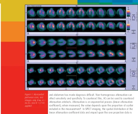
An obese 43 year-old female who had experienced left sided chest pain underwent an exercise tolerance test (with ECG); no ST depression was noted. The origin of her cardiac related pain was required and, consequently, the patient underwent stress and rest myocardial perfusion imaging (MPI; see fi gure 1). The images shown are rest only; row A is not attenuation corrected (AC); row B is with CT AC applied. On comparing rows A and B, the attenuation induced photon defi cient artefact (on the non AC images) is corrected on the CT AC images, so ruling out potential myocardium infarction (the stress study demonstrated similar photon defi cient areas on the non AC images). This is an interesting case as the resting CT AC images rule out any problem in the inferior wall but pinpoint the problem in the anterior wall. This case illustrates that the use of attenuation correction in MPI should only be used as a tool to support decision making in relation to establishing the presence of attenuation artefacts3.
For certain SPECT studies non-homogeneous photon attenuation within the chest
and abdomen has made diagnosis diffi cult. Non-homogenous attenuation can affect sensitivity and specifi city. To counteract this, AC can be used to counteract attenuation artefacts. Attenuation is an exponential process (linear attenuation coeffi cient); when measured, the value depends upon the proportion of scatter included in the measurement4. In SPECT imaging, the spatial distribution of the
linear attenuation coeffi cient data and impact upon the raw projection data is unknown, and additional information (such as the information provided from CT) is required to correct for the effects of attenuation and/or scatter3.
Evidence suggests a necessity to interpret attenuation corrected studies as part of an overall quality control process3. This may include various steps but the
added value of attenuation corrected images should be considered as part of the complete imaging process, and to aid the overall clinical decision making process. In terms of clinical studies which have utilised AC for cardiac imaging, the majority include small to medium sample sizes. Although Ficaro et al’s study in 19955
[image:2.595.339.808.22.410.2]included a limited number of patients (n=10), lateral-to-posterior and basal-to-apical wall ratios of near unity value were demonstrated using AC. The use of AC is further supported in more recent research, also undertaken by Ficaro et al in 20026.
Additional studies by Prvulovich et al in 1997 and Hendel et al in 1999 demonstrated improvements in diagnostic specifi city in patients with follow-up angiography, in comparison with non-attenuation corrected images7,8. It should,
however, be noted that these studies were all conducted using radioisotope and transmission based attenuation correction units, not CT. In terms of the impact of attenuation correction on diagnostic sensitivity of MPI, Duvernoy et al’s study on 28 patients with left main coronary artery disease in 20009 showed this to be present
on 64 per cent of AC images versus seven per cent of uncorrected images.
In the 1980s, the use of external transmission sources such as Ba-133 and Gd-153 were utilised to provide attenuation correction data. However, issues related to poor statistical quality has resulted in a limited use of such sources within modern clinical practice. Nevertheless, in clinical instances whereby only AC is being performed, as in nuclear cardiology for example, the use of a transmission-based system may be justifi ed. Acquiring the required AC data using a transmission based system such as Gd-153 takes longer than a CT based method due to the large difference in available photon fl ux and the fact that transmission based systems have a limited fi eld of view coverage3.
CT data may be used to create linear attenuation coeffi cients based upon anatomical detail specifi c to each patient. This data (attenuation map) may be used to correct for the effects of photon attenuation within the subject and, if only AC is being undertaken (eg myocardial perfusion imaging), the CT may be acquired with a lower statistical quality. Although this reduces the spatial resolution, this technique reduces the radiation dose to the patient.
With reference to oncology-based applications, CT AC has particular values. SPECT imaging is increasingly used to quantify the uptake of a particular radiopharmaceutical within tumours/organs. Such targets are quantifi ed in SPECT as volumes of interest (VOI); these are defi ned around the region to integrate the number of counts, which is directly proportional to the activity (expressed as MBq or mCi) in the volume defi ned10.
Accurate quantitative measurement of radioactivity from a SPECT study can be compromised by the effects of attenuation (and scatter). Such factors may be corrected by the use of CT AC data. The use of SPECT-CT is beginning to create a new role for nuclear medicine in terms of accurately quantifying radionuclide uptake within targets, compared to measurements undertaken with SPECT alone. Results from clinical studies11,12 have demonstrated the value of SPECT-CT in terms of measuring
response to radiotherapy treatment for patients with non-Hodgkin’s lymphoma and radioimmunotherapy. The use of SPECT-CT within oncology is beginning to replicate clinical experience with PET-CT, in terms of improving localisation and extent of disease, and differentiation of physiological and pathological uptake.
Localisation
CT (low resolution or diagnostic quality) can be used in conjunction with SPECT to help localise cold or hot areas seen on SPECT imaging. This has particular value in cases where there are limited or no anatomical landmarks present on SPECT imaging and/or when such landmarks are present but greater anatomical spatial resolution is required. Examples with the registration of SPECT with CT for localisation purposes could include: to aid defi nitive diagnosis and to give more precise lesion localisation as part of surgical work up, such as sentinel lymph nodes13, radiotherapy treatment planning14 or initial patient treatment work up15
(eg neuroendocrine tumours (NET)).
Advancements in computing power and software algorithms have also impacted upon the ability to compensate for the errors in SPECT imaging. In particular the inclusion of an iterative reconstruction model (eg OSEM) for the correction of attenuation with SPECT data and providing quantitative information is becoming more common within clinical practice10. Numerous studies have identifi ed the
improved diagnostic accuracy of SPECT/CT over conventional SPECT imaging and the empirical evidence points toward the following areas of clinical practice which have benefi ted from the emerging hybrid imaging technology, for example:
Lymphoma16
Lung Cancer16
Primary and secondary malignant bone disease17
Infection and infl ammation18
Abdominal disease19
Endocrinology15,20
A comprehensive list of potential clinical applications for SPECT-CT has previously been published10 and the majority of applications relate to the anatomical
localisation of tumours using various radiopharmaceuticals. Published research by Koral et al identifi ed the added value of SPECT-CT in terms of providing valuable tumour organ uptake and dosimetry data, enabling accurate patient workup prior to treatment for lymphoma11,12. This research involved a range of professional fi elds
and provides evidence of multiprofessional working.
Case study
An eight-year-old male presented with a painful right tibia; the clinical question surrounded whether this patient had an osteoid osteoma or leukaemia. A whole body bone scan was conducted (anterior and posterior) and a focal area of uptake was noted within the right femur. Subsequently, localised SPECT images of pelvis and femora were acquired; the poor resolution could not differentiate the high uptake between cortex and medulla and CT imaging was undertaken (see fi gure 2). Correlation with plain fi lms and a SPECT-CT acquisition of the area show a localised area of thickening and sclerosis of the medial cortex of the upper right
femoral shaft, and a small central lucent nidus with a surrounding sclerotic rim. The features are consistent with an osteoid osteoma.
Surgical resection of certain pathologies often requires quite precise localisation of lesions prior to surgery. This helps the surgeon locate the area to be removed more quickly; it also helps the surgeon plan more accurately which areas need
to be removed. Better intelligence prior to and during surgery results in increased probability of removing the affected tissue and reduced operating time. Both can have an impact on post-operative recovery and, ultimately, patient outcome.
[image:4.595.30.799.106.453.2]Low dose/resolution and high dose/resolution can both play a part in localising lesions seen on SPECT studies, and the precise overlay of regional anatomy, together with co-registered SPECT-CT (structural and functional aspects) of the lesion make for better surgical localisation, possible radiotherapy treatment or, even, extraction of tumour tissue. For example, SPECT-CT of brain tumours using a range of Tc99m based radiopharmaceutical agents, such as Sestamibi and Tetrofosmin and In-111 Pentetreotide, may be utilised to provide accurate functional and anatomical diagnosis, and monitor patients’ responses to Figure 2.
An educational
strategy is
radiotherapy/chemotherapy treatment. CT used in isolation cannot always distinguish tumour progression from radiotherapy damage/necrosis, even up to several months after the patient has received treatment14.
Various empirical studies and critical reviews have demonstrated the additional value of SPECT-CT over stand alone SPECT systems, particularly in cases such as lymphoma21,22, infection and bone disease17. However, there appears to be a
debate relating to the appropriate use of low dose CT for attenuation correction and/or basic localisation purposes (which were the parameters of a low performance x-ray scanning device) and higher quality CT data for improved anatomical image quality. Roach et al’s study in 200623 was conducted using
a multislice SPECT-CT unit which permitted greater spatial resolution for the anatomical data. Although the overall fi nal diagnosis and reporter confi dence was improved using a multislice SPECT-CT unit, the increase over fi rst generation low-performance SPECT-CT units was minimal.
Diagnosis using diagnostic quality CT
SPECT-CT has a role in diagnosis, particularly when ‘targeted’ SPECT imaging reveals lesions with no surrounding discernable anatomical landmarks (for localisation) and also when the internal structure of the SPECT lesion needs additional
radiological (high resolution CT) scrutiny to determine its nature. An example could be differentiated thyroid cancer, using whole body imaging with iodine 123 or 131. Here, the precise localisation of lesions is often not possible because of the absence of anatomical landmarks in nuclear medicine data. Co-registered SPECT-CT allows for differentiation between artefactual and normal uptake, and pathological uptake.
Research conducted by Roach et al highlighted the value of SPECT-CT in terms of diagnostic accuracy and reporter confi dence within clinical practice23. This
evaluation of the impact of SPECT/CT on common areas of clinical practice, such as bone scintigraphy, infection imaging (Gallium-67), Indium-111 octreotide scans, I-123/1-131 MIBG scans/treatment monitoring, and Tc-99m Sestamibi parathyroid scans, refl ected a typical nuclear medicine department workload. Overall, the utilisation of SPECT-CT added extra confi dence to the fi nal diagnosis, and reporter confi dence was also increased in particular cases where anatomical landmarking would have been an issue without the CT data. The following case study demonstrates the clinical value of diagnostic accuracy using SPECT-CT.
Case Study
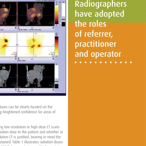
A 78 year old female had known multiple liver metastases from a carcinoid tumour and Yttrium-90 therapy was being considered. An In-111 octreotide SPECT-CT scan was conducted (see fi gure 3). As can be seen, the images demonstrate liver metastases, incidental adrenal gland fi ndings and hydronephrosis. The
morphological appearance of the metastases can be clearly located on the diagnostic quality CT image series, giving heightened confi dence for areas of radiopharmaceutical uptake.
One of the biggest considerations of using low resolution or high dose CT scans in addition to SPECT is the additional radiation dose to the patient and whether or not the extra dose from using high-resolution CT is justifi ed, bearing in mind the extra clinical information that may be obtained. Table 1 illustrates radiation doses that a patient will typically receive from SPECT, low-resolution and high-resolution CT, and plain radiography for reference. As can be seen, the CT component adds a signifi cant amount to the total dose the patient receives; this is particularly true for high resolution CT. It is interesting to compare plain fi lm and high resolution CT and the dose differential. This brings into sharp focus the need to consider carefully whether CT or plain fi lm imaging would give the same radiological information and, if so, whether the lower dose alternative (plain fi lm) would be better justifi ed.
Professional responsibilities and legislative
considerations
Depending upon local circumstances in clinical imaging departments, radiographers can be referrers, practitioners and operators within the context of the Ionising Radiation (Medical Exposure) Regulations2 (IR(ME)R). Within nuclear medicine and with regard
[image:5.595.349.649.29.276.2]to radiopharmaceuticals, this is not the case. Radiopharmaceuticals are regulated Figure 3. Radiation dose from CT
Radiographers
have adopted
the roles
Imaging and Dose (mSv)
SPECT [i] Low-Res CT[ii] High-Res CT[iii] Plain Film[iv]
Pulmonary Nodes Tc-99m Depreotide 6 Chest 1 Chest 5.8 Chest 0.02
Lumber Spine Metastases
Tc-99m phosphonate 5 Abdo + pelvis 1.5 Abdo + pelvis
7.1 Lumber Spine
1.3
Myocardial Imaging Tc-99m sestamibi 4 Chest 1 Chest 5.8 Chest 0.02
under both medicines and ionising radiation legislation and the former mandates that only a registered dental or medical practitioner may direct the clinical service and, as such, provide the clinical justifi cation required under IR(ME)R. For nuclear medicine examinations, with regard to IR(ME)R, only the ARSAC licence holder may justify clinical examinations. Radiographers working within nuclear medicine and with specifi c reference to radiopharmaceuticals may only act as operators.
Within nuclear medicine departments, legislative arrangements permit radiographers to act as practitioners and referrers for x-ray examinations and acting as referrer for plain x-ray examinations, in association with nuclear medicine procedures, has become common practice. Using the same legislative arrangements, radiographers can, indeed should, act as referrer and practitioner for the CT component of SPECT-CT. There is logic for both as SPECT-CT has limited routine applications. For many patients, the decision to make a CT exposure (low or high resolution) will depend on factors such as the clinical background, physical make up and the nuclear medicine images themselves. Hence, the decision to refer for CT may only be made at the point of care within the nuclear medicine department. Therefore, the radiographer is ideally placed to fulfi l the role of referrer. In full knowledge of the evidence base and the clinical background, a radiographer can act as practitioner, thereby protecting the patient from unnecessary x-radiation exposure. On discussion with several nuclear medicine departments, it was found that radiographers have already adopted formally the roles of referrer, practitioner and operator for the CT component of the SPECT-CT studies.
Business, practical and educational considerations
when purchasing a SPECT-CT system
A department should consider the physical footprint required for a hybrid SPECT-CT system. This is especially true if the existing gamma camera has a small footprint. Some clinical departments within the United Kingdom are
replacing fi rst generation SPECT systems with SPECT-CT units, which generally require a larger physical space. The space required for a SPECT-CT system will depend on the type of unit being installed and the nature of the examinations conducted. The quoted minimum room size for a GE Hawkeye/Hawkeye-4 is 1Hawkeye/Hawkeye-4 feet x 16 feet and the amount of lead shielding required for this environment may be less than a dedicated CT unit. Some departments utilise mobile lead shielding devices for the GE Hawkeye devices which permits some fl exibility with the organisation of the imaging environment. The exposure rate from low performance CT units is approximately 20 times less than that from a multislice CT unit24 and the use of mobile shields are common in departments
with these units.
A larger physical room environment (minimum 15 feet x 24 feet) is required for SPECT-CT systems employing a dedicated multislice CT unit, and thicker protective shielding is required for the use of multislice SPECT/CT units. The weight bearing parameters of the fl oor should also be considered24, especially if the unit is not
being installed on the ground level within a hospital. Separate operator console environments are also becoming common within SPECT-CT rooms, although this does remove an element of patient interaction normally associated with nuclear medicine.
Currently the cost of the high end CT components (eg 64 slice) may exceed the cost of the SPECT device and the justifi cation for such units will depend upon the clinical workload of a nuclear medicine department. With current shifts in imaging tools for certain conditions (eg pulmonary embolism), a SPECT-CT unit with multislice capabilities may be positioned to undertake contrast enhanced CT scans, such as pulmonary angiography and the assessment of coronary calcifi cation10,25
and, potentially, providing a ’one stop‘ approach for patients undergoing their diagnostic CT and physiological scans, if required.
Table 1.
[i] Notes for Guidance on the Clinical Administration of Radiopharmaceuticals and Use of Sealed Radioactive Sources Health Protection Agency, Administration of Radioactive Substances Advisory Committee, March 2006 (Revised 20 April 2006).
[ii] Effective doses to patients from CT acquisitions on the GE Infi nia Hawkeye: a comparison of calculation methods. Sawyer, L et al. Nuclear Medicine Communications. 29(2):144-149, February 2008.
Nuclear medicine departments may wish to evaluate the feasibility of undertaking CT examinations during periods where access to radiopharmaceuticals is limited (eg early morning/late afternoon), or utilising the CT component of the hybrid unit for out-of-hours work to alleviate some of the demands on the radiology department’s main CT unit. This may require some degree of cross pollination of training requirements26 and the development of appropriate educational curriculum
within the workplace and at academic institutions. Educational programmes for nuclear medicine technologists in North America and Australia provide focused academic and clinical training specifi cally related to the utilisation of CT within the nuclear medicine environment.
In the USA, working relationships between the Society of Nuclear Medicine Technologists and the American Society for Radiologic Technologists are considered to be a good example of a synergistic approach to the training requirements between professions. The University of Sydney in Australia also offers a dedicated distance based CT module aimed at nuclear medicine technologists who are involved in using CT within a nuclear medicine environment. In the UK, there is limited scope for the provision of a dedicated hybrid imaging programme. However, short courses for technologists related to the safe use of CT, and hybrid imaging modules of study have emerged from two academic institutes over the last couple of years. Currently, it is unclear what CT training requirements are needed offi cially by a technologist working within the hybrid nuclear medicine fi eld, especially if the CT unit has a multislice confi guration and the imaging parameters are interchangeable. A clear educational strategy is required for the safe and optimal use of CT within a hybrid nuclear medicine environment, to minimise patient dose and optimise disease detection, aiding patient management.
Conclusion
Following the inception of SPECT-CT hybrid technology, there appears to be an evolution in the useful applications associated with this modality. Initial ‘low-end’ systems introduced over a decade ago provided a platform for improved anatomical localisation and high photon-fl ux attenuation co-effi cient data, potentially improving the sensitivity and specifi city of existing imaging techniques. Beyond the existing techniques, SPECT-CT has the potential for mirroring the success of PET-CT in terms of radiotherapy planning, dosimetry calculations and multi-imaging approaches, which include coronary artery calcium scoring and myocardial perfusion imaging.
As detector technology also continues to evolve, the overall design and utilisation of future SPECT/CT units may further develop. The use of solid state materials such as cadmium-zinc-telluride will provide a single detector interface and, potentially, improve the functional count rate and spatial resolution further. The training and
There is
a debate
about the
appropriate
use of low
dose CT
education of the hybrid imaging workforce is crucial to the future utilisation of this important area of clinical practice.
References
Schillaci O, Danieli R, Manni C, Simonetti G. (2004) Is SPECT/CT with a hybrid camera useful to improve scintigraphic imaging interpretation? Nuclear Medicine Communications, Volume 25, pp 705-710.
Department of Health (1999) Ionising Radiation (Medical Exposure) Regulations 2000, London, Department of Health.
Bateman TM, Cullom SJ. (2005) Attenuation correction Single Photon Emission Computed Tomography Myocardial Perfusion Imaging, Seminars in Nuclear Medicine, Volume 35, pp 37-51.
Sorenson SA, Phelps ME. (2003) Physics in Nuclear Medicine, 3rd Edition, Philadelphia, Saunders.
Ficaro EP, Fessler JA, Ackermann RJ. (1995) Simultaneous transmission-emission thallium-201 cardiac SPECT: Effect of attenuation correction on myocardial tracer distribution, Journal of Nuclear Medicine, Volume 36, pp 921.
Ficaro EP. (2002) Controversies – Should SPET attenuation correction be more widely employed in routine clinical practice? – For, European Journal of Nuclear Medicine, Vol 29, 3, pp 409-411.
Prvulovich EM, Lonn AHR, Bomanji JB. (1997) Effect of attenuation correction on myocardial thallium-201 distribution in patients with a low likelihood of coronary artery disease, European Journal of Nuclear Medicine, pp 266-275.
Hendel RC, Berman DS, Cullom SJ. (1999) Mulicenter clinical trial to evaluate the effi cacy of correction for photon attenuation and scatter in SPECT myocardial perfusion imaging, Circulation, Volume 99, pp 2742-2749.
Duvernoy CS, Ficaro EP, Karabajakian MZ. (2000) Improved detection of left main coronary artery disease with attenuation corrected SPECT, Journal of Nuclear Cardiology, Volume 7, pp 639-648.
Seo Y, Mari C, Hasegawa B. (2008) Technological development and advances in SPECT/CT, Seminars in Nuclear Medicine, Volume 38, pp 177-198.
Koral KF, Dewaraja Y. (2000) Initial results for hybrid SPECT – conjugate-view tumor dosimetry in I-131 anti-B1 antibody therapy of previously untreated patients with lymphoma, Journal of Nuclear Medicine, Volume 41, pp 1579-1586.
Koral KF, Dewaraja Y. (2003) Update on hybrid conjugate view SPECT tumour dosimetry and response in I-131 tositumomab therapy of previously untreated lymphoma patients, Journal of Nuclear Medicine, Volume 44, pp 457-464. Van der Ploeg I, Renato A, Valdés Olmos B, Kroon R, Emiel J, Rutgers T, Omgo N, (2009) The hidden sentinel node and SPECT/CT in breast cancer patients, European Journal of Nuclear Medicine and Molecular Imaging, Volume 36, pp 6-11.
Schillaci O, Filippi L, Manni C, Santoni R, (2007) Single-Photon Emission Computed Tomography / Computed Tomography in Brain Tumours, Seminars in Nuclear Medicine, Volume 37, pp 34-47.
Krausz Y, Israel O, (2006) SPECT/CT in Endocrinology, Seminars in Nuclear Medicine, Volume 36, pp 267-274.
Schillaci O, (2006) SPECT/CT in lung cancer and malignant lymphoma, Seminars in Nuclear Medicine, Volume 36, pp 275-285.
Tomography/Computed Tomography in Benign and Malignant Bone Disease, Seminars in Nuclear Medicine, Volume 36, pp 286-294.
Bunyaviroch T, Aggarwal A, Oates ME, (2006) Optimized Scintigraphic Evaluation of infection and infl ammation: Role of SPECT/CT fusion imaging, Seminars in Nuclear Medicine, Volume 36, pp 295-311.
Schillaci O, Filippi L, Danieli R, Simonetti G. (2007) Single-Photon Emission Computed Tomography/Computed Tomography in Abdominal Diseases. Seminars in Nuclear Medicine Volume 37, pp 48-61.
Ozer S, Dobrozemsky G, Kienast O (2004) Value of combined SCT/SPECT technology for avoiding false positive planar I-123 MIBG scintigraphy, Nuklearmedizin, Volume 43, pp 164-170.
Bar-Shalom R, Yefremov N, Haim N, (2003) Camera-based FDG PET and Gallium-67 SPECT in evaluation of lymphoma: comparative study, Radiology, Volume 227, pp 353-350.
Palumbo B, Sivolella S, Palumbo I, (2005) Gallium-67 SPECT /CT with a hybrid system in the clinical management of lymphoma, European Journal of Nuclear Medicine and Molecular Imaging, Volume 32, pp 1011-1017.
Roach PJ, Schembri GP, Ho Shen IA, Bailey EA, Bailey DL, (2006) SPECT using a spiral CT Scanner for anatomical localisation: impact on diagnostic accuracy and reporter confi dence in clinical practice, Nuclear Medicine Communications, Volume 27, pp 977-987.
O’Connor MK, Kemp BJ, (2006) Single-photon emission computed tomography/ Computed tomography: basic instrumentation and innovations, Seminars in Nuclear Medicine, Volume 36, pp 258–266.
Schepis T, Gaemperli O, Koepfl i P, (2007) Use of coronary calcium score scans from stand alone multislice computed tomography for attenuation correction of myocardial perfusion SPECT, European Journal of Nuclear Medicine and Molecular Imaging, Volume 34, pp 11-19.
Rose T, (2005) Educating the Hybrid Technologist, Radiologic Technology, Vol 77, 1, pp 15–18.
This article was compiled by Peter Hogg and Marc Griffi ths. Peter is a professor of radiography at the University of Salford. His role includes being programme leader for the MSc Nuclear Medicine. Marc is the subject group leader for radiography and programme leader of the MSc Nuclear Medicine at the University of the West of England, Bristol.
Acknowledgements
[image:8.595.38.331.34.363.2]Figures 1 and 3: Dr Kat Dixon, principal clinical scientist, Nuclear Medicine Department, Poole Hospital NHS Trust.
Figure 2: Phil Facey, superintendent radiographer, nuclear medicine, University Hospital of Wales, Cardiff & Vale NHS Trust.
18.
19.
20.
21.
22.
23.
24.
25.