Morphometric analysis of the brain base arteries
in fallow deer (Dama dama)
W. Brudnicki
University of Technology and Life Sciences, Faculty of Animal Breeding and Biology, Bydgoszcz, Poland
ABSTRACT: This paper describes the course and variation in the brain base arteries in fallow deer. The metrical features of the brain base arteries were determined with an image analysis system. The main sources of blood supply to the brain in fallow deer are internal carotid arteries; vertebral arteries rarely participate in blood supply. The brain base arteries in fallow deer show variation both in their course and in the way of descent of particular vessels. The highest variation was observed in the way of the opening of caudal cerebral arteries. The volume of the arterial circle of brain in fallow deer is similar to the volume of the basilar artery. Considering the correlation between specific parameters, it can be concluded that the volume of the basilar artery is highly correlated with the volume of the posterior part of the arterial circle of the brain, i.e., the volume of the caudal communicating arteries.
Keywords: artery; brain; morphometric analysis; fallow deer
Blood supply to the brain, despite the long time that has elapsed since the first description of that vascular region, remains a current area of research with regard to cognitive and practical considera-tions. There are abundant sources in the literature which describe the brain base arteries in various spe-cies of ruminants. Besides these traditional studies, such as those of Hoffman (1900) and Jenke (1919), the structure and variation in the brain base arteries has been studied in sheep (Jablonski and Wiland, 1973), and in goats (Brudnicki, 2000). In addition, Wegrzyn et al. (1983) investigated the brain base arteries in European bison, Ding et al. (2007) in yaks and Frackowiak and Jakubowski (2008) studied these arteries in giraffes. The course and variation of the brain base arteries has also been determined in the bovine foetus (Brudnicki and Gielecki, 1996) and in representatives of Cervidae (Godynicki and Wiland, 1970, 1971; Jablonski, 2005). Interestingly, Ashwini et al. (2008) provided a comparative analysis of the brain base arteries in humans, cattle, sheep, goats and pigs. Not only were the structure and course of
the brain base arteries evaluated, but also the degree of variation and comparative metrical features of these anatomical structures were determined.
The present paper describes the course as well as the morphological and morphometric variation in the brain base arteries in fallow deer. The struc-ture and the variation in the vascular region were compared with the cerebral arteries of different mammal species.
MATERIAL AND METHODS
im-ages were then analysed for artefacts. If none were present, then the uncompressed TIFF images were analysed with the Image 1.44 programme for digital image analysis (Rasband and Image, 1997–2011). The length, the average diameter and the capacity of the vessels creating the arterial circle of the brain were determined. The same parameters were de-scribed for the basilar artery. For the arterial circle of the brain, the parameters for its anterior part, for the rostral cerebral arteries as well as for the posterior part, the caudal communicating arteries as well as, independently, the parameters of the left and the right part of the arterial circle of the brain, were separately calculated. The results were statistically analysed, taking into consideration the brain weight of the individuals.
RESULTS
The brain in fallow deer is supplied with blood by internal carotid arteries the intracranial segment of which is regenerated from the rostral epidural rete mirabile, made of vessels which are branches of the external carotid artery and maxillary artery. The ex-tracranial segment of the internal carotid artery un-dergoes atrophy after birth. Having passed through the dura mater, the internal carotid artery is separated on both sides into the rostral cerebral artery and the caudal communicating artery. Rostral cerebral
arter-ies create the anterior-lateral part of the arterial circle of the brain, whereas caudal communicating arteries create its posterior part. Having anastomosed, they give off the basilar cerebral artery.
Rostral cerebral arteries in fallow deer run with a wide arch around the optic nerves, heading to-wards the piriform lobe. A rostral choroidal artery descends from the initial part of each artery.
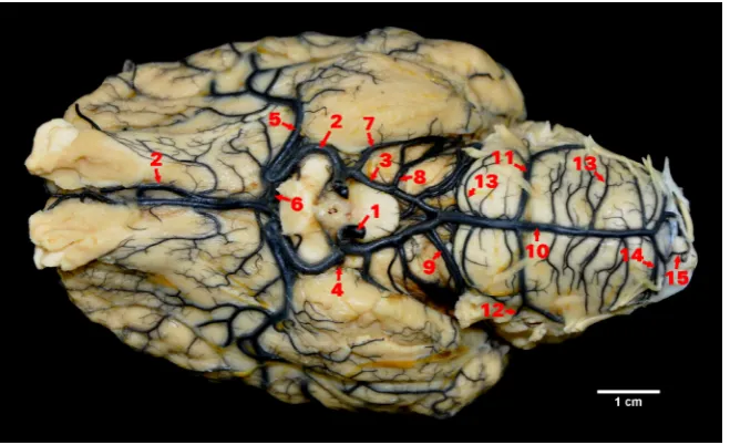
[image:2.595.147.477.485.686.2]At the rostral level of the margin of the crossing of the optic nerves, the rostral cerebral artery gives off a thick arterial vessel, the middle cerebral artery. The middle cerebral artery in its initial part runs parallel to the rostral cerebral artery; however, it runs in the opposite direction or goes in an arch and then heads towards the lateral cerebral fissure. Before reaching it or in its area, the main trunk of the middle cerebral artery gets divided into numerous cortical branches. The main stream of blood going through the rostral cerebral artery is collected by the middle cerebral artery, and so a section of the rostral cerebral artery above its descent is clearly thinner. The symmetrical rostral cerebral arteries in that part head towards the longitudinal cerebral fissure, then along the medial olfactory tract towards the olfactory bulbs. Before reaching the median fissure, the symmetrical rostral cerebral arteries are anastomosed by the rostral com-municating artery. The callosal artery is frequently separated from the rostral communicating artery. It may also depart directly from one of the rostral cer-ebral arteries (Figure 1).
The posterior part of the arterial circle of the brain is made up of caudal communicating arter-ies. They run on brain branches medially from the oculomotor nerve and then get anastomosed, giv-ing off the basilar artery. Along their course, caudal communicating arteries give off symmetrically the caudal cerebral artery, caudal choroidal artery and the rostral cerebellar artery. Medially from caudal communicating arteries there descend branches to the cruses of the brain and phypothalamus.
The basilar artery begins in the posterior part of the interpeduncular fossa and then runs caudally across the pons and then in the median fissure of the medulla oblongata. The diameter of the basi-lar artery decreases gradually in the caudal direc-tion. In its initial part, the basilar artery gives off many branches running towards the pons. These are mostly minor vessels, the number of which varies. Below the abducent nerve, there are cau-dal cerebellar arteries departing from the basilar artery. Initially, they run at the height of the pos-terior margin of the pons and then they run later-ally and superiorly. Having reached the cerebellum, they give off branches to its caudal and dorsal sur-face. The branches of the caudal cerebellar arteries are labyrinthine arteries. Below the departure of the caudal cerebellar arteries there are numerous branches departing from the basilar artery towards the medulla oblongata. In its final part, the basilar artery separates into poorly-developed vertebral arteries.
The arteries running on the brain base in fallow deer demonstrate variation in the course and way of departure of individual vessels. In 23 (76.7%) individuals, the division of the internal carotid ar-tery followed the perforation of the dura mater. In seven (23.3%) individuals the internal carotid artery was separated symmetrically above the dura mater. Formed as a result of the division, the rostral cer-ebral arteries and caudal communicating arteries pass through the dura mater independently.
The rostral part of the circle created by the rostral cerebral arteries, coming with a wide curve around the optic nerves, assumes the shape of an ellipse. Rostral cerebral arteries along their route give off arterial branches, first the rostral choroidal artery. The next and strongest branch of the rostral artery was the middle cerebral artery. In 10 (33.3%) brains the middle cerebral artery ran in an arc and then headed towards the Sylvian sulcus. In 14 (46.7%) individuals the middle cerebral artery departed from the parent vessel at an acute angle caudally
and then ran in front of the piriform lobe. In six (20%) individuals that departure displayed a mixed pattern. One vessel departed with a curve, whilst the vessel on the other side created an acute angle with the rostral cerebral artery.
The rostral part of the arterial circle of the brain in fallow deer showed variation in the descent of vessels. In 22 (73.3%) brains the rostral commu-nicating artery was found from which the callosal artery descended. In two (6.6%) brains the termi-nal sections of the rostral cerebral arteries crossed running to the opposite hemisphere. In the other individuals those sections ran along the margin of the median fissure of the brain. The posterior part of the arterial circle of the brain assumed the shape of an isosceles triangle with the line passing at the point of the opening of the caudal communicating arteries as the base of the triangle.
The caudal communicating arteries were quite well-developed and no clear change in the diameter was found in their pattern.
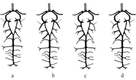
The highest variation in was observed in the way of descent of the caudal cerebral arteries. In seven (23.3%) individuals they departed already from the rostral cerebral arteries. In four (13.3%) individuals those vessels descended at the point of the division of the internal carotid artery. In 14 (46.7%) brains those vessels departed from caudal communicating arteries. In the other five (16.7%) individuals the descent of the caudal cerebral arteries was asym-metric (Figure 2). In all cases, however, the sub-sequent course and vessel division was typical for ruminants.
[image:3.595.309.533.589.721.2]Caudal cerebral arteries were seen to depart bi-laterally to rostral cerebral arteries – (a) caudal cer-ebral arteries depart at the point of the division of the internal carotid artery into the rostral cerebral artery and caudal communicating artery; (b) caudal
cerebral arteries depart from caudal communicat-ing arteries; (c) there is an asymmetrical departure of caudal cerebral arteries, the right-sided artery departs from the caudal communicating artery, while the left-sided artery departs from the rostral cerebral artery.
The caudal choroidal arteries in all cases depart-ed from the caudal communicating artery with a single trunk which divided, after a short course on the cerebral cruses, into two or three vessels.
From the terminal section of the caudal communi-cating arteries there descended symmetrically ros-tral cerebellar arteries. These spread along the rosros-tral margin of the pons. In eight (26.7%) brains the ros-tral cerebellar arteries at the point of the opening created vascular islands. In those cases one of the branches departed from the caudal communicating artery, while the second one departed from the basi-lar artery to communicate with a common trunk.
The departure of the caudal cerebellar arteries was usually symmetrical. In two (6.6%) individu-als only a slight asymmetry was observed in the departure of those vessels on both sides.
Metrical features of the arterial circle of the brain
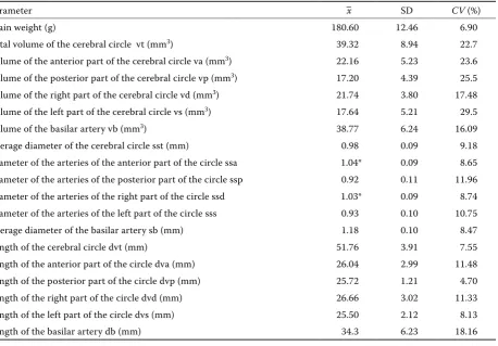
Table 1 presents data on the metrical features of the brain base arteries in fallow deer. The to-tal volume of the arterial circle of the brain was 39.32 ± 8.94 mm3. Significant differences between
[image:4.595.74.533.426.745.2]the volume of the anterior and posterior part of the arterial circle were observed. The anterior part of the arterial circle of the brain created by the rostral cerebral arteries and the rostral communicating artery showed a higher volume than the posterior part formed by the caudal communicating arteries. Similarly significant differences were observed be-tween the volume of the right and the left part of the arterial circle of the brain. The arteries creating the right part of the arterial circle of the brain indicated a significantly greater volume. Both the length and the diameter of the arteries forming the right part of the arterial circle demonstrated higher values, however, the differences were non-significant. In the case of the length of arteries creating the arte-rial circle of the brain in fallow deer, no significant
Table 1. Summary of the parameters examined
Parameter –x SD CV (%)
Brain weight (g) 180.60 12.46 6.90
Total volume of the cerebral circle vt (mm3) 39.32 8.94 22.7
Volume of the anterior part of the cerebral circle va (mm3) 22.16 5.23 23.6
Volume of the posterior part of the cerebral circle vp (mm3) 17.20 4.39 25.5
Volume of the right part of the cerebral circle vd (mm3) 21.74 3.80 17.48
Volume of the left part of the cerebral circle vs (mm3) 17.64 5.21 29.5
Volume of the basilar artery vb (mm3) 38.77 6.24 16.09
Average diameter of the cerebral circle sst (mm) 0.98 0.09 9.18 Diameter of the arteries of the anterior part of the circle ssa 1.04* 0.09 8.65 Diameter of the arteries of the posterior part of the circle ssp 0.92 0.11 11.96 Diameter of the arteries of the right part of the circle ssd 1.03* 0.09 8.74 Diameter of the arteries of the left part of the circle sss 0.93 0.10 10.75 Average diameter of the basilar artery sb (mm) 1.18 0.10 8.47 Length of the cerebral circle dvt (mm) 51.76 3.91 7.55 Length of the anterior part of the circle dva (mm) 26.04 2.99 11.48 Length of the posterior part of the circle dvp (mm) 25.72 1.21 4.70 Length of the right part of the circle dvd (mm) 26.66 3.02 11.33 Length of the left part of the circle dvs (mm) 25.50 2.12 8.13 Length of the basilar artery db (mm) 34.3 6.23 18.16
differences were identified between the anterior and posterior parts of the arterial circle.
At the height of the anterior margin of the pons the diameter of the basilar artery was 1.6 ± 0.07 mm, at the height of the posterior margin of the pons it was 1.1 ± 0.11 mm. At the point of the departure of the vertebral arteries the diameter was 0.9 ± 0.09 mm. The volume of the basilar artery was 38.77 mm3. The mean diameter of the arteries of the right and the left part of the circle was significantly different than the mean diameter of the anterior and posterior part of the arterial circle of the brain. The diameter of the basilar artery decreased cau-dally. At the height of the anterior margin of the pons it was 1.6 ± 0.07 mm, while at the height of the posterior margin of the pons it measured 1.1 ± 0.11 mm and at the point of departure of the ver-tebral arteries it was 0.9 ± 0.09 mm.
The value of the coefficient of the volume of the arterial circle of the brain to the brain weight (Vt × 100/m) was 21.77. The value of the coefficient of the volume of the basilar artery to the brain weight (Vb × 100/m) was 21.47.
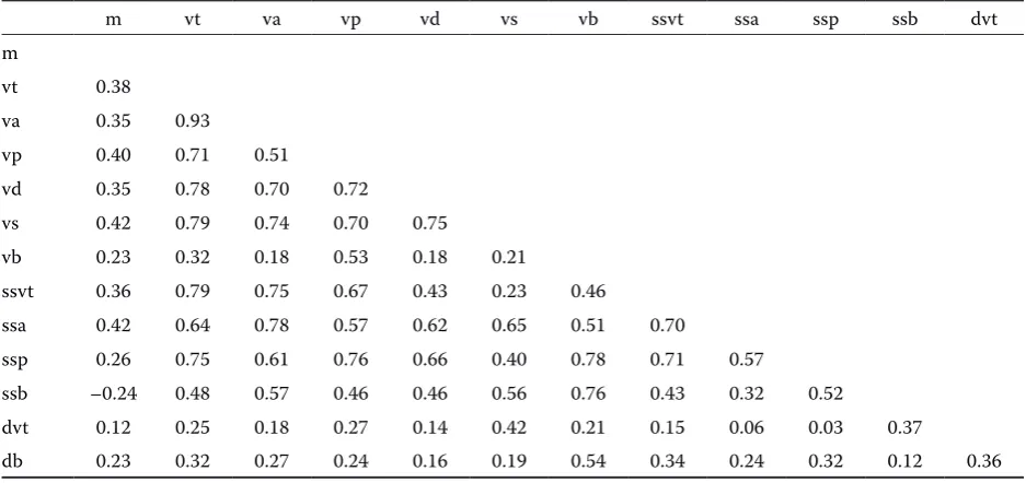
Table 2 presents the results of the correlation analysis between the parameters. The parameters of the arteries of the arterial circle of the brain and the basilar artery correlate slightly with the brain weight. The diameter of the basilar artery and the length of the posterior part of the arterial circle of the brain are in fact negatively correlated with
the brain weight. The total volume of the arterial circle of the brain is most highly correlated with the volume of specific segments of the circle and the diameter of the arteries forming the circle is, however, slightly correlated with their length. The volume of the basilar artery is correlated with the volume of the posterior part of the arterial circle of the brain. It is also correlated with the mean diameter of the basilar artery and its length.
DISCUSSION
Describing the anatomy and variation in the brain base arteries in mammals, Wiland (1974) indicates that the final shape of the vascular tree is influenced by genetic and environmental factors. According to the author, this variation does not, however, exceed some borders defined by the phylogenetic development of the particular systematic group of mammals. The structure, course and the variation in the brain base arteries in fallow deer fall within those borders. The shape of the arterial circle of the brain was characteristic for Cervidae; however, its posterior part, as compared with roe deer or deer, was more regular in shape. The main stream of blood from the intracranial part of the internal carotid arteries heads towards the rostral cerebral arteries, which is seen from the diameter of those vessels. Middle cerebral arteries depart
symmetri-Table 2. Correlation of parameters of the brain-base arteries in fallow deer
m vt va vp vd vs vb ssvt ssa ssp ssb dvt m
vt 0.38
va 0.35 0.93
vp 0.40 0.71 0.51
vd 0.35 0.78 0.70 0.72
vs 0.42 0.79 0.74 0.70 0.75
vb 0.23 0.32 0.18 0.53 0.18 0.21
ssvt 0.36 0.79 0.75 0.67 0.43 0.23 0.46
ssa 0.42 0.64 0.78 0.57 0.62 0.65 0.51 0.70
ssp 0.26 0.75 0.61 0.76 0.66 0.40 0.78 0.71 0.57
ssb –0.24 0.48 0.57 0.46 0.46 0.56 0.76 0.43 0.32 0.52
dvt 0.12 0.25 0.18 0.27 0.14 0.42 0.21 0.15 0.06 0.03 0.37
db 0.23 0.32 0.27 0.24 0.16 0.19 0.54 0.34 0.24 0.32 0.12 0.36
[image:5.595.63.532.102.323.2]cally at an acute angle in 46.2% cases. A similar way of departure of the middle cerebral arteries prevailed in cattle, yaks and in European bison. In 19% of cases the departure was mixed, while in 33% of the brains examined middle cerebral arteries ran with a wide curve, similarly as in roe deer and deer. According to Ruedi (1922), the arcs and bends of arterial vessels decrease laterally the negative effect of the pulse wave on the delicate brain tissue. As very dynamic animals, deer and roe deer are pro-vided with such a security mechanism, whereas in representatives of Bovidae, which exhibit weaker motion dynamics, the departure of the middle cer-ebral artery has developed in a different way.
A smaller range of variation was observed in the brain base arteries in fallow deer in comparison with deer and roe deer. The shape of the arterial circle of the brain and the proportions of its seg-ments are most similar to the arterial circle in goats. In five (16.7%) individuals there was noted a divi-sion of the internal carotid artery above the dura mater. In those cases the rostral cerebral artery and the caudal communicating artery penetrated under the dura mater independently. Such a varia-tion was also reported in fallow deer by Godynicki (1971), who investigated the blood supply to the head. In 72.6% cases in fallow deer it was found that rostral cerebral arteries were anastomosed by the rostral communicating artery. The highest vari-ation in its departure was observed for the caudal cerebral artery. A similar way of departure of caudal cerebral arteries was observed in goats in 22.22% cases. This vascular variation was also described in deer by Godynicki and Wiland (1970, 1971). Only in 14 (46.7%) individuals was the departure typi-cal for other ruminant species; the caudal cerebral arteries departed from the caudal communicating arteries.
The estimation of the volume of the arterial circle of the brain and the basilar artery gives an insight into quantitative dependencies in the vascular sys-tem of the brain. Gielecki et al. (1996) suggests that it is the most sensitive measure of ontogen-esis changes. The volume of the arterial circle of the brain in fallow deer is similar to the volume of the basilar artery. The volume of the arterial circle of the brain in fallow deer is considerably influenced by the volume of the anterior part of the circle formed by the rostral cerebral arteries. Interestingly, the diameter of the basilar artery decreases considerably in the caudal direction. At the height of the anterior margin of the pons the
diameter of the basilar artery was 1.6 ± 0.07 mm, at the height of the posterior margin of the pons it measured 1.1 ± 0.11 mm and at the point of de-parture of vertebral arteries it was 0.9 ± 0.09 mm. A decreasing diameter of the basilar artery in the caudal direction in cattle, sheep and goats was de-scribed by Ashwini et al. (2008). The analysis of correlation between the specific parameters shows that the volume of the basilar artery is highly cor-related with the volume of the posterior part of the arterial circle of the brain, namely the volume of the caudal communicating arteries, which confirms the suggestions that in ruminants the main sources of blood supply to the basilar artery are the caudal communicating arteries. According to Ashwini et al. (2008), the basilar artery is the branch of the posterior part of the arterial circle of the brain in ruminants and it shows some dependence on the parameters of the arterial circle of the brain. This dependence between the parameters of the arterial circle of the brain and the basilar artery, however, was not observed in polar foxes (Goscicka et al., 1991). Upon analysis of the metrical features it was concluded that the arterial circle of the brain and the basilar artery are independent vascular systems. According to Rogers (1947) and Hale (1960), simi-larly the arterial circle of the brain and the basilar artery in humans are independent vascular systems. The species in which the relationship between the arterial circle of brain and the basilar artery has been investigated includes chinchilla; in these ani-mals a very high correlation between the volume and the diameter of the basilar artery and the ar-terial circle of the brain was reported (Gielecki et al., 1996).
Corresponding Author:
Witold Brudnicki, University of Technology and Life Sciences, Faculty of Animal Breeding and Biology, Department of Animal Morphology and Hunting, ul. Bernardynska 6, 85-029 Bydgoszcz, Poland E-mail: [email protected]
e.g. the pathological narrowing or even the total lack of flow in internal carotid arteries or vertebral arteries.
REfERENCES
Ashwini CA, Shubha R, Jayanthi KS (2008): Comparative anatomy of the circle of Willis in Man, cow, Steep, goat, and pig. Neuroanatomy 7, 54–85.
Brudnicki W (2000): Basilar arteries of the brain in do-mestic goat (Capra hircus L). Electronic Journal of Polish Agricultural University Veterinary Medicine 3, 1–6.
Brudnicki W, Gielecki J (1996): Digital image analysis of the brain base In bosine foetuses. I. Variability of basi-lar brain arteries. Zeszyty Naukowe ATR w Bydgoszczy, Zootechnika 28, 53–62.
Ding Y, Shao B, Wang J (2007): The arterial supply to the brain of the yak (Bos grunniens). Annals of Anat-omy 189, 31–38.
Frąckowiak H, Jakubowski H (2008): Arterial vasculari-zation in the giraffe brain. Annales Zoologici Fennici 45, 1–7.
Gielecki J, Brudnicki W, Nowacki M (1996): Digital-image Analysis of the brain-base arteries in chinchilla, Chinchilla laniger(Molina). Anatomia, Histologia, Embryologia 25, 117–119.
Godynicki S, Wiland C (1970): Basilar arteries in red deer (in Polish). Roczniki Wyzszej Szkoly Rolniczej w Poznaniu 49, 45–52.
Goscicka D, Gielecki J, Brudnicki W, Jablonski R (1991): Analysis of the volume of cerebral circle and basal ar-tery in blue fox. Polskie Archiwum Wear-terynaryjne 31, 83–90.
Godynicki S (1971): Arteries of the head in fallow deer
(Dama dama L.) Polskie Archiwum Weterynaryjne
15, 851–864.
Godynicki S, Wiland C (1971): Basilar arteries in roe deer (in Polish). Roczniki Wyzszej Szkoly Rolniczej w Poznaniu LIV, 47–54.
Hale AR (1960): Circle of Willis functional concepts old and new. American Heart Journal 60, 491.
Hofmann M (1900): Zur Vergleichenden Anatomie der Gehiren und Ruckenmarksarterien der Vertebraten. Zeitschrift fur Morphologie und Anthropologie 2, 247–320.
Jablonski R (2005): Variation in the pattern of arteries of the encephalon base in roe deer. Folia Biologica (Krakow) 53, 31–34.
Jablonski R, Wiland C (1973): Variation of the arteries of the base of the brain in sheep. Folia Morphologica, Warszaw 32, 3, 339–347.
Jenke WKK (1919): Die Gechinarterien des Pferdes, Hundes, Rindes und Schweines vergleichen mit denen des Menschen. [Inaugural Dissertation.] Dresden. Rasband WS, Image J (1997–2011): U.S. National
Insti-tutes of Health, Bethesda, Maryland, USA. http:// imagej.nih.gov/ij/.
Rogers L (1947): The function of the circulus arteriosus of Willis. Journal of Neurology 70, 17.
Ruedi M (1922): Topographie und Function der Arteria carotis interna des Pferdes. Veterinary Medicine, Zu-rich.
Wegrzyn M, Roskosz T, Makowiecka M (1983): Brain arteries of the European bison, Bison bonasus L.1758. Annals of Warsaw Agricultural University SGGW-AR, Veterinary Medicine 11, 9–16.
Wiland C (1974): Factors inducing the mutability of ar-teries at the basis of encephalon in Mammals. Przegląd Zoologiczny 18, 400–418.