4,4
000-Bipyridine–dimethylglyoxime (1/1)
Yan Yang, Ziping Huang, Haitang Lv and Aixia Han*
College of Chemical Engineering,Qinghai University, Xining 810016, People’s Republic of China
Correspondence e-mail: yyan217@163.com
Received 14 November 2011; accepted 17 December 2011
Key indicators: single-crystal X-ray study;T= 298 K; mean(C–C) = 0.002 A˚;
Rfactor = 0.043;wRfactor = 0.133; data-to-parameter ratio = 17.6.
In the title compound, C10H8N2C4H8N2O2, both the dimethyl-glyoxime and the 4,40-bipyridine molecules have
crystal-lographicCisymmetry. The molecules stack along the a-axis direction with a dihedral angle of 20.4 (8) between their
planes. In the crystal, the components are linked by O—H N hydrogen bonds into alternating chains along [120] and [120].
Related literature
For the coordination modes of dimethylglyoxime, see: Malinovskiiet al.(2004); Coropceanuet al.(2009). For its use in mediate magnetic interactions, see: Cerveraet al.(1997).
Experimental
Crystal data
C10H8N2C4H8N2O2
Mr= 272.31 Monoclinic,P21=c a= 8.7247 (17) A˚ b= 7.1486 (14) A˚
c= 11.502 (2) A˚ = 99.44 (3)
V= 707.6 (2) A˚3
Z= 2
MoKradiation
= 0.09 mm1
T= 298 K
0.200.180.15 mm
Data collection
Bruker SMART APEX CCD diffractometer
Absorption correction: multi-scan (SADABS; Bruker, 2001) Tmin= 0.982,Tmax= 0.987
9684 measured reflections 1636 independent reflections 1265 reflections withI> 2(I) Rint= 0.040
Refinement
R[F2> 2(F2)] = 0.043
wR(F2) = 0.133
S= 1.05 1636 reflections
93 parameters
H-atom parameters constrained
max= 0.19 e A˚ 3 min=0.13 e A˚3
Table 1
Hydrogen-bond geometry (A˚ ,).
D—H A D—H H A D A D—H A
O1—H1 N1i
0.82 1.94 2.7459 (17) 169
Symmetry code: (i)xþ1;y1 2;zþ
1 2.
Data collection:SMART(Bruker, 2001); cell refinement:SAINT (Bruker, 2001); data reduction:SAINT; program(s) used to solve structure: SHELXTL (Sheldrick, 2008); program(s) used to refine structure:SHELXTL; molecular graphics:SHELXTL; software used to prepare material for publication:SHELXTL.
This work was supported by the Qinghai Province Inter-national Science and Technology Cooperation Plan Projects (2011-H-808).
Supplementary data and figures for this paper are available from the IUCr electronic archives (Reference: LD2038).
References
Bruker (2001).SMART,SAINTandSADABS. Bruker AXS Inc., Madison,-Wisconsin, USA.
Cervera, B., Ruiz, R., Lloret, F., Julve, M., Cano, J., Faus, J., Bois, C. & Mrozinski, J. (1997).J. Chem. Soc. Dalton Trans.pp. 395–401.
Coropceanu, E., Croitor, L., Gdaniec, M., Wicher, B. & Fonari, M. (2009). Inorg. Chim. Acta,362, 2151–2158.
Malinovskii, S. T., Bologa, O. A., Coropceanu, E. B., Luboradzki, R. & Gerbeleu, N. V. (2004).Russ. J. Coord. Chem.30, 339–345.
Sheldrick, G. M. (2008).Acta Cryst.A64, 112–122. Acta Crystallographica Section E
Structure Reports
Online
supporting information
Acta Cryst. (2012). E68, o242 [doi:10.1107/S1600536811054341]
4,4′-Bipyridine–dimethylglyoxime (1/1)
Yan Yang, Ziping Huang, Haitang Lv and Aixia Han
S1. Comment
Dimethylglyoxime (H2dmg) with its two oximate group (═N–O–) is a suitable scaffold to construct metal-containing
building blocks for extended supramolecular architectures. Several complexes of transition metals with this ligand and its
derivatives have been reported (Malinovskii et al., 2004; Coropceanu et al., 2009). Moreover, the NO oxime group has a
remarkable efficiency to mediate magnetic interactions when it acts as a bridging ligand (Cervera et al., 1997).
Starting from Mn(CH3COO)2 and H2dmg, and using 4,4′-dpy as a bridging ligand, we have aimed to prepare a complex
with superior magnetic properties. However, the reaction resulted in a stoichiometric (1:1) molecular complex of
di-methylglyoxime-4,4′-bipyridine.
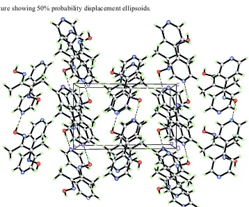
In this structure, the molecules of H2dmg and 4,4′-dpy are linked through O—H···N hydrogen bonds into alternating
chains (Fig. 2).
S2. Experimental
Mn(CH3COO)2.4H2O (0.025 g, 0.1 mmol) in 5 ml of water and CH3COONa(0.016 g, 0.2 mmol) were added to a mixture
of H2dmg (0.024 g, 0.2 mmol) and 4,4′-dpy in 10 ml of methanol. The reaction mixture was boiled in a crucible for ~10
min. The solvent was then evaporated and colorless crystals of the title compound were obtained.
S3. Refinement
Methyl H atoms were placed in calculated position with C—H=0.96 Å, and torsion angles were refined,
Uiso(H)=1.5Ueq(C). The position of the O-bound H-atom was determined from a difference Fourier map and then
geometrically restrained with O—H=0.82 Å, and Uiso(H)=1.5Ueq(O). Aromatic H atoms were placed in calculated
Figure 1
Figure 2
Heterosoric stacks of the molecules.
4,4′-Bipyridine–dimethylglyoxime (1/1)
Crystal data
C10H8N2·C4H8N2O2
Mr = 272.31 Monoclinic, P21/c Hall symbol: -P 2ybc
a = 8.7247 (17) Å
b = 7.1486 (14) Å
c = 11.502 (2) Å
β = 99.44 (3)°
V = 707.6 (2) Å3
Z = 2
F(000) = 288 707.6(2)
Dx = 1.278 Mg m−3
Mo Kα radiation, λ = 0.71073 Å Cell parameters from 1636 reflections
θ = 2.4–27.5°
µ = 0.09 mm−1
T = 298 K Block, colourless 0.20 × 0.18 × 0.15 mm
Data collection
Bruker SMART APEX CCD diffractometer
Radiation source: fine-focus sealed tube Graphite monochromator
φ and ω scans
Absorption correction: multi-scan (SADABS; Bruker, 2001)
Tmin = 0.982, Tmax = 0.987
9684 measured reflections 1636 independent reflections 1265 reflections with I > 2σ(I)
Rint = 0.040
θmax = 27.5°, θmin = 2.4°
h = −11→11
k = −9→9
l = −14→14
Refinement
Refinement on F2 Least-squares matrix: full
R[F2 > 2σ(F2)] = 0.043
wR(F2) = 0.133
S = 1.05 1636 reflections 93 parameters 0 restraints
Primary atom site location: structure-invariant direct methods
Secondary atom site location: difference Fourier map
Hydrogen site location: inferred from neighbouring sites
H-atom parameters constrained
w = 1/[σ2(F
o2) + (0.062P)2 + 0.1229P] where P = (Fo2 + 2Fc2)/3
(Δ/σ)max = 0.021 Δρmax = 0.19 e Å−3 Δρmin = −0.13 e Å−3
Special details
Geometry. All e.s.d.'s (except the e.s.d. in the dihedral angle between two l.s. planes) are estimated using the full covariance matrix. The cell e.s.d.'s are taken into account individually in the estimation of e.s.d.'s in distances, angles and torsion angles; correlations between e.s.d.'s in cell parameters are only used when they are defined by crystal symmetry. An approximate (isotropic) treatment of cell e.s.d.'s is used for estimating e.s.d.'s involving l.s. planes.
Refinement. Refinement of F2 against ALL reflections. The weighted R-factor wR and goodness of fit S are based on F2, conventional R-factors R are based on F, with F set to zero for negative F2. The threshold expression of F2 > σ(F2) is used only for calculating R-factors(gt) etc. and is not relevant to the choice of reflections for refinement. R-factors based on F2 are statistically about twice as large as those based on F, and R- factors based on ALL data will be even larger.
Fractional atomic coordinates and isotropic or equivalent isotropic displacement parameters (Å2)
x y z Uiso*/Ueq
O1 0.72158 (14) 0.26180 (17) 0.14282 (10) 0.0699 (4) H1 0.7740 0.1714 0.1296 0.105* N1 0.14118 (15) 0.43625 (18) 0.41432 (12) 0.0600 (4) N2 0.62500 (14) 0.31488 (17) 0.03926 (12) 0.0556 (4) C5 −0.04603 (17) 0.1905 (2) 0.38632 (14) 0.0557 (4) H5 −0.1364 0.1429 0.3421 0.067* C6 0.55111 (16) 0.4662 (2) 0.05327 (13) 0.0518 (4) C3 0.21126 (19) 0.3451 (2) 0.50901 (16) 0.0652 (5) H3 0.2997 0.3982 0.5527 0.078* C2 0.16017 (18) 0.1766 (2) 0.54594 (15) 0.0591 (4) H2 0.2137 0.1190 0.6130 0.071* C4 0.01328 (18) 0.3583 (2) 0.35553 (15) 0.0605 (4) H4 −0.0391 0.4208 0.2899 0.073* C7 0.5661 (2) 0.5712 (3) 0.16681 (15) 0.0701 (5) H7A 0.5414 0.4897 0.2275 0.105* H7B 0.4958 0.6754 0.1578 0.105* H7C 0.6707 0.6158 0.1881 0.105*
Atomic displacement parameters (Å2)
U11 U22 U33 U12 U13 U23
C1 0.0428 (7) 0.0475 (7) 0.0486 (7) 0.0018 (5) 0.0062 (5) −0.0049 (6) O1 0.0727 (8) 0.0700 (8) 0.0608 (7) 0.0253 (6) −0.0076 (6) −0.0006 (5) N1 0.0600 (8) 0.0521 (7) 0.0671 (8) −0.0065 (6) 0.0081 (6) −0.0017 (6) N2 0.0527 (7) 0.0553 (7) 0.0560 (7) 0.0087 (5) 0.0001 (5) 0.0000 (5) C5 0.0513 (8) 0.0572 (8) 0.0550 (8) −0.0064 (6) −0.0020 (6) 0.0017 (7) C6 0.0473 (7) 0.0539 (8) 0.0529 (8) 0.0050 (6) 0.0042 (6) −0.0030 (6) C3 0.0583 (9) 0.0607 (9) 0.0723 (11) −0.0125 (7) −0.0024 (8) −0.0045 (8) C2 0.0548 (8) 0.0562 (9) 0.0614 (9) −0.0035 (7) −0.0049 (7) 0.0018 (7) C4 0.0621 (9) 0.0570 (9) 0.0596 (9) −0.0021 (7) 0.0020 (7) 0.0063 (7) C7 0.0760 (11) 0.0732 (11) 0.0567 (9) 0.0180 (9) −0.0026 (8) −0.0097 (8)
Geometric parameters (Å, º)
C1—C5 1.384 (2) C6—C6ii 1.474 (3) C1—C2 1.391 (2) C6—C7 1.493 (2) C1—C1i 1.484 (3) C3—C2 1.376 (2) O1—N2 1.3941 (17) C3—H3 0.9300 O1—H1 0.8200 C2—H2 0.9300 N1—C4 1.329 (2) C4—H4 0.9300 N1—C3 1.329 (2) C7—H7A 0.9600 N2—C6 1.2831 (18) C7—H7B 0.9600 C5—C4 1.376 (2) C7—H7C 0.9600 C5—H5 0.9300
N2—O1—H1 109.5 C1—C2—H2 120.0 C4—N1—C3 116.54 (14) N1—C4—C5 123.68 (15) C6—N2—O1 111.59 (12) N1—C4—H4 118.2 C4—C5—C1 120.16 (14) C5—C4—H4 118.2 C4—C5—H5 119.9 C6—C7—H7A 109.5 C1—C5—H5 119.9 C6—C7—H7B 109.5 N2—C6—C6ii 114.82 (16) H7A—C7—H7B 109.5 N2—C6—C7 124.04 (14) C6—C7—H7C 109.5 C6ii—C6—C7 121.13 (16) H7A—C7—H7C 109.5 N1—C3—C2 123.61 (14) H7B—C7—H7C 109.5 N1—C3—H3 118.2
C2—C1—C5—C4 1.8 (2) N1—C3—C2—C1 0.1 (3) C1i—C1—C5—C4 −177.95 (15) C5—C1—C2—C3 −1.7 (2) O1—N2—C6—C6ii 178.74 (15) C1i—C1—C2—C3 178.06 (16) O1—N2—C6—C7 −0.5 (2) C3—N1—C4—C5 −1.3 (2) C4—N1—C3—C2 1.4 (3) C1—C5—C4—N1 −0.3 (3)
Symmetry codes: (i) −x, −y, −z+1; (ii) −x+1, −y+1, −z.
Hydrogen-bond geometry (Å, º)
D—H···A D—H H···A D···A D—H···A
O1—H1···N1iii 0.82 1.94 2.7459 (17) 169