An incarcerated inguinal hysterocele in a pregnant
bitch: a case report
G. Serin
1, R. Yaygingul
2, T. Tarimcilar
1, M. Sarierler
2Faculty of Veterinary Medicine, Adnan Menderes University, Aydin, Turkey
ABSTRACT: A 3.5-year-old Boxer breed bitch was referred to our clinic with a history of progressive and painless
inguinal mass on the left side with no significant general symptoms. Ultrasonographic examination of the inguinal mass and abdomen revealed a hypo echoic area covered with a hyper echoic capsule in inguinal mass and also two 40-day-old live foetuses, respectively. During ventral midline laparotomy, an incarcerated left uterus horn was detected in an inguinal hernia. A successful herniorraphy and ovariohysterectomy was performed in one session. A dead foetus was detected histopathologically in the herniated uterine horn. The main differential in this case was a severe incarceration and foetal death in the herniated uterus horn with no significant clinical and ultrasonographic findings. This condition did not affect either the general health condition of the bitch or the foetuses localized abdominally.
Keywords: inguinal hernia; pregnancy abnormality; bitch
Acquired inguinal hernias are infrequently docu-mented in female dogs and are most often diagnosed in intact, middle aged bitches. In carnivores, canalis inguinalis is open physiologically. Bitches are the only animals that have processus vaginalis. There is a cord which goes posteriorly below the processus vaginalis and connects with the ligamentum teres uteri. The uterus might be herniated in the cana-lis inguinacana-lis (Dean et al., 1990; Formston, 1990). Besides the anatomy of the canalis inguinalis, high oestrogen levels (oestrus and/or pregnancy) and the increasing of intra abdominal pressure (obesity and/or pregnancy) are other potential risk factors for this disease (Read and Bellenger, 2003).
At diagnosis, it can be confused with malignant mammary neoplasm, mastitis or a local abscess (Noakes, 2001). In the case of a non-reducible and/or incarcerated uterus horn in inguinal hernia, herniorraphy following ovariohysterectomy opera-tion has been proposed as treatment for the hernia (Noakes, 2001).
Case report
A 3.5-year-old, 12 kg, Boxer breed bitch present-ed to the Clinics of Adnan Menderes University,
Faculty of Veterinary Medicine with a 48-hour his-tory of progressive inguinal mass on the left side. The owners reported that the dog had an approxi-mately 1-year history of a small and flaccid mass in the same region but that during the preceding 48 hours it had rapidly become larger. No informa-tion about her cyclic phase (the last pregnancy and/ or the last proestrus haemorrhage) was available.
in the hernial content. During the abdominal ul-trasonography, two live foetuses were seen on the right side of the horn. Foetometry was performed to determine the gestational age, as the owners were not aware of the pregnancy. Gestational age was calculated as 40 days based on the dimension parameters (CRL and BPD) of the foetuses. Foetal heart rates were within normal ranges (206 and 218 bpm).There were no pathological features detected in these foetuses and their vesicles in ultrasonog-raphy.
After the clinical and ultrasonographic examina-tions, it was decided to repair the inguinal hernia surgically. Dissociative anaesthesia was applied with 1.1 mg/kg xylazine hydrochloride
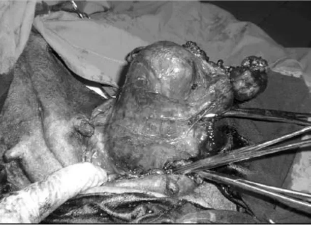
(Alfazine-Alfasan) and 10 mg/kg ketamine hydrochloride (Alfamine-Alfasan) intramuscularly after premedi-cation with 0.04 mg/kg atropine sulphate (Atropan-Vetas) subcutaneously. The patient was placed in dorsal recumbency, and the ventral abdomen was aseptically prepared in a standard fashion. After the first incision of the skin on the inguinal mammary gland (Figure 2) it was observed that the hernial sac was tightly adhered to muscles of the abdominal wall (Figure 3). Therefore, it was decided to per-form a ventral midline laparotomy. During the ven-tral midline laparotomy, an incarcerated, brownish left uterus horn in an inguinal hernia was detected (Figure 4). The right uterus horn was normal and totally localized in the abdominal cavity. Bilateral ovariohysterectomy was performed because of the advanced incarceration and the owners’ request for spaying (Figure 5). Following ovariohysterectomy, the peritoneal cavity was lavaged with warmed sterile 0.9% sodium chloride and herniorraphy was performed in a horizontal mattress pattern. Postoperatively, antibacterial therapy was applied with 400 000 IU penicillin (Iecilline®; IE Ulagay)
daily for five days and an Elizabethan collar was used until removing skin sutures. During the first week after surgery, the bitch was clinically normal. There was no recurrence over the next six months post surgery.
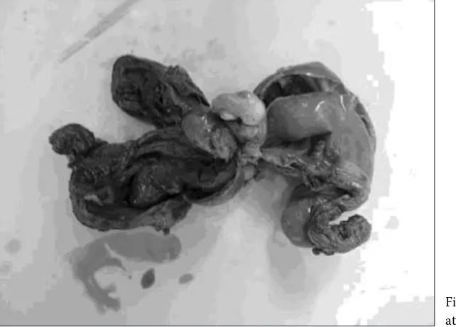
[image:2.595.65.291.83.261.2]The herniated uterus horn contained brown col-our- necrotic tissue and no foetal tissue was detected macroscopically (Figure 6). Histopathologic analysis of the uterus revealed trophoblast cells and foetal tissue residues in the incarcerated left horn.
Figure 1. Ultrasonographic appearance of inguinal mass. Hypo echoic area covered by hyper echoic fibrous capsule
[image:2.595.65.372.534.756.2]DISCUSSION
Hernia graviditatis (hysterocele) is a specific form of inguinal hernia (De Bois, 1978). Although some inguinal hernia cases related to either healthy or infected uteruses have been reported (Sander, 1970; Munro and Stead, 1993; Kassay, 2003; Nak et al., 2004; Vivek et al., 2005; Byers et al., 2007), there are limited reports regarding gravid uterus in an inguinal hernia in bitches (De Bois, 1978; Seyrek-Intas and Seyrek-Seyrek-Intas, 1993). In the present case, a pregnant left uterus horn was herniated thor-ough the canalis inguinalis and incarcerated se-verely. Noakes (2001) reported a herniated mass
as painless, reducible and causing no systemic disturbance unless incarcerated. In this case, be-cause of impaired blood supply to the herniated part of this horn, the foetus and/or foetuses un-derwent resorption. Interestingly, this herniation was acutely progressive over 48 hours, but death of the foetus and/or foetuses was seen earlier than herniation based on the histopathological findings. Furthermore, this incarceration case did not affect either the general health condition of the bitch or the foetuses localized abdominally.
Besides the anatomy of the canalis inguinalis in bitches, pregnancy and oestrus stage are other risk factors related to the aetiology of inguinal
[image:3.595.63.378.83.318.2]hyste-Figure 3. Adhesion of the hernial sac to muscles of the abdominal wall
[image:3.595.64.375.530.761.2]rocele. Some hernias are diagnosed during oestrus stage or pregnancy as estrogens may alter the con-nective tissue function (Noakes, 2001; Read and Bellenger, 2003). Moreover, high intra abdominal pressure during pregnancy also predisposes to herniation of the uterus. In advanced herniation, because of the increasing size and tension of the swelling, reducing the hernia graviditatis is impos-sible. The majority of cases are presented when pregnancy has advanced to about 30 days (Noakes, 2001). With reference to the literature, gestational age was calculated as being on average 40 days based on the dimension of CRL and BPD from live
Figure 5. Macroscopic appearance of the uterus after ovariohysterectomy
foetuses in this case and reduction of the hernia was not possible.
Read and Bellenger (2003) note that incarcerated hernias present a significant diagnostic challenge, as palpation may not yield a definitive diagnosis. Furthermore, this disease is rare and is accompa-nied with depression and local inflammation symp-toms. Although high local temperatures along with adhesion to skin were observed, the only evidence of systemic disturbance was mild anorexia.
Some researchers have reported that a herni-ated gravid or infected uterus is easily detected by plain radiography and ultrasonography according
[image:4.595.65.386.526.756.2]to the appearance of the foetus (Munro and Stead, 1993; Seyrek-Intas and Seyrek-Intas, 1993; Read and Bellenger, 2003). However ultrasonographic exams could not facilitate differential diagnosis in this case. There was only a hypo echoic area like a haematoma covered by a hyper echoic capsule, and no detected organs (jejunum, uterus, foetus, fat tis-sue etc.) in the ultrasonography of the hernial mass. Therefore, the determination of the hernial content could not have been possible before surgery.
Although the inguinal swelling formation had developed only in the last 48 hours based on her anamnesis, it was observed that the herniated preg-nant horn adhered to skin and abdominal muscles firmly during operation. Furthermore, foetal tissue residues were only detected during histopathologi-cal examination.
REFERENCES
Byers C.G., Williams J.E., Saylor D.K. (2007): Pyometra with inguinal herniation of the left uterine horn and omentum in a Beagle dog. Journal of Veterinary Emer-gency and Critical Care, 17, 86–92.
Dean P.W., Bojrab M.J., Costantinescu G.M. (1990): In-guinal hernia repair in the dog. In: Bojrab M.J. (ed.): Current Techniques in Small Animal Surgery. Lea-Fe-biger, Philadelphia. 439–442.
De Bois C.H.W. (1978): Hernia graviditatis. In: Richter J., Gotze R., Rosenberger G., Tillmann H. (eds.): Tier-geburtshilfe. Paul Parey Verlag, Berlin. 209–213.
Formston C. (1990): Inguinal hernia in dogs. Journal of Small Animal Practice,31, 212.
Kassay V. (2003): Pyometra and inguinal hernia combined in a dog: case study. KisallatPraxis, 4, 130–133. Munro E., Stead C. (1993): Ultrasonographic diagnosis
of uterine entrapment in an inguinal hernia. Journal of Small Animal Practice, 34, 139–141.
Nak Y., Misirlioglu D., Nak D., Tuna B., Kumru I.H., Ala-sonyalilar A. (2004): Findings of focal adenomyosis in a case of inguinal hysterocele accompanied with mam-mary tumour in a bitch. Uludag Üniversitesi, Veteriner Fakültesi Dergisi, 23, 99–102.
Noakes D.E. (2001): Maternal dystocia: Causes and treat-ment. In: Noakes D.E., Parkinson T.J., England G.C.W. (eds.): Arthur’s Veterinary Reproduction and Obstet-rics. WB Saunders, Philadelphia. 240–241.
Read R.A., Bellenger C.R. (2003): Hernias. In: Slatter D. (ed.): Textbook of Small Animal Surgery. WB Saun-ders, Philadelphia. 446–470.
Sander W. (1970): Inguinal hysterocele in a bitch. Monatsh Veterinarmed, 25, 441.
Seyrek-Intas K., Seyrek-Intas D. (1997): Hernia inguina-lis utero gravido in a bitch. Veteriner Cerrahi Dergisi, 3, 36–39.
Vivek M., Kumar D., Sheshman R.P. Pandey, Singh B. (2005): Inguinal hysterocele in a bitch. Indian Journal of Veterinary Surgery, 26, 59.
Received: 2009–08–11 Accepted: 2009–09–02
Corresponding Author:
Gunes Serin, Ph.D., Adnan Menderes University, Faculty of Veterinary Medicine, Department of Obstetrics and Gynaecology, Aydin, Turkey