Cytenamide–butyric acid (1/1)
Andrea Johnston,aAlastair J. Florence,a* Francesca J. A. Fabbiani,bKenneth Shanklandcand Colin T. Bedfordd
a
Solid-State Research Group, Strathclyde Institute of Pharmacy and Biomedical Sciences, The John Arbuthnott Building, University of Strathclyde, Glasgow G4 0NR, Scotland,b
GZG, Department of Crystallography, University of Go¨ttingen, D-37077 Go¨ttingen, Germany,cISIS Facility, Rutherford Appleton Laboratory, Chilton, Didcot,
Oxon OX11 0QX, England, anddDepartment of Chemistry, University College
London, London WC1H 0AJ, England
Correspondence e-mail: [email protected]
Received 14 May 2008; accepted 13 June 2008
Key indicators: single-crystal X-ray study;T= 160 K; mean(C–C) = 0.002 A˚;
Rfactor = 0.041;wRfactor = 0.089; data-to-parameter ratio = 18.0.
Cytenamide forms a 1:1 solvate with butyric acid [systematic name: 5H-dibenzo[a,d ]cycloheptatriene-5-carboxamide–buta-noic acid (1/1)], C16H13NOC4H8O2. The title compound crystallizes with one molecule of cytenamide and one of butyric acid in the asymmetric unit; these molecules are linked by N—H O and O—H O hydrogen bonds to form an
R2 2
(8) heterodimer motif. Pairs of adjacent motifs are further connected via N—H O interactions to form a discrete centrosymmetric assembly.
Related literature
For details on experimental methods used to obtain the title solvate, see: Davis et al.(1964); Florence et al.(2003); Flor-ence, Johnston, Fernandes et al. (2006). For literature on cytenamide and related molecules, see: Florence, Bedford et al.(2008); Cyret al.(1987); Fleischmanet al.(2003); Florence, Johnston, Price et al. (2006); Florence, Leech et al. (2006); Bandoliet al.(1992); Harrisonet al.(2006); Leechet al.(2007); Florence, Shanklandet al.(2008). For other related literature, see: Etter (1990) ; Desiraju & Steiner (1999).
Experimental
Crystal data
C16H13NOC4H8O2
Mr= 323.39
Monoclinic,P21=n
a= 5.9351 (2) A˚ b= 16.3595 (5) A˚ c= 17.6738 (4) A˚
= 98.046 (2)
V= 1699.15 (9) A˚3
Z= 4
MoKradiation
= 0.09 mm1
T= 160 K
0.350.150.12 mm
Data collection
Oxford Diffraction Gemini diffractometer
Absorption correction: multi-scan (CrysAlis RED; Oxford Diffraction, 2007) Tmin= 0.91,Tmax= 0.99
18979 measured reflections 4069 independent reflections 2928 reflections withI> 2(I) Rint= 0.031
Refinement
R[F2> 2(F2)] = 0.040
wR(F2) = 0.088
S= 0.95 4069 reflections 226 parameters 3 restraints
H atoms treated by a mixture of independent and constrained refinement
max= 0.38 e A˚3
min=0.27 e A˚3
Table 1
Hydrogen-bond geometry (A˚ ,).
D—H A D—H H A D A D—H A
N1—H11 O2 0.860 (14) 2.348 (14) 2.8761 (15) 120.0 (10) N1—H12 O2i
0.898 (13) 2.146 (13) 3.0167 (15) 163.2 (13) O3—H311 O1i
0.879 (17) 1.698 (17) 2.5658 (13) 168.8 (16)
Symmetry code: (i)xþ2;yþ1;zþ1.
Data collection: CrysAlis CCD (Oxford Diffraction, 2007); cell refinement: CrysAlis RED (Oxford Diffraction, 2007); data reduc-tion:CrysAlis REDandSORTAV(Blessing, 1997); program(s) used to solve structure:SIR92(Altomareet al., 1994); program(s) used to refine structure: CRYSTALS (Betteridge et al., 2003); molecular graphics: Mercury (Macrae et al., 2006) and ORTEP-3 (Farrugia, 1997); software used to prepare material for publication:publCIF
(Westrip, 2008).
The authors thank the Basic Technology programme of the UK Research Councils for funding this work under the project Control and Prediction of the Organic Solid State (http:// www.cposs.org.uk).
Supplementary data and figures for this paper are available from the IUCr electronic archives (Reference: GK2147).
References
Altomare, A., Cascarano, G., Giacovazzo, G., Guagliardi, A., Burla, M. C., Polidori, G. & Camalli, M. (1994).J. Appl. Cryst.27, 435.
Bandoli, G., Nicolini, M., Ongaro, A., Volpe, G. & Rubello, A. (1992).J. Chem. Crystallogr.22, 177–183.
Betteridge, P. W., Carruthers, J. R., Cooper, R. I., Prout, K. & Watkin, D. J. (2003).J. Appl. Cryst.36, 1487.
Blessing, R. H. (1997).J. Appl. Cryst.30, 421–426.
Cyr, T. D., Matsui, F., Sears, R. W., Curran, N. M. & Lovering, E. G. (1987).J. Assoc. Off. Anal. Chem.70, 836–840.
Davis, M. A., Winthrop, S. O., Thomas, R. A., Herr, F., Charest, M.-P. & Gaudry, R. (1964).J. Med. Chem.7, 88–94.
Desiraju, G. R. & Steiner, T. (1999).The Weak Hydrogen Bond in Structural Chemistry and Biology.Oxford University Press.
Etter, M. C. (1990).Acc. Chem. Res.23, 120–126. Farrugia, L. J. (1997).J. Appl. Cryst.30, 565.
Fleischman, S. G., Kuduva, S. S., McMahon, J. A., Moulton, B., Walsh, R. D. B., Rodriguez-Hornedo, N. & Zaworotko, M. J. (2003).Cryst. Growth Des.3, 909–919.
organic compounds
Acta Cryst.(2008). E64, o1295–o1296 doi:10.1107/S1600536808018059 Johnstonet al.
o1295
Acta Crystallographica Section EStructure Reports
Online
Florence, A. J., Baumgartner, B., Weston, C., Shankland, N., Kennedy, A. R., Shankland, K. & David, W. I. F. (2003).J. Pharm. Sci.92, 1930– 1938.
Florence, A. J., Bedford, C. T., Fabbiani, F. P. A., Shankland, K., Gelbrich, T., Hursthouse, M. B., Shankland, N., Johnston, A. & Fernandes, P. (2008). CrystEngComm.doi:10.1039/b719717a.
Florence, A. J., Johnston, A., Fernandes, P., Shankland, N. & Shankland, K. (2006).J. Appl. Cryst.39, 922–924.
Florence, A. J., Johnston, A., Price, S. L., Nowell, H., Kennedy, A. R. & Shankland, N. (2006).J. Pharm. Sci.95, 1918–1930.
Florence, A. J., Leech, C. K., Shankland, N., Shankland, K. & Johnston, A. (2006).CrystEngComm,8, 746–747.
Florence, A. J., Shankland, K., Gelbrich, T., Hursthouse, M. B., Shankland, N., Johnston, A., Fernandes, P. & Leech, C. K. (2008).CrystEngComm,10, 26– 28.
Harrison, W. T. A., Yathirajan, H. S. & Anilkumar, H. G. (2006).Acta Cryst. C62, o240–o242.
Leech, C. K., Florence, A. J., Shankland, K., Shankland, N. & Johnston, A. (2007).Acta Cryst.E63, o675–o677.
Macrae, C. F., Edgington, P. R., McCabe, P., Pidcock, E., Shields, G. P., Taylor, R., Towler, M. & van de Streek, J. (2006).J. Appl. Cryst.39, 453–457. Oxford Diffraction (2007). CrysAlis CCD and CrysAlis RED. Oxford
supporting information
sup-1
Acta Cryst. (2008). E64, o1295–o1296supporting information
Acta Cryst. (2008). E64, o1295–o1296 [doi:10.1107/S1600536808018059]
Cytenamide
–
butyric acid (1/1)
Andrea Johnston, Alastair J. Florence, Francesca J. A. Fabbiani, Kenneth Shankland and Colin T.
Bedford
S1. Comment
Cytenamide (CYT) is an analogue of carbamazepine (CBZ), a dibenzazepine drug used to control seizures (Cyr et al.,
1987). CYT-butyric acid solvate was produced during an automated parallel crystallization study (Florence, Johnston,
Fernandes et al., 2006) of CYT as part of a wider investigation that couples automated parallel crystallization with crystal
structure prediction methodology to investigate the basic science underlying the solid-state diversity in CBZ (Florence,
Johnston, Price et al., 2006; Florence, Leech et al., 2006) and its closely related analogues, CYT (Florence, Bedford et
al., 2008), 10,11-dihydrocarbamazepine (Bandoli et al., 1992; Harrison et al., 2006; Leech et al., 2007) and cyheptamide
(Florence, Shankland et al., 2008). The sample was identified as a new form using multi-sample foil transmission X-ray
powder diffraction analysis (Florence et al., 2003). Subsequent manual recrystallization from a saturated butyric acid
solution by slow evaporation at 278 K yielded a sample suitable for single-crystal X-ray diffraction (Fig. 1).
The compound crystallizes in the monoclinic space group P21/n with one CYT and one solvent molecule in the
asymmetric unit. The molecules adopt a hydrogen-bonded arrangement similar to that observed in CBZ-butyric acid
solvate (1/1) (Fleischman et al., 2003) whereby the CYT and butyric acid molecules are connected via N—H···O and O—
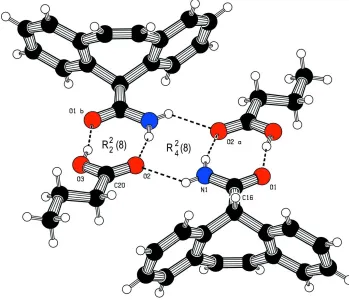
H···O hydrogen bonds to form an R22(8) dimer motif (Etter, 1990). Adjacent dimers are linked via a third contact (N1—
H1···O2; Fig 2) to form an R42(8) centrosymmetric double motif arrangement. The O1···O3 distance of 2.566 (1) Å lies
within the expected range for strong hydrogen bonds (2.5 - 3.2 Å; Desiraju and Steiner, 1999).
CYT-butyric acid solvate structure reported here is essentially isostructural with both CBZ-formic acid and CBZ-acetic
acid solvates (Fleischman et al., 2003).
S2. Experimental
A sample of cytenamide was synthesized according to a modification of the published method (Davis et al., 1964). A
single-crystal sample of cytenamide-butyric acid was grown form a saturated butyric acid solution by isothermal solvent
evaporation at 278 K.
S3. Refinement
H-atoms were found on a difference Fourier map and were initially refined with soft restraints on the bond lengths and
angles to regularize their geometry and Uiso(H) (in the range 1.2–1.5 times Ueq of the parent atom), after which the
positions were refined with riding constraints. The positions of H-atoms involved in H-bonding were refined subject to
Figure 1
The molecular structure of CYT–butyric acid (1/1), showing 50% probablility displacement ellipsoids.
Figure 2
The hydrogen bonded R22(8) motifs of CYT-butyric acid joined in a centrosymmetric arrangement via an R42(8) motif.
[image:4.610.128.477.320.623.2]supporting information
sup-3
Acta Cryst. (2008). E64, o1295–o12965H-dibenzo[a,d]cycloheptatriene-5-carboxamide–butanoic acid (1/1)
Crystal data
C16H13NO·C4H8O2
Mr = 323.39
Monoclinic, P21/n
Hall symbol: -P 2yn a = 5.9351 (2) Å b = 16.3595 (5) Å c = 17.6738 (4) Å β = 98.046 (2)° V = 1699.15 (9) Å3
Z = 4
F(000) = 688 Dx = 1.264 Mg m−3
Melting point: 216.2 K
Mo Kα radiation, λ = 0.71073 Å Cell parameters from 6486 reflections θ = 3–29°
µ = 0.09 mm−1
T = 160 K Block, colourless 0.35 × 0.15 × 0.12 mm
Data collection
Oxford Diffraction Gemini diffractometer
Radiation source: Enhance (Mo) X-ray Source Graphite monochromator
Detector resolution: 15.9745 pixels mm-1
ω scans
Absorption correction: multi-scan
(CrysAlis RED; Oxford Diffraction, 2007) Tmin = 0.91, Tmax = 0.99
18979 measured reflections 4069 independent reflections 2928 reflections with I > 2σ(I) Rint = 0.031
θmax = 28.7°, θmin = 2.6°
h = −7→7 k = 0→21 l = 0→23
Refinement
Refinement on F2
Least-squares matrix: full R[F2 > 2σ(F2)] = 0.040
wR(F2) = 0.088
S = 0.95 4069 reflections 226 parameters 3 restraints
Primary atom site location: structure-invariant direct methods
Hydrogen site location: geom+difmap H atoms treated by a mixture of independent
and constrained refinement
Method = Modified Sheldrick w = 1/[σ2(F2) +
(0.03P)2 + 0.5P],
where P = [max(Fo2,0) + 2Fc2]/3
(Δ/σ)max = 0.001
Δρmax = 0.38 e Å−3
Δρmin = −0.27 e Å−3
Fractional atomic coordinates and isotropic or equivalent isotropic displacement parameters (Å2)
x y z Uiso*/Ueq
C1 0.3604 (2) 0.32253 (7) 0.41898 (7) 0.0261
C2 0.1718 (2) 0.36489 (8) 0.38294 (7) 0.0323
C3 0.1256 (2) 0.36854 (9) 0.30423 (8) 0.0391
C4 0.2687 (2) 0.33058 (9) 0.25996 (7) 0.0392
C5 0.4591 (2) 0.28990 (8) 0.29445 (7) 0.0344
C6 0.5061 (2) 0.28359 (7) 0.37441 (7) 0.0279
C7 0.6996 (2) 0.23388 (8) 0.40716 (7) 0.0304
C8 0.7115 (2) 0.18488 (8) 0.46816 (7) 0.0301
C9 0.5383 (2) 0.17092 (8) 0.51782 (6) 0.0269
C10 0.5251 (2) 0.09343 (8) 0.55037 (7) 0.0341
C11 0.3603 (2) 0.07491 (9) 0.59538 (8) 0.0398
C12 0.2069 (2) 0.13407 (9) 0.61005 (8) 0.0389
C14 0.3844 (2) 0.23123 (7) 0.53434 (6) 0.0258
C15 0.4034 (2) 0.31760 (7) 0.50530 (6) 0.0257
C16 0.6242 (2) 0.35710 (7) 0.54306 (7) 0.0276
C17 0.3158 (3) 0.57421 (11) 0.16558 (9) 0.0568
C18 0.5184 (3) 0.60108 (10) 0.22080 (8) 0.0455
C19 0.5957 (2) 0.53835 (9) 0.28103 (8) 0.0371
C20 0.8040 (2) 0.56040 (8) 0.33497 (7) 0.0295
O1 0.70946 (16) 0.33413 (6) 0.60746 (5) 0.0369
O2 0.84351 (15) 0.53413 (6) 0.39986 (5) 0.0346
N1 0.7091 (2) 0.41860 (7) 0.50777 (6) 0.0335
O3 0.94149 (17) 0.61035 (6) 0.30567 (5) 0.0435
H151 0.2828 0.3488 0.5249 0.0293*
H21 0.0713 0.3916 0.4145 0.0377*
H81 0.8456 0.1498 0.4786 0.0333*
H71 0.8293 0.2332 0.3790 0.0372*
H191 0.4766 0.5266 0.3115 0.0486*
H192 0.6295 0.4889 0.2564 0.0473*
H131 0.1155 0.2536 0.5914 0.0383*
H101 0.6291 0.0526 0.5389 0.0392*
H41 0.2338 0.3317 0.2057 0.0465*
H121 0.0935 0.1222 0.6419 0.0464*
H51 0.5637 0.2636 0.2641 0.0395*
H111 0.3507 0.0214 0.6150 0.0470*
H182 0.4770 0.6515 0.2459 0.0609*
H181 0.6503 0.6120 0.1946 0.0611*
H31 −0.0099 0.3971 0.2794 0.0463*
H172 0.2712 0.6162 0.1279 0.0868*
H173 0.1858 0.5625 0.1933 0.0867*
H171 0.3504 0.5240 0.1402 0.0875*
H12 0.834 (2) 0.4432 (9) 0.5320 (8) 0.0446*
H11 0.652 (2) 0.4352 (9) 0.4630 (8) 0.0433*
H311 1.058 (3) 0.6243 (11) 0.3395 (9) 0.0689*
Atomic displacement parameters (Å2)
U11 U22 U33 U12 U13 U23
C1 0.0283 (6) 0.0234 (6) 0.0265 (6) −0.0013 (5) 0.0032 (5) −0.0008 (5)
C2 0.0315 (7) 0.0305 (7) 0.0348 (7) 0.0030 (5) 0.0040 (5) 0.0027 (5)
C3 0.0366 (8) 0.0396 (8) 0.0383 (7) 0.0032 (6) −0.0041 (6) 0.0075 (6)
C4 0.0476 (8) 0.0422 (8) 0.0259 (6) −0.0032 (7) −0.0010 (6) 0.0030 (6)
C5 0.0414 (8) 0.0345 (8) 0.0279 (6) −0.0020 (6) 0.0075 (6) −0.0025 (5)
C6 0.0297 (6) 0.0258 (7) 0.0279 (6) −0.0019 (5) 0.0031 (5) −0.0021 (5)
C7 0.0287 (6) 0.0318 (7) 0.0315 (6) 0.0018 (5) 0.0065 (5) −0.0068 (5)
C8 0.0270 (6) 0.0295 (7) 0.0321 (6) 0.0056 (5) −0.0016 (5) −0.0067 (5)
C9 0.0273 (6) 0.0284 (7) 0.0227 (6) −0.0004 (5) −0.0051 (5) −0.0026 (5)
C10 0.0388 (7) 0.0291 (7) 0.0313 (6) 0.0021 (6) −0.0057 (6) −0.0015 (5)
C11 0.0507 (9) 0.0310 (8) 0.0347 (7) −0.0078 (6) −0.0051 (6) 0.0073 (6)
supporting information
sup-5
Acta Cryst. (2008). E64, o1295–o1296C13 0.0298 (7) 0.0379 (8) 0.0275 (6) −0.0022 (6) 0.0010 (5) −0.0008 (5)
C14 0.0264 (6) 0.0277 (7) 0.0215 (5) −0.0016 (5) −0.0027 (5) −0.0024 (5)
C15 0.0270 (6) 0.0256 (7) 0.0252 (6) 0.0034 (5) 0.0055 (5) −0.0028 (5)
C16 0.0334 (7) 0.0245 (6) 0.0252 (6) 0.0009 (5) 0.0059 (5) −0.0041 (5)
C17 0.0518 (10) 0.0666 (12) 0.0470 (9) 0.0033 (8) −0.0109 (7) −0.0040 (8)
C18 0.0467 (9) 0.0430 (9) 0.0435 (8) −0.0033 (7) −0.0052 (7) 0.0042 (7)
C19 0.0398 (8) 0.0347 (8) 0.0361 (7) −0.0062 (6) 0.0026 (6) −0.0017 (6)
C20 0.0366 (7) 0.0246 (7) 0.0279 (6) −0.0004 (5) 0.0066 (5) −0.0014 (5)
O2 0.0421 (5) 0.0326 (5) 0.0290 (5) −0.0029 (4) 0.0045 (4) 0.0035 (4)
N1 0.0402 (7) 0.0304 (6) 0.0291 (5) −0.0056 (5) 0.0024 (5) 0.0018 (5)
O3 0.0457 (6) 0.0529 (7) 0.0297 (5) −0.0205 (5) −0.0027 (4) 0.0077 (4)
O1 0.0441 (5) 0.0373 (5) 0.0271 (4) −0.0124 (4) −0.0027 (4) 0.0021 (4)
Geometric parameters (Å, º)
C1—C2 1.3924 (17) C12—H121 0.956
C1—C6 1.4018 (17) C13—C14 1.3860 (17)
C1—C15 1.5133 (15) C13—H131 0.965
C2—C3 1.3811 (18) C14—C15 1.5129 (17)
C2—H21 0.975 C15—C16 1.5282 (17)
C3—C4 1.380 (2) C15—H151 0.981
C3—H31 0.980 C16—N1 1.3204 (16)
C4—C5 1.378 (2) C16—O1 1.2376 (14)
C4—H41 0.952 C17—C18 1.504 (2)
C5—C6 1.4054 (17) C17—H172 0.968
C5—H51 0.976 C17—H173 0.988
C6—C7 1.4591 (17) C17—H171 0.971
C7—C8 1.3373 (18) C18—C19 1.5034 (19)
C7—H71 0.974 C18—H182 0.984
C8—C9 1.4601 (18) C18—H181 0.980
C8—H81 0.977 C19—C20 1.4956 (18)
C9—C10 1.3988 (18) C19—H191 0.967
C9—C14 1.4026 (17) C19—H192 0.954
C10—C11 1.3778 (19) C20—O2 1.2165 (14)
C10—H101 0.950 C20—O3 1.3114 (15)
C11—C12 1.378 (2) N1—H12 0.899 (14)
C11—H111 0.946 N1—H11 0.860 (14)
C12—C13 1.3848 (19) O3—H311 0.880 (14)
C2—C1—C6 119.25 (11) C14—C13—H131 119.0
C2—C1—C15 119.95 (11) C9—C14—C13 119.42 (12)
C6—C1—C15 120.79 (10) C9—C14—C15 120.31 (11)
C1—C2—C3 121.06 (12) C13—C14—C15 120.22 (11)
C1—C2—H21 118.6 C1—C15—C14 112.44 (10)
C3—C2—H21 120.4 C1—C15—C16 115.54 (10)
C2—C3—C4 120.03 (13) C14—C15—C16 110.29 (10)
C2—C3—H31 120.5 C1—C15—H151 107.5
C3—C4—C5 119.87 (12) C16—C15—H151 104.4
C3—C4—H41 119.9 C15—C16—N1 118.47 (11)
C5—C4—H41 120.2 C15—C16—O1 119.24 (11)
C4—C5—C6 121.01 (12) N1—C16—O1 122.14 (12)
C4—C5—H51 121.0 C18—C17—H172 110.8
C6—C5—H51 118.0 C18—C17—H173 110.1
C5—C6—C1 118.72 (11) H172—C17—H173 108.7
C5—C6—C7 118.30 (11) C18—C17—H171 110.2
C1—C6—C7 122.92 (11) H172—C17—H171 109.7
C6—C7—C8 127.14 (12) H173—C17—H171 107.2
C6—C7—H71 116.0 C17—C18—C19 113.39 (13)
C8—C7—H71 116.7 C17—C18—H182 108.1
C7—C8—C9 128.04 (12) C19—C18—H182 108.9
C7—C8—H81 117.0 C17—C18—H181 111.4
C9—C8—H81 114.7 C19—C18—H181 105.9
C8—C9—C10 118.27 (11) H182—C18—H181 109.0
C8—C9—C14 123.29 (11) C18—C19—C20 115.46 (11)
C10—C9—C14 118.45 (11) C18—C19—H191 110.9
C9—C10—C11 121.46 (13) C20—C19—H191 107.3
C9—C10—H101 118.2 C18—C19—H192 108.6
C11—C10—H101 120.2 C20—C19—H192 106.7
C10—C11—C12 119.67 (13) H191—C19—H192 107.6
C10—C11—H111 120.0 C19—C20—O2 123.28 (12)
C12—C11—H111 120.3 C19—C20—O3 113.77 (11)
C11—C12—C13 119.87 (13) O2—C20—O3 122.95 (12)
C11—C12—H121 120.4 C16—N1—H12 117.5 (10)
C13—C12—H121 119.7 C16—N1—H11 123.2 (10)
C12—C13—C14 121.09 (13) H12—N1—H11 119.3 (14)
C12—C13—H131 119.9 C20—O3—H311 111.5 (12)
C6—C1—C2—C3 0.48 (19) C14—C9—C10—C11 2.43 (18)
C15—C1—C2—C3 −178.34 (12) C8—C9—C14—C13 177.89 (11)
C2—C1—C6—C5 1.17 (17) C8—C9—C14—C15 −4.73 (17)
C2—C1—C6—C7 −176.25 (11) C10—C9—C14—C13 −1.96 (17)
C15—C1—C6—C5 180.00 (12) C10—C9—C14—C15 175.42 (10)
C15—C1—C6—C7 2.57 (18) C9—C10—C11—C12 −1.2 (2)
C2—C1—C15—C14 114.81 (12) C10—C11—C12—C13 −0.6 (2)
C2—C1—C15—C16 −117.37 (12) C11—C12—C13—C14 1.0 (2)
C6—C1—C15—C14 −63.99 (14) C12—C13—C14—C9 0.28 (18)
C6—C1—C15—C16 63.83 (14) C12—C13—C14—C15 −177.10 (11)
C1—C2—C3—C4 −0.7 (2) C9—C14—C15—C1 64.61 (14)
C2—C3—C4—C5 −0.8 (2) C9—C14—C15—C16 −65.93 (13)
C3—C4—C5—C6 2.5 (2) C13—C14—C15—C1 −118.03 (12)
C4—C5—C6—C1 −2.67 (18) C13—C14—C15—C16 111.43 (12)
C4—C5—C6—C7 174.87 (12) C1—C15—C16—O1 −156.56 (11)
C1—C6—C7—C8 36.0 (2) C1—C15—C16—N1 27.97 (15)
C5—C6—C7—C8 −141.46 (14) C14—C15—C16—O1 −27.68 (15)
supporting information
sup-7
Acta Cryst. (2008). E64, o1295–o1296C7—C8—C9—C10 147.22 (13) C17—C18—C19—C20 177.01 (13)
C7—C8—C9—C14 −32.6 (2) C18—C19—C20—O2 152.97 (13)
C8—C9—C10—C11 −177.43 (12) C18—C19—C20—O3 −27.50 (17)
Hydrogen-bond geometry (Å, º)
D—H···A D—H H···A D···A D—H···A
N1—H11···O2 0.86 (1) 2.35 (1) 2.8761 (15) 120 (1)
N1—H12···O2i 0.90 (1) 2.15 (1) 3.0167 (15) 163 (1)
O3—H311···O1i 0.88 (2) 1.70 (2) 2.5658 (13) 169 (2)