Turner, D. (2013) Characterisation of three bacteriophages infect-ing serovars of salmonella enterica. PhD, University of the West of England.
We recommend you cite the published version. The publisher’s URL is:
https://eprints.uwe.ac.uk/secure/22112/
Refereed: No (no note)
Disclaimer
UWE has obtained warranties from all depositors as to their title in the material deposited and as to their right to deposit such material.
UWE makes no representation or warranties of commercial utility, title, or fit-ness for a particular purpose or any other warranty, express or implied in respect of any material deposited.
1
CHARACTERISATION OF THREE BACTERIOPHAGES INFECTING SEROVARS OF
SALMONELLA ENTERICA
DANN TURNER BSc. MSc.
A thesis submitted in partial fulfilment of the requirements of the University of the West of England, Bristol for the degree of Doctorate of Philosophy.
Department of Applied Sciences, University of the West of England, Bristol
i
Author’s Declaration
ii
Abstract
A collection of three newly isolated Salmonella bacteriophages, vB_SenS-Ent1, vB_SenS-Ent2 and vB_SenS-Ent3 was established. These bacteriophages were characterised by electron microscopy, host range, sensitivity to restriction enzymes and profiles of structural proteins on SDS-PAGE gels. The complete genome sequences of each bacteriophage were established to greater than 30x
coverage and bioinformatics analysis identified the functions of a number of coding sequences.
Since the last update of virus taxonomy by the ICTV a number of additional genome sequences for bacteriophages infecting the genus Salmonella have been reported in the literature. To date, all but one of the Siphoviridae comprising the Salmonella bacteriophages with fully sequenced genomes remain unclassified by the ICTV. Comparative genomic analysis reveals that a number of these phages form a coherent group within the Siphoviridae and supports the establishment of a new genus, the “Setp3likeviruses”. The proposed genus includes 5 bacteriophages infecting Salmonella; SETP3, vB_SenS-Ent1, SE2, wksl3 and SS3e, and 5 infecting Escherichia; K1G, K1H, K1ind1, K1ind2 and K1ind3. This group share identical virion morphology, have terminally redundant, circularly permuted
genomes ranging between 42-45 kb in size and are characterised by high nucleotide sequence similarity, shared homologous proteins and conservation of gene order.
iii
Acknowledgements
It would not have been possible to complete this doctoral thesis without the humour, friendship, and support of those around me. I am indebted to my supervisor, Dr. Darren Reynolds for his support and instruction throughout this project and for providing an open and positive environment in which to work. I would like to extend my thanks to the members of my project supervisory team, Professor
Vyv Salisbury, Dr. Shona Nelson and Dr. Gareth Robinson for all their guidance, support and patience over the years of this project.
Many thanks are also due to Pablo Ledezma, Dr. Ben Taylor, David and Laura Corry, Dr. Robin Thorn and Dr. Natasha McGuire for their friendship, support in and outside of the laboratory and many a
shared evening of beer and laughter! Special thanks go to my girlfriend Jenny, mostly for putting up with me but also for her unending support and encouragement.
I am most grateful to Professor Steve Abedon of Ohio State University for providing invaluable discussions about plaques and copies of published and unpublished work. Further thanks are due to
Professor Hans Wolfgang Ackermann for his kind permission to reproduce figures and electron micrographs and also to Seth Bunton-Stashyshn, Professor Don Seto and Dr. Padmanabhan Mahadevan for their help and support in developing the CoreGenes3.5 batch submission tool.
I would like to acknowledge the financial, academic and technical support of the University of the West of England, Bristol and its staff. This studentship was funded by the Higher Education Funding
Council for England.
iv
Table of Contents
Author’s Declaration ... i
Abstract ... ii
Acknowledgements ... iii
Table of Contents ... iv
List of Figures ... ix
List of Tables ... xii
Abbreviations ... xiii
Chapter 1 Introduction ... 1
1.1 Overview ... 1
1.2 The Bacteriophages ... 2
1.2.1 Morphology forms the basis for classification ... 2
1.2.2 Bacteriophage lifecycles ... 5
1.2.3 Bacteriophage structure ... 7
1.2.4 Capsid assembly ... 8
1.2.5 The Bacteriophage tail ... 10
1.3 The Salmonellae ... 11
1.4 Bacterial Bioluminescence ... 16
1.4.1 Biochemical characteristics of Bacterial Luciferases ... 18
1.4.2 Luciferases of bioluminescent bacteria... 20
1.4.3 Bioluminescent bacterial reporters ... 21
1.4.4 Applications of bioluminescent bacterial reporters ... 22
1.4.5 Bioluminescence imaging ... 23
Chapter 2 Materials and Methods ... 28
2.1 Bacterial strains and plasmids ... 28
2.2 Enumeration of bacteria ... 30
2.3 Determination of bacteriophage titres ... 30
2.4 Isolation of bacteriophages ... 31
v
2.6 Morphological examination of bacteriophages by transmission electron microscopy ... 32
2.7 Determination of adsorption rate constants ... 32
2.8 One-step growth curves of bacteriophage isolates ... 33
2.9 Single-burst experiments ... 33
2.10 Bacteriophage stability assays ... 33
2.10.1 Temperature ... 33
2.10.2 Long-term storage ... 34
2.10.3 Bacteriophage host range: Spot plate assay ... 34
2.10.4 Bacteriophage host range: Efficiency of plating ... 35
2.11 Extraction of genomic DNA from CsCl purified bacteriophages ... 35
2.11.1 Concentration, purity and yield of bacteriophage genomic DNA ... 36
2.11.2 Estimation of bacteriophage genome size by pulsed field gel electrophoresis ... 36
2.11.3 Restriction digests of phage genomic DNA and gel electrophoresis ... 37
2.11.4 Determination of phage genomic termini ... 37
2.12 Genome sequencing and bioinformatics analysis of bacteriophages ... 37
2.13 Comparative genomics ... 38
2.14 Bacteriophage structural proteins ... 39
2.14.1 Quantification of protein ... 39
2.14.2 Extraction and analysis of bacteriophage structural proteins by SDS-PAGE ... 39
2.15 Transformation of Salmonella with the luxCDABE operon ... 39
2.15.1 Calibration of bioluminescence and cell density ... 40
2.15.2 Calibration of light emission and growth in batch culture ... 40
2.15.3 Measurement of bioluminescence spectra ... 40
2.15.4 Microplate broth lysis assay ... 41
2.16 Bioluminescence imaging ... 41
2.16.1 EMCCD system characterisation ... 41
2.16.2 Limits of detection for bioluminescent bacteria using EMCCD imaging ... 42
2.17 Calibration of colony forming units and light emission in overlay agar ... 42
vi
2.19 Processing and analysis of bioluminescence images ... 43
2.20 Imaging of Plaques by CSLM and Live:Dead staining ... 44
2.21 Biocontrol of Salmonella Enteritidis in foodstuffs ... 45
2.21.1 Stability of bioluminescent reporters ... 45
2.21.2 Food samples ... 45
2.21.3 Addition of Salmonella Enteritidis and bacteriophages ... 45
2.21.4 Determination of bacterial and phage counts ... 46
Chapter 3 Characterisation of the vB_SenS-Ent Salmonella Bacteriophages ... 47
3.1 Introduction ... 47
3.2 Results ... 47
3.2.1 Isolation of the vB_SenS-Ent phages ... 47
3.2.2 Propagation and Purification ... 48
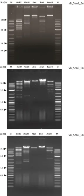
3.2.3 Restriction analysis and estimation of genome size ... 49
3.2.4 Virion morphology ... 51
3.2.5 Adsorption and one step growth ... 51
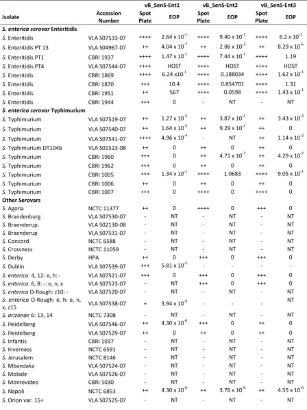
3.2.6 Host range and efficiency of plating ... 53
3.2.7 Genome properties and architecture ... 56
3.2.8 Packaging, morphogenesis and structural proteins ... 59
3.2.9 Regulatory proteins ... 62
3.2.10 The vB_SenS-Ent DNA replication module contains mobile elements ... 63
3.2.11 Host lysis and the late Gene Cluster ... 64
3.2.12 Promoters ... 64
3.2.13 Terminators ... 66
3.2.14 Physical genome ends ... 70
Discussion ... 71
Chapter 4 A proposed new genus of bacteriophage: the “Setp3likevirus” ... 72
4.1 Introduction ... 72
4.1.1 Bacteriophage taxonomy and mosaicism ... 72
vii
4.3 Comparative genomics of the Salmonella bacteriophages ... 76
4.4 The Setp3likevirus ... 80
4.5 Members of the proposed genus ... 80
4.5.1 SETP3 ... 80
4.5.2 vB_SenS-Ent1, vB_SenS-Ent2 and vB_SenS-Ent3 ... 80
4.5.3 SS3e ... 81
4.5.4 SE2 ... 81
4.5.5 wksl3 ... 81
4.5.6 Escherichia coli phages K1G, K1H, K1ind1, K1ind2 and K1ind3 ... 81
4.6 Virion morphology ... 82
4.7 Host specificity ... 83
4.8 Genome structure ... 87
4.8.1 Immunity and regulatory region ... 88
4.8.2 DNA maintenance and replication ... 88
4.8.3 Structural and morphogenesis regions ... 90
4.8.4 Lysis cluster ... 92
4.8.5 Regulatory sequences ... 93
4.9 CoreGenes ... 93
4.10 Phylogenetic analysis ... 95
4.11 Discussion ... 99
4.12 Addendum ... 101
Chapter 5 Measuring bacteriophage-mediated lysis of Salmonella using bioluminescence ... 102
5.1 Introduction ... 102
5.2 Results ... 104
5.2.1 Calibration curves... 104
5.2.2 Bioluminescence emission spectra ... 109
5.2.3 Growth of bioluminescent bacteria ... 110
5.2.4 Multiplicity of infection ... 112
viii
Chapter 6 Control of Salmonella in raw and ready-to-eat foods by the vB_SenS-Ent bacteriophages 123
6.1 Introduction ... 123
6.2 Results ... 128
6.2.1 Long term storage of bacteriophages ... 128
6.2.2 Thermostability of bacteriophages ... 128
6.2.3 Stability of lux plasmid retention by Salmonella at 4oC ... 129
6.2.4 Inoculation and recovery of bacteriophages and bacteria ... 129
6.2.5 Food Matrix 1 – Bean sprouts ... 132
6.2.6 Food Matrix 2 – Mixed salad ... 134
6.2.7 Food Matrix 3 – Cooked skinless chicken breast ... 136
6.2.8 Food Matrix 4 – Raw skinless chicken breast ... 138
6.3 Discussion ... 140
Chapter 7 Bioluminescence Imaging of Bacteriophage Plaque Expansion ... 142
7.1 Introduction ... 142
7.2 Results ... 144
7.2.1 Calibration of cell density and light emission ... 144
7.2.2 Plaque morphology ... 145
7.2.3 Plaque development ... 146
7.2.4 Examination of plaques with confocal microscopy ... 149
7.2.5 Plaque enlargement ... 151
7.2.6 Resistant bacteria ... 155
7.3 Discussion ... 155
7.4 Acknowledgements ... 161
Chapter 8 Discussion ... 162
References... 167
Appendix I: Published material ... 190
Appendix II: Supplementary Material ... 238
ix
List of Figures
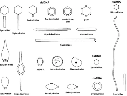
Figure 1. Schematic diagram of the different morphologies of prokaryotic viruses. ... 3 Figure 2. Representatives of the three families of the order Caudovirales, the tailed dsDNA bacteriophages. ... 4
Figure 3. Schematic representation of the possible outcomes of infection of bacteria by filamentous and tailed bacteriophages. ... 7 Figure 4. Enhanced satellite image of a bioluminescent milky sea projected onto the Blue Marble (NASA). ... 17
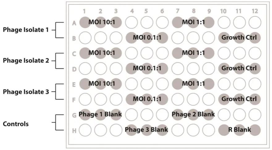
Figure 5. Ribbon cartoon of the quaternary structure of Vibrio harveyii bacterial luciferase... 18 Figure 6. Simplified kinetic models of the bacterial luciferase reaction (A) in vitro and (B) in vivo. 19 Figure 7. Arrangement of log-fold dilutions of bacteriophage upon spot plate assays of host range.34 Figure 8. Score card for the visual assessment of plaques. ... 35 Figure 9. Layout of samples on 96 well microtitre plates. ... 41
Figure 10. Detection of plaque formation by bioluminescence imaging. ... 44 Figure 11. Propagation of bacteriophages vB_SenS-Ent1, Ent2 and Ent3 measured by optical density at 540nm. ... 48 Figure 12. Bacteriophage bands recovered after isopynic CsCl density gradient centrifugation. ... 49 Figure 13. Sensitivity of the vB_SenS_Ent bacteriophage genomic DNA to restriction enzymes. .. 50
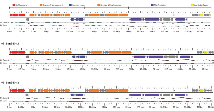
Figure 14. Transmission electron micrograph of A) Ent1, B) Ent2 and C) vB_SenS-Ent3 stained using 2 % aqueous uranyl acetate. ... 51 Figure 15. Adsorption of vB_SenS-Ent1 (A), vB_SenS-Ent2 (B) and vB_SenS-Ent3 (C) to cells of Salmonella Enteritidis shown as the fraction of free phages remaining over time. ... 52 Figure 16. One step growth curve for vB_SenS-Ent1 using S. Enteritidis PT4 as host. ... 53 Figure 17. Percentage of Salmonella isolates forming plaques by spot plate assay with decreasing concentrations of bacteriophage. ... 54 Figure 18. Linear map of the vB_SenS-Ent1, vB_SenS-Ent2 and vB_SenS-Ent3 genomes prepared using
GView. ... 58 Figure 19. vB_SenS-Ent phage structural proteins resolved by 1D and 2D SDS-PAGE. ... 60 Figure 20. Late gene cluster of the vB_SenS-Ent phages. ... 64 Figure 21. Weblogo of consensus motif from sequences identified using MEME and aligned with ClustalW for the vB_SenS-Ent phages. ... 66
x
Figure 24. Hierarchical clustering of 40 bacteriophages by numbers of shared proteins determined by CoreGenes3.0. ... 79 Figure 25. Morphology of the Setp3likevirus. Images represent electron micrographs of bacteriophages Jersey (1 & 2), vB_SenS-Ent1 (3) and Heidelberg typing phage 2 (4)... 83 Figure 26. ClustalX2 alignments of the N-terminal region of the Setp3likevirus tailspike proteins. 84 Figure 27. Sequence identity amongst the P22-like tailspikes of the Salmonella bacteriophages. 85 Figure 28. Condensed Neighbour joining tree of the P22-like tailspikes. ... 86 Figure 29. Clustering of the related SETP3-like Salmonella phage by nucleotide sequence similarity. ... 87 Figure 30. Presence and location of inteins in the DNA polymerases of the Setp3likevirus. ... 90 Figure 31. Synteny of gene order and function in the structural and immunity regions of the SETP3-like phage genomes. ... 91
Figure 32. CGView Comparison Tool map comparing vB_SenS-Ent1 to other bacteriophages belonging to the predicted Setp3likevirus genus. ... 95 Figure 33. Neighbour-joining phylogenetic tree of major capsid proteins from the Setp3likevirus and representatives of other bacteriophage genera. ... 96 Figure 34. Neighbour-joining phylogenetic tree of the large terminase subunit from the Setp3likevirus and representatives of other bacteriophage genera. ... 97 Figure 35. Neighbour-joining phylogenetic tree of the DNA polymerases from the Setp3likevirus and representatives of other bacteriophage genera. ... 98 Figure 36. Calibration curves of optical density at 540 nm, light emission and colony forming units. ... 105
Figure 37. Calibration curves of light emission versus bacterial cell concentrations performed on batch cultures of S. Enteritidis and S. Typhimurium expressing the luxCDABE operon. ... 107 Figure 38. Growth of wild-type and bioluminescent S. Enteritidis and S. Typhimurium in batch culture. ... 108
Figure 39. Bioluminescence spectra across 400 to 750 nm for bioluminescent reporters S. Enteritidis and S. Typhimurium. ... 109 Figure 40. Growth of bioluminescent Salmonella serovarsEnteritidis (top) and Typhimurium (bottom) over time in batch cultures measured by colony counts, absorbance at 540nm and relative light emission. ... 111
xi
Figure 43. Bioluminescence for S. Enteritidis at high density (107 cfu ml-1) incubated in the presence and absence of vB_SenS-Ent bacteriophages. ... 118 Figure 44. Stability of bacteriophage titres stored at 4oC in SM buffer over a 12 month period
measured by overlay plaque assay. ... 128 Figure 45. Thermal stability profile for vB_SenS-Ent1. ... 129 Figure 46. Typical colonies of Salmonella Enteritidis PT4 grown on XLD agar. ... 130 Figure 47. Example of identification of bioluminescent S. Enteritidis in mixed cultures recovered from fresh mixed salad on plate count agar supplemented with 10 ug ml-1 kanamycin. ... 131
Figure 48. Recovery of Salmonella Enteritidis and bacteriophages from artificially contaminated raw beansprouts. ... 133 Figure 49. Recovery of Salmonella Enteritidis and bacteriophages from artificially contaminated mixed salad leaves. ... 135
Figure 50. Recovery of Salmonella Enteritidis and bacteriophages from artificially contaminated cooked chicken breast. ... 137 Figure 51. Recovery of Salmonella Enteritidis and bacteriophages from artificially contaminated raw chicken breast. ... 139 Figure 52. Calibration curve of log-fold dilutions of bioluminescent bacteria immobilised in overlay
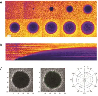
agar using a microplate luminometer. ... 144 Figure 53. Bioluminescence imaging of a growing lawn of S. Enteritidis absent of bacteriophages.145 Figure 54. Photographs of plaques formed by Felix O1 and vB_SenS-Ent1. ... 146 Figure 55. Thresholding of plaques. ... 147 Figure 56. Profiles of bioluminescence from stationary phase lawns (24 hours old) after application of
xii
List of Tables
Table 1. Differential biochemical characteristics of species and subspecies of Salmonella. ... 13
Table 2. Sources and characteristics of Luciferases in nature. ... 16
Table 3. Selected microbiological applications of bioluminescence imaging. ... 26
Table 4. Bacterial strains used in this study. ... 29
Table 5. Plasmids used in this study. ... 30
Table 6. Host range of the vB_SenS-Ent bacteriophages. ... 55
Table 7. Locations and DNA sequences of a MEME-identified motif in the vB-SenS-Ent1 genome.65 Table 8. Locations and DNA sequences of a MEME-identified motif in the vB-SenS-Ent2 genome.65 Table 9. Locations and DNA sequences of a MEME-identified motif in the vB-SenS-Ent3 genome.65 Table 10. Putative rho-independent terminators identified in the vB_SenS-Ent1 genome. ... 67
Table 11. Putative rho-independent terminators identified in the vB_SenS-Ent2 genome. ... 68
Table 12. Putative rho-independent terminators identified in the vB_SenS-Ent2 genome. ... 69
Table 13. Caudovirales infecting the genus Salmonella with complete genome sequences held in GenBank. ... 74
Table 14. Genome properties and aliases of phages K1G, K1H, K1ind1, K1ind2 and K1ind3. ... 82
Table 15. Number and percentage of shared proteins between the Setp3likevirus relative to the proposed type species, SETP3. ... 94
xiii
Abbreviations
Abbreviation Definition
aa Amino acid(s).
ATCC American Type Culture Collection, USA. ATP Adenosine 5’-triphosphate.
BIM Bacteriophage insensitive mutant. BLI Bioluminescence imaging.
bp Base pair(s).
CFRA Campden Food Research Association. cfu Colony-forming units.
CCD Charge-coupled device.
CDS Coding sequence.
CMOS Complementary metal oxide semiconductor. CsCl Cesium chloride.
dsDNA Double-stranded DNA.
DSM German Collection of Microorganisms and Cell Cultures. EMCCD Electron multiplying charge-coupled device.
EOP Efficiency of plating.
FDA Food and Drug Administration, USA. FMN Flavin mononucleotide.
g Gravity.
Gm Gentamycin.
iCCD Intensified charge-coupled device.
ICTV International committee on the Taxonomy of Viruses.
ID Identity.
(k)bp (Kilo)-base pairs. (k)Da (Kilo)-Daltons.
Km Kanamycin.
LB Luria Bertani Miller. LDA Lithoum dodecyl sulphate LPS Lipopolysaccharide. MCP Major capsid protein. MOI Multiplicity of infection. MTP Major tail protein.
MW Molecular weight.
MWCO Molecular weight cut-off value.
NADP Nicotinamide adenine dinucleotide phosphate. NCIMB National Collection of Industrial Bacteria, UK. NCTC National Collection of Type Cultures, UK.
nt Nucleotides.
ORF(s) Open reading frame(s).
PBSA Dulbecco’s phosphate buffered saline. PFGE Pulsed field gel electrophoresis. pfu Plaque forming units.
RBP Receptor binding protein.
xiv
RS Ringer’s solution.
RTE Ready-to-eat.
SDS Sodium dodecyl sulphate
SDS-PAGE Sodium dodecyl sulphate polyacrylamide gel electrophoresis. SM Sodium-magnesium buffer.
SPI-1 Salmonella pathogenicity island 1. SPI-2 Salmonella pathogenicity island 2. TAE Tris-Acetate EDTA buffer.
TE Tris-Chloride EDTA buffer.
TEM Transmission electron microscopy. TMP Tape measure protein.
tRNA Transfer RNA.
TSP Tailspike protein. TTP Tail tip protein
1
Chapter 1
Introduction
1.1
Overview
Members of the genus Salmonella continue to represent significant aetiological agents of disease in humans and animals. With the continued rise in antibiotic resistance it is imperative to identify and
investigate alternative strategies for the development of novel antimicrobials for use as clinical therapeutics or for the control of bacterial pathogens in industry. The bacteriophages, obligate parasites of bacteria, represent one such potential source of antimicrobial agents.
The present study was carried out to further understand the biology of phages infecting the genus Salmonella and the potential of such phages to act as biological control agents. To achieve this, the specific aims of this thesis were five-fold:
1. To isolate, select and characterise bacteriophages specific for Salmonella. 2. To sequence and annotate the genomes of the isolated bacteriophages
3. To investigate the chosen phages for their ability to function as biological control agents 4. To investigate the use of bioluminescent bacterial reporters as a sensitive and
non-destructive method to monitor bacteriophage-mediated lysis of Salmonella.
5. To employ bioluminescent imaging as a method to follow the expansion of plaques in overlay agar.
This thesis is composed of eight chapters. Chapter one provides a general introduction to the
bacteriophages, the genus Salmonella and bacterial bioluminescence. Chapter two describes the methodologies employed to achieve the aims of this project. Chapter three details the isolation, microbiological characterisation, sequencing and annotation of three bacteriophages specific for Salmonella: vB_SenS-Ent1, vB_SenS-Ent2 and vB_SenS-Ent3. Chapter four summarises a comparative genomics study of 42 Salmonella bacteriophages whose complete genome sequences are available within the international nucleotide sequence database and a proposal for the formation of a novel genus within the family Siphoviridae. Chapter five describes the use of Salmonella serovars transformed to express a bioluminescent phenotype to monitor the effects of co-incubation of bacteria and bacteriophage in liquid media. Chapter six investigates the use of the vB_SenS-Ent bacteriophages as biological control agents for the reduction of Salmonella on a variety raw and ready to eat food matrices. Chapter seven describes the application of bioluminescence imaging to monitor the formation and expansion of plaques on host bacterial lawns in overlay agar plates. Lastly, chapter eight presents the main conclusions of the studies presented in this thesis and
2
1.2
The Bacteriophages
The word virus stems from the Latin meaning ‘poison’ or ‘venom’. Every domain of life is affected by
the actions of viruses. The Bacteria are no exception to this rule, providing a nice analogy that the
bacteria can catch colds too. Discovered independently by Frederick Twort (1915) and Felix D’Herelle
(1917), the bacteriophages (phages) are obligate intracellular parasites of bacteria, that is, they are unable to reproduce without the presence of the intact molecular machinery constituting the host
cell. Despite the independent reports and that Twort did not pursue his finding, it is d’Herelle who is
accredited with the discovery of these viruses and who coined the name for viruses of bacteria: the
bacteriophages. ‘Phage’ derives from the Greek ‘phagos’ literally translating as ‘to eat’, ‘devour’ or ‘consume’, hence bacteriophages are the ‘eaters of bacteria’. It was suggested early on that the
bacteriophages could be employed to prevent or treat bacterial infections. Many of the early phage therapy trials were reported to be successful and several preparations were marketed by companies
such as Eli Lily and L’Oreal. However, the advent of antibiotics diverted interest in the United States
and Western Europe away from the study of bacteriophages as therapeutic agents. Rather, research in the West focussed upon a few model phages and led to fundamental discoveries which formed the backbone of modern molecular biology. In contrast, the use of bacteriophages as therapeutic agents was continued in Eastern Europe and the former Soviet Union where several institutions were
founded, including the Eliava Institute in Tbilisi, Georgia. Subsequently, with the current rise of antibiotic resistance possibly representing the advent of a new ‘pre-antibiotic’ era, the exploitation
of bacteriophages as therapeutic and biological control agents is subject to renewed interest.
1.2.1 Morphology forms the basis for classification
Phage classification began in earnest with the definition of six morphotypes, based upon virion
morphology and type of nucleic acid (Bradley, 1967). This system was revised by Ackermann and Eisenstark (1974) and has subsequently been reviewed and updated over time (Ackermann, 1987, Ackermann, 1996, Ackermann, 2001, Ackermann, 2007a, Ackermann and Prangishvili, 2012). The taxonomic classification of bacteriophages is coordinated by an international body, the International Committee for the Taxonomy of Viruses (ICTV). For bacterial viruses, in the ninth report on virus
taxonomy the ICTV recognise one order, the Caudovirales comprising the tailed bacteriophages and seven other families (King et al., 2011). Bacteriophages are classified into families on the basis of up to 70 criteria (Ackermann, 2009a) but most consideration is given to physical characteristics including virion shape (tailed, polyhedral, filamentous or pleomorphic), type and structure of nucleic acid and genomic information, if available. In a recent update Ackermann summarised the morphological
3
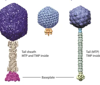
have very few members (Ackermann, 2007a). The tailed dsDNA phages, classified in the order Caudovirales are represented by three families (Figure 2), the Siphoviridae (non-contractile tails), Myoviridiae (contractile tails) and Podoviridae (very short tails). This literature review is limited exclusively to the Caudovirales as all the phages isolated during the course of this work belonged to this order (Chapter 3).
[image:19.595.106.540.151.479.2]4
Figure 2. Representatives of the three families of the order Caudovirales, the tailed dsDNA bacteriophages. The capsid enclosing the virion nucleic acid is coloured purple for each family. The tail is composed of the major tail (MTP) and tapemeasure (TMP) proteins and extends from the capsid, which for Myoviridae is enclosed by the tail sheath. The tail terminates at the baseplate to which the apparatus (not shown) required for adsorption and infection of host cells is attached. Reproduced with permission from Veesler and Cambillau (2011).
The classification of bacteriophages is subject to continuous discussion and debate, particularly with
the increasing volume of available genomic and proteomic data. New approaches to phage taxonomy are complicated as no single candidate gene analogous to the 16S rRNA gene used for the classification of bacteria exists in all phage families. Additionally comparative genomic studies have demonstrated that phage genomes are mosaic, that is, are subject to horizontal transfer events at
relatively high frequency, further complicating approaches for classification. As such, there is no single criterion for the demarcation of bacteriophage species and genera. Instead a polypthetic approach is favoured, where clades are delineated by a set of properties, some of which may be absent among related members. A number of alternative taxonomic approaches have been proposed in the literature. These include classification based upon terminase amino acid sequence similarity
[image:20.595.159.494.59.344.2]5
Recently a systematic method of nomenclature has been proposed (Kropinski et al., 2009). This system allows the rapid identification of bacteriophage family, lifestyle and host genus for a specific phage isolate.
1.2.2 Bacteriophage lifecycles
The first stage in infection of a host cell is the primary adsorption event. Adsorption refers to reversible and irreversible binding events between a phage receptor binding protein and a bacterial cell surface structure such as LPS, one or more outer membrane proteins, fimbriae or flagella. Primary adsorption is mediated by phage protein structures including tailspikes and tail fibres while irreversible binding tends to be a function of secondary binding proteins localised at the baseplate.
Irreversible adsorption results in the transfer of the virion genome to the host cell cytoplasm, a unidirectional process termed ejection or injection. Quite how the phage genome travels across the bacterial cell wall and inner membrane remains unclear, although a conformational change is likely transmitted along the tail in order to open the closed phage capsid upon irreversible binding. It is probable that the bacteriophages have evolved a number of different methods to facilitate this
translocation event.
Bacteriophages possess several lifecycles; lytic (virulent), lysogenic (temperate), pseudolysogenic and chronic. The lytic cycle is where phages infect and rapidly replicate within a host bacteria. The lytic lifecycle results in the release of newly formed progeny virions and death of the host cell through
lysis, mediated by phage-encoded proteins which enzymatically degrade the cell wall. Lytic infection results in clear plaques on the respective host bacterial lawns. The period encompassing phage adsorption to genome replication, production of progeny virions and lysis of the host cell is described as the latent period. The duration from adsorption to the time of formation of progeny virions, when intracellular virions can be detected by plaque assay after prematurely lysing the host cell, is
described as the eclipse phase (Ellis and Delbrück, 1939).
Temperate phages are those which possess a lytic life cycle and a lysogenic life cycle. In the lysogenic life cycle, the infecting phage does not directly kill the host cell but instead either integrates into the host bacterial chromosome by transposition or site-specific recombination or resides as a plasmid
within the host cell cytoplasm. In the integrated state, the phage DNA is termed a prophage and through gene expression can confer new or additional properties to the bacterial cell when expressed, a process termed lysogenic conversion (Brüssow et al., 2004). The prophage can stably reside within the host cell for many generations until appropriate environmental or physiological conditions trigger the lytic lifecycle, a process termed induction. Genome sequencing has revealed
6
profile or repressor specificity or by alteration of the cell surface epitope used for adsorption. Through various mutation events, the ability of a prophage to form functional virions upon activation of the lytic cycle may be lost whereby such prophages are termed cryptic prophages. Temperate
phages tend to form cloudy plaques on bacterial lawns as the probability of infection leading to lysogeny is greater than for lytic infection.
A further state, pseudolysogeny, has been proposed and is defined as a replicative and non-productive state following a successful adsorption and genome transfer event (Ripp and Miller, 1997). Pseudolysogeny is thought to occur in the presence of adverse conditions which prevent
either genome integration or replication, perhaps caused by host cell starvation. This strategy has the potential to enhance bacteriophage survival, as the phage genome is protected from environmental
conditions by ‘sheltering’ within the host cell (Miller and Ripp, 2002). A final lifestyle, termed chronic infection, is observed for some archaeal viruses and filamentous phage. In this scenario, progeny virions are slowly and continuously shed or extruded from the cell surface rather than being released
as a single burst event (Lopez and Webster, 1983).
An infection event which results in either integration or replication of the virion genome may be termed a successful infection. A productive infection is one which results in the assembly and release of progeny virions competent for subsequent infection of host bacteria. Infection does not always
7
Figure 3. Schematic representation of the possible outcomes of infection of bacteria by filamentous and tailed bacteriophages. The diagram depicts the interactions between phage and host and the survival outcome for each. Adsorption to and release from bacterial cells are shown as short dashed lines. Infections leading to death of the bacterial cell are shown as long dashed lines. Reproduced with permission from Hyman and Abedon (2010).
1.2.3 Bacteriophage structure
A bacteriophage virion consists of a single or double stranded DNA or RNA encapsulated within either a protein or lipoprotein coat. For members of the Caudovirales the nucleic acid is exclusively double-stranded DNA and ranges in size from 22.2 kb for Lactococcus phage c2 to 497.5 kb for Bacillus phage G. Irrespective of the target host organism, the virion structure is designed to contain and protect the viral chromosome, facilitating its safe transfer to a host cell to allow subsequent replication. The binary symmetry, or head to tail structure, of the Caudovirales is unique in virology and phages of this order are also differentiated from animal viruses in that the delivery of their genetic material occurs by an ejection mechanism, rather than by an un-coating process, following
8
force microscopy (AFM) and 3D reconstruction from cryo-electron microscopy has yielded additional insights into the structure and location of these proteins in situ (Guerrero-Ferreira and Wright, 2013, Kuznetsov et al., 2011, Parent et al., 2012).
1.2.4 Capsid assembly
The head or capsid forms the protective container for the condensed bacteriophage chromosome and the formation of this closed shell for genome packaging requires the recruitment and organisation of multiple copies of protein subunits. Recent studies using 3D reconstructions from cryo-electron microscopy have shown that DNA resides within the mature capsid as a tightly wound spool (Cerritelli et al., 2003, Parent et al., 2012). It is estimated that the pressure within the capsid is as much as 20 atmospheres (Cordova et al., 2003) and as such, the mature capsid lattice must be sufficiently robust to withstand the pressure of the condensed chromosome. In general, the size of the phage capsid is correlated to the size of the genome being packaged.
All members of the Caudovirales have capsids with icosahedral (20 sides/12 vertices) symmetry or prolate derivatives thereof, providing a characteristic appearance under the electron microscope. The icosahedral structure forms with known triangulation numbers T=4, 7, 13, 16 and 52, assembled from many copies of one or two major coat proteins (MCP). The triangulation number system was introduced by Caspar and Klug (1962) to describe the relationship between the number of pentameric and hexameric subunits giving rise to the quasi-symmetry of the capsid shell. Each corner
of the icosahedron is generally made up of pentamers of the MCP, while the rest of each side is made up of hexamers of the same or a similar protein.
Three gene products are critical for capsid formation; the portal, scaffold and MCP. Several other factors, including accessory proteins, chaperones and proteases are also required. The first stage in
capsid formation is the production of assembly intermediate, precursor structures called procapsids. The scaffold proteins co-assemble with the MCP subunits, forming a core inside the prohead. The scaffold is provided either by a separately encoded protein, as for P22 (Greene and King, 1994), or from a region of the major coat protein itself as for HK97 (Huang et al., 2011).
The true T=7 symmetry of the capsid is broken locally at one of the five-fold vertices by the portal
protein complex. The portal vertex is formed as a dodecameric ring of portal protein subunits and serves as the gateway through which DNA enters the pro-capsid during particle assembly, and exits during infection (Johnson and Chiu, 2007). The portal also forms part of the junction or neck connecting the capsid and tail. Like other phage structural proteins, the portal proteins from
9
With the start of DNA packaging the scaffold is removed signalling the start of maturation. Two mechanisms for removal of the scaffold have been identified. For phages T4, HK97 and λ scaffold
proteins are proteolytically cleaved by a phage-encoded protease (Liu and Mushegian, 2004), while
for P22 and ɸ29 the scaffold proteins exit the procapsid (Eppler et al., 1991). With maturation the procapsid undergoes a structural transition, enlarging, becoming more angular and taking on the characteristic isometric shape.
Most characterised tailed dsDNA phages assemble the capsid structure from multiple copies of one MCP. The exceptionally well characterised myovirus T4 is an exception, encoding two capsid
proteins: gp24, forming the pentameric vertex capsomers and gp23, forming hexameric capsomers (Olson et al., 2001). The MCPs of the tailed phages show extreme amino acid sequence variation. Despite such disparate sequences, the 3D structure of MCPs examined at high resolution by X-ray crystallography exhibit the presence of HK97-like fold, named after the bacteriophage in which this structure was first described (Wikoff et al., 2000). High resolution structures of MCPs have been resolved for phages T4 (Fokine et al., 2005); ɸ29 (Morais et al., 2005), ɛ15 (Jiang et al., 2006), T7 (Agirrezabala et al., 2007), λ (Lander et al., 2008), BPP-1 (Dai et al., 2010) and P22 (Jiang et al., 2003) all of which possess the HK97-like fold. These data are indicative of a common, very ancient evolutionary ancestor; it has long been recognised that 3D structure tends to be more highly
conserved over much greater timespans than either nucleotide or amino acid sequence (Rossmann et al., 1974). Moreover, Baker et al., (2005) demonstrated similarities between the HK97-like fold of MCPs of the Caudovirales with those of herpesviruses and suggested that the existence of related viruses infecting different domains of life might have arisen from a progenitor virus predating the
separation of the different domains of life or one that arose from a later adaptation event.
Once the capsid is packaged with DNA, the terminase complex is substituted by neck proteins, variously described as connector, tail terminator or head completion proteins, which together with the portal form successive rings with the bottom ring being termed the gatekeeper complex (Orlova et al., 2003). For bacteriophage SPP1 the gatekeeper has been demonstrated to prevent premature exit or leakage of DNA (Lhuillier et al., 2009).
Some bacteriophages have been found to recruit additional accessory proteins to the capsid, termed head decoration or capsid stabilisation proteins. Studies of the Dec protein of bacteriophage L (Gilcrease et al., 2005) found that this protein increased tolerance to EDTA of the closely related P22 virion. The authors suggested that a loss of divalent cations by the chelating activity of EDTA causes
10 1.2.5 The Bacteriophage tail
The tail functions to facilitate adsorption and attachment of the phage to the host cell surface and provide a conduit for genome ejection and it is this feature which distinguishes the three families of the Caudovirales. Analogous to genes encoding capsid assembly components, genes encoding elements for tail structure and assembly show great variation in sequence composition. However, the order of genes tends to be conserved, providing valuable information for the prediction of gene
function. Genes tend to follow a ‘body plan’ of consecutive open reading frames beginning with the major tail protein (MTP) and ending with genes encoding the baseplate and adsorption apparatus (Veesler and Cambillau, 2011). Tail assembly occurs separately to the capsid and occurs by the
addition of components in a strict sequential order (Aksyuk and Rossmann, 2011).
The architecture of the long, flexible siphovirus tail is relatively simple, reflected in the use of the Greek siphon, meaning tube. The tail is based upon three main components: the tape measure protein, the major tail protein and the tail terminator protein. In contrast, the Myoviridae (Greek myos, muscle) are characterised by a complex rigid tail apparatus. For this family of phages the tail tube is enclosed by an outer contractile sheath. This added layer of complexity is reflected by the number of genes encoding the tail structure and assembly components; at least 22 genes are involved in assembly of the T4 bacteriophage tail (Miller et al., 2003). The Podoviridae (Greek podos, meaning foot) possess no real tail, rather the adsorption apparatus is connected directly to the neck
region.
The tail tube is formed by the polymerisation of multiple copies of the MTP and there is evidence for structural conservation of this protein in both contractile and non-contractile tails (Pell et al., 2009a). The length of the tail is determined by the tape measure protein (TMP), located inside the tail shaft. All long-tailed phages sequenced to date possess a large gene, usually greater than 2 kbp in length,
encoding the TMP. This family of proteins is characterised by the presence of long hydrophobic α -helices and the relationship between tail length and TMP length has been shown by insertion and deletion studies to be precisely correlated, with an approximate length of 0.15 nm per amino acid residue for phage λ (Katsura, 1990, Katsura and Hendrix, 1984). However, the position and precise molecular mechanism governing tail length determination by these proteins has not been
unequivocally determined. Binding of the tail terminator protein halts polymerisation of the tail tube, completing the tail assembly. The tail terminator also interacts with the capsid to facilitate joining of the head and tail (Pell et al., 2009b, Zhao et al., 2003). In addition to the MTP and TMP, tailed phages encode a number of tail assembly chaperones and completion proteins. One common feature of
11
in a fixed abundance ratio between gpG and gpGT. These proteins are thought to form a spiral scaffold with dimensions similar to the internal diameter of the tail tube (Xu et al., 2004). Notably, functional and sequence comparisons of the tail tube and head-tail connector proteins suggest that
the tail proteins of Myoviridae and Siphoviridae arose from a single ancestral gene (Cardarelli et al., 2010).
The apparatus facilitating host adsorption is located at the distal end of the phage tail. Binding to the host cell surface occurs as a result of interactions with one or more host receptors by one or more phage receptor binding proteins. The recognition event is highly specific and facilitates rapid and
efficient attachment to the host cell. Bacteriophages are capable of binding to a wide range of cell surface structures including proteins, LPS and components of the cell wall and this ability is reflected by a stunning diversity of distal tail morphologies. Phage receptor binding proteins are generally either described as tail fibres or tail spikes. Tail fibres refer to long, thin, flexible structures composed of one or more proteins, which extend from the top of the tail tip assembly in siphoviruses and to the
baseplate of myoviruses. Tailspikes, in contrast, tend to be much shorter and appear as globular or tear-shaped structures. Transmission and cryo-electron microscopy have amply illustrated the diversity of tail-tip/baseplate complexes among the bacteriophages. Despite low levels of sequence identity, many of the characterised tail fibres and tailspikes of the Caudovirales form homotrimers rich in β structure. The N-terminal domain is exclusively associated with attachment of the RBP to the phage tail, either directly or through interaction with an adaptor protein, while the C-terminal domain is responsible for receptor binding.
1.3
The
Salmonella
e
The Salmonellae remain significant aetiological agents of zoonotic and food-borne disease world-wide. Members of the genus are Gram-negative, facultatively anaerobic, non-sporulating bacilli that are the causative agents of typhoid fever, gastroenteritis and enteric fever in both humans and animals. With the exception of S. Gallinarum, serovars (serotypes) of Salmonella are motile, possessing peritrichous flagellae.
The first successful clinical definition of typhoid fever was undertaken in1851by Sir William Jenner who differentiated the disease from Typhus, a vector borne disease caused by transmission of Rickettsia typhi or Rickettsia prowazekii by ticks and lice. In 1880, Karl Joseph Eberth isolated a bacillus suspected of causing typhoid from spleen sections and mesenteric lymph nodes, and in 1884
Georg Gaffky confirmed Eberth’s findings. The bacillus reported by Eberth and Gaffky was first
identified as a separate genus by Dr Theobald Smith, a researcher investigating swine cholera under the auspices of the USDA Bureau of Animal Industry. Despite identifying the bacteria, Salmon, as
Smith’s administrator, took priority on the research paper and hence the new bacterium was named
12
The nomenclature of Salmonella is complex and continues to be frequently revised. The differentiation of Salmonella isolates was initially performed using clinical evidence, serological and biochemical tests. Serological analysis of Salmonella dates back to the beginning of the 20th century with a study of antigenic components in motile and non-motile strains of S. Choleraesuis (Smith and Reagh, 1903). Phase variation of flagella was subsequently discovered in S. Typhimurium by Andrewes (1922). The first systematic serological analysis of Salmonella was initiated by White (1926) and continued and extended by Kauffmann (1961). Prior to the introduction of the Kauffmann-White scheme in 1946 by the World Health Organisation (W.H.O.) serovar names were
allocated according to clinical manifestation, geographic location or source from which the serovar was first isolated. Subsequent DNA-DNA hybridisation analysis demonstrated that the Salmonella serovars form a single hybridisation group sharing between 70 % and 90 % DNA content, demonstrating that relatedness between different serovars existed at the species level
13
Table 1. Differential biochemical characteristics of species and subspecies of Salmonella. Reproduced from Grimont and Weill (2007).
Species S. enterica S. bongori
Subspecies enterica (I) salamae (II) arizonae (IIIa) diarizonae (IIIb) houtenae (IV) indica (VI)
Dulcitol + + - - - d +
ONPG (2 h) - - + + - d +
Malonate - + + + - - -
Gelatinase - + + + + + -
Sorbitol + + + + + - +
Growth with KCN - - - - + - +
L(+)-tartrate(a) + - - - -
Galacturonate - + - + + + +
γ-glutamyltransferase + (
*) + - + + + +
β-glucuronidase d d - + - d -
Mucate + + + - (70 %) - + +
Salicine - - - - + - -
Lactose - - - (75 %) + (75 %) - d -
Lysed by phage O1 + + - + - + d
Usual habitat Warm-blooded
animals Cold-blood animals and the environment
(a) = d-tartrate. (*) = Typhimurium d, Dublin -. + = 90 % or more positive reactions. - = 90 % or more negative reactions. d = different reactions given by different serovars.
The current consensus is that the genus Salmonella consists of two species, S. enterica and S. bongori. S. enterica is divided into five subspecies based upon biochemical tests (Table 1) and serotyping is used for the differentiation of isolates beyond the subspecies level. Serovars are distinguished on the basis of the somatic (O) and flagellar (H1 and H2) antigens. A capsular polysaccharide, the Vi antigen, is also present on a limited number of serovars including Typhi and Dublin. The list of designated serovars and their antigenic formulae, now known as the Le
Minor-Kauffmann White Scheme, is maintained by the WHO Collaborating Centre for Reference and Research on Salmonella at the Pasteur Institute in Paris (Grimont and Weill, 2007). The total number of reported serovars for all subspecies currently stands at 2,610 and continues to grow (Guibourdenche et al., 2010). Serovars belonging to subspecies I have retained their common names, due to their familiarity with clinical practitioners, and are formally presented as Salmonella enterica subspecies enterica serovar Enteritidis. Those serovars belonging to subspecies other than enterica are designated by antigenic formulae; (i) subspecies numeral, (ii) O antigen followed by a colon, (iii) Phase 1 H antigen followed by a colon and (iv) Phase 2 H antigen (e.g. S. IIIa 50:g,z51:-). In practice,
many scientists, clinicians and physicians continue to use a simplified nomenclature omitting the
subspecies epithet as the majority of infections in both humans and animals are caused by serovars belonging to subspecies enterica.
14
exclusively human pathogens, S. Typhi and S. Paratyphi. Typhoid is characterised by an array of clinical manifestations including fever, abdominal pain and transient diarrhoea. The pathological features of typhoid fever are mononuclear cell infiltration and hypertrophy of the
reticulo-endothelial systems including the intestinal Peyer’s patches, mesenteric lymph nodes, spleen and
bone marrow arising from the intracellular invasion and dissemination of the bacillus. In the absence of treatment, mortality ranges between 10 and 15 % (Parry et al., 2002). Non-typhoidal infections by Salmonella serovars other than Typhi and Paratyphi are generally self-limiting with clinical manifestations ranging from mild to severe gastroenteritis. Like many other infections by pathogenic
bacteria, the susceptibility, severity and predisposition to serious complications upon infection by Salmonella are dependent upon a number of intrinsic host factors. Associated risk factors include age, immunosuppressive drugs, the presence underlying medical states (morbidities) and an altered endogenous bowel microflora resulting either from antibiotic therapy or from surgical procedures
(Acheson and Hohmann, 2001). The incubation period varies depending upon these host factors, the causative serovar and size of the infectious dose ingested (Blaser and Newman, 1982). Patients typically present with headache, nausea, vomiting, fever, diarrhoea and abdominal cramps. In a small proportion of cases further complications arise including bacteraemia, gastrointestinal bleeding and focal infections (Acheson and Hohmann, 2001). The development bacteraemia, potentially leading to
bacterial endarteritis and or endocarditis, is associated almost exclusively with the presence of underlying co-morbidities (Laupland et al., 2010). Focal infections may occur at virtually any anatomical site, arising from dissemination by intra-cellular invasion or bacteraemia. Specifically, focal infections may lead to osteomyelitis, polyarticular reactive joint disease, splenic abscesses, CNS infection, meningitis and urinary tract infection (Acheson and Hohmann, 2001). Whilst most
non-typhoidal Salmonella infections are transient and self-limiting, it has been demonstrated that the bacteria may be detected for a period after the cessation of symptoms. The median duration of faecal shedding is reported to be about one month in adults and over seven weeks in children under
five years of age (Buchwald and Blaser, 1984).
Transmission of Salmonella to humans is primarily associated with the ingestion of a wide variety of contaminated foods but may also arise by contact with animals, contaminated water and infected individuals (Hanning et al., 2009). Upon entry to the gastrointestinal tract, Salmonella preferentially
15
encoded within Salmonella Pathogenicity Island-2 (Hensel et al., 1998, Cirillo et al., 1998). After uptake by CD18+ phagocytes or macrophages, the acidic environment of the phagosome causes the induction of regulatory systems promoting intracellular survival including surface remodelling and
induction of the SPI-2 T3SS. The SPI2 T3SS effectors are translocated across the phagosomal membrane and result in remodelling of the phagosome, cytoskeletal rearrangements, alterations to gene expression and mitigation of the oxidative burst. These protein allow Salmonella to survive within the host cell, where the phagosome is termed the Salmonella-containing vacuole (Haraga et al., 2008). A further action of the SPI2 T3SS effectors is the inhibition of antigen presentation by dendritic cells to naïve CD4+ T-cells. (Tobar et al., 2006), thereby limiting the host innate (adaptive) immune response (van Diepen et al., 2005). Systemic infection is characterised by dissemination and colonisation of Salmonella to the liver, spleen and lymph nodes. A further aspect of Salmonella pathogenesis is the establishment of an asymptomatic carrier state that serves as a reservoir of infection. Asymptomatic carriers of S. Typhi discontinuously shed the bacilli thereby facilitating the dissemination of this pathogen into the environment.
Serovars of Salmonella can be coarsely differentiated as host-adapted, host-restricted or promiscuous based upon their ability to colonise and cause disease in related and unrelated species (Uzzau et al., 2000). Host-restricted serotypes typically cause systemic disease in a limited number of related species. Those serotypes frequently associated with a specific host and infrequently in unrelated hosts are termed host-adapted, whilst promiscuous serotypes are defined as those capable of causing disease in a wide range of unrelated host species. Over the past decade a number of Salmonella genomes have been sequenced and from subsequent comparisons, the underlying genetic and phenotypic characteristics contributing to host specificity and promiscuity are starting to be elucidated. Anjum et al., (2005) summarise the genus Salmonella as comprising a composite gene pool, within which distinctive variants (serovars) have arisen, possessing different degrees of specialisation by the acquisition, loss or mutation of different genes.
Epidemiological data shows that the majority of non-typhoidal infections by Salmonella are limited to relatively few serovars, belonging predominantly to subspecies enterica and serogroups A, B, C1, C2, D
and E (ECDC, 2010, Uzzau et al., 2000). In the US, the annual economic burden associated with medical care and lost productivity due to Salmonellosis is estimated at several billion dollars (Voetsch et al., 2004). In 2009 over 49,000 laboratory confirmed cases of Salmonella in humans were reported in the US while 109,885 confirmed cases were reported in the EU for the same period. The increased
16
1.4
Bacterial Bioluminescence
The ethereal qualities of bioluminescence has always attracted and intrigued human kind, evidenced by a wealth of historical records spanning some 3000 years (Lee, 2008, Newton, 1957). The light
emitting enzymes responsible for bioluminescence, termed luciferases by the French physiologist Raphael Dubois, have been characterised in both eukaryotic and prokaryotic organisms. The term "luminescenz" was first coined by Elihard Wiedemann (Harvey, 1957) and literally, bioluminescence
means “living light” comprising the Greek words bios for “living” and lumen for “light”. Luciferases
from different phyla have evolved independently, made evident by markedly different tertiary and
quaternary structures, substrate requirements, reaction kinetics and wavelength of emitted light. The single common requirement of all luciferase systems is oxygen (O2), a characteristic first
demonstrated in the 17th century by Robert Boyle (1672), long before the discovery of bacteria. Although many luminescent species exist in nature (Widder, 2010, Wilson and Hastings, 1998),
luciferases from six sources have been widely studied (Table 2).
Table 2. Sources and characteristics of Luciferases in nature.
Source Requirements Peak Emission Structure
Bacterial
Various
Flavin mononucleotide Fatty acid aldehyde O2
490 nm Heterodimer, 77 kDa
Firefly
Photinus pyralis
D-Luciferin ATP O2
562 nm Monomer, 62 kDa
Click Beetle Pyrophorus plagiophthalamus D-luciferin ATP O2
Green: 537 nm Red: 613 nm
Monomer, 62 kDa
Ostracod crustacean
Cypridinia noctiluca
Cypridina luciferin 465 nm Monomer, 62 kDa
Cocepod crustacean
Gaussia princeps
Coelenterazine O2
460 nm Monomer, 35 kDa
Sea pansy
Renilla reniformis
Coelenterazine O2
480 nm Monomer, 19 kDa
Of the different luciferases, the bacterial system is best understood both in terms of regulation and
17
predation (through flashing and counter-illumination), defence (using light as a decoy), communication and the attraction of prey, the latter exemplified by the Photobacterium-containing lure of the deep sea Angler fish (Haddock et al., 2010). In many cases the bacteria colonise specialised light emitting organs, termed photophores. Many marine organisms possess photophores containing bioluminescent bacteria including members of Sepiolid and Lolliginid squid and various fish.
Bioluminescence caused by free-living bacteria in the sea can encompass vast areas given appropriately high concentrations of bacteria, a phenomenon termed milky seas. Recently, Miller et al., (2005) corroborated reports from merchant ships off the east coast of Africa with satellite imagery, providing the first geographic identification of this phenomenon from space (Figure 4).
Figure 4. Enhanced satellite image of a bioluminescent milky sea projected onto the Blue Marble (NASA). Reproduced with permission from Haddock et al., (2010).
Luminescence in bacteria is conferred by a contiguous sequence of DNA, the luxCDABE operon (Meighen, 1991) and the order of lux genes is conserved within all characterised bacterial systems. The most widely studied of these luciferases originate from the species Vibrio harveyi, Aliivibrio fischeri (previously Vibrio fischeri), Photobacterium phosphereum, Photobacterium leiognathi and Photorhabdus luminescens. The luxAB genes encode the two subunits; α (40.11 kDa) and β
18
acid reductase, the multi-enzyme complex responsible for biosynthesis and turnover of the fatty acid aldehyde substrate is encoded by three genes, luxC, luxD and luxE. These three genes code for a reductase, a transferase and a synthetase of molecular weights 54 kDa, 42 kDa and 32 kDa
respectively (Boylan et al., 1988). Nucleotide sequences of the luxA and luxB genes have been determined for V. harveyi, A. fischeri and P. phosphorum, P. leigionathi and P. luminescens (Cohn et al., 1985, Johnston et al., 1986, Miyamoto et al., 1986, Johnston et al., 1990, Xi et al., 1991).
Bioinformatics evidence suggests that the α and β subunits arose by gene duplication within a
common ancestral species, as approximately 30 % nucleotide sequence homology exists between the
two subunits independent of the bacterial source (Cohn et al., 1985, Johnston et al., 1986, Szittner and Meighen, 1990). The 3-D structure of A. fischeri luciferase has been determined at low (2.7 Å) and high (1.5 Å) resolution by x-ray crystallography (Fisher et al., 1996). These data show that the α and β subunits are non-covalently bound and that each folds into a single domain (β/α)8 barrel
conformation (Figure 5).
Figure 5. Ribbon cartoon of the quaternary structure of Vibrio harveyii bacterial luciferase. The alpha subunit is shown in blue and the beta subunit in purple. The image was rendered with Jmol Version 12.2.15 using Protein Databank Entry: 1LUC.
1.4.1
Biochemical characteristics of Bacterial LuciferasesBacterial luciferase catalyses the oxidation of reduced flavin mononucleotide (FMNH2) using
molecular O2 and a long chain aliphatic aldehyde, yielding FMN, carboxylic acid and a photon of
blue-green light (Equation 1). The reaction is highly exergonic with the primary energy source for light supplied by conversion of aldehyde to fatty acid (60 kcal Enstein-1).
Equation 1. Bacterial luciferase reaction.
19
Two assays have been described for the quantification luciferase enzymatic activity which differ in the method of FMN reduction (Hastings et al., 1978). Due to the rapid oxidation of flavin in these assays and slow decay of the EFO-A complex, the luciferase enzyme undergoes only a single turnover
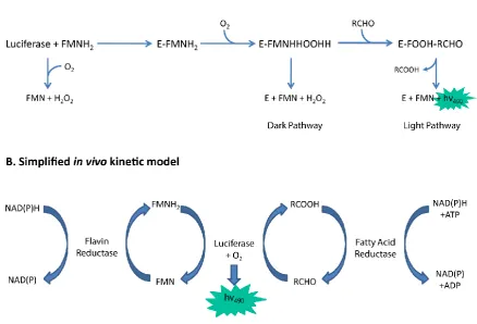
under these assay conditions, a feature which has allowed detailed investigation of the kinetics of these enzymes (Gibson and Hastings, 1962). The complex bioluminescence reaction (Figure 6) depends upon the sequential formation of enzyme-flavin intermediates (Hosseinkhani et al., 2005). Firstly, luciferase (E) reversibly binds FMNH2 via the alpha subunit to form the Enzyme-FMNH2 (EF)
complex. The enzyme-flavin complex then reacts rapidly with O2 forming a C4a-peroxy-flavin (EFO)
(intermediate II). The EFO complex may either decompose to yield oxidized FMN and H2O2 or interact
with an aliphatic aldehyde (A), supplied by the LuxCDE fatty acid reductase complex, to form the tetrahenal intermediate (EFO-A). Decay of the EFO-A complex occurs by either dissociation of the aldehyde followed by dark pathway decomposition or upon completion of the reaction to yield
[image:35.595.95.534.370.668.2]blue-green light with an emission maximum at 490 nm, an aliphatic carboxylic acid and oxidised flavin. The dark- and light-side reactions possess distinct rate constants, termed kD and kL, respectively (Hastings and Balny, 1975).
Figure 6. Simplified kinetic models of the bacterial luciferase reaction (A) in vitro and (B) in vivo. Blue arrows denote the conversion of substrate to product. Light emission is shown within the green star.Adapted from Campbell and Baldwin (2009).
20
(Ulitzur and Hastings, 1979, Wall et al., 1984). The net reaction involves reduction of fatty acid with the oxidation of NADPH and cleavage of ATP to AMP and PPi, catalysed by the luxCDE gene products. Biosynthesis of the long chain aldehyde is catalysed by the action of the luxCDE gene products. LuxC and LuxE are required for the reduction of myristic acid to myristic aldehyde while LuxD transports the fatty acid between the synthase and reductase complexes (Meighen, 1991).
The second substrate require for bioluminescence is Flavin mononucleotide (FMN). FMN belongs to the family of flavoproteins whose members all contain a nucleic acid derivative of riboflavin. Reduced FMN is provided by species specific oxidoreductases; Frp in A. harveyi, Frase-I in V. fischeri and Fre in Escherichia coli (Campbell and Baldwin, 2009). These enzymes show broad substrate specificity but little sequence homology despite their functional similarities. Flavin mononucleotide plays an important role in electron transport since it can accept and donate one or two electrons and is involved in initial electron transfer with bacterial complex I leading to the oxidation of NADH with reduction of coenzyme Q (Friedrich et al., 1995). Two models are proposed for the mechanism of transfer of the flavin substrate from oxidoreductase to luciferase. The first proposes that a stable complex is formed between the oxidoreductase and luciferase (Lei and Tu, 1998). However, formation of a stable complex would necessitate the presence of specific recognition domains in the luciferase structure. The flavin oxidoreductase of E. coli shows little homology with that of A. fischeri yet luciferase activity is adequately expressed in transformed cells, apparently discounting the interaction model. A second model proposes that transfer of the flavin substrate occurs by free diffusion (Campbell and Baldwin, 2009).
1.4.2 Luciferases of bioluminescent bacteria
Luciferases encoded by different bacteria have been differentiated by turnover rate, thermostability and rates of decay over time from in vitro assays of the purified enzymes. Luciferases from the genus Photobacterium are characterised as possessing a rapid decay rate whereas those from Vibrio and Photorhabdus exhibit slow decay rates. The structural region responsible for this trait has been identified through the creation of chimeric luciferases. Chimeric luciferases were prepared by substituting regions of luxA in one bacterium with the same region from another. Substitution of a 67 amino acid sequence in the central region of LuxA from P. luminescens with the corresponding sequence from P. phosphoruem resulted in a chimera exhibiting a significantly greater decay rate than the wild-type enzyme (Valkova et al., 1999). Hosseinkhani et al., (2005) furthered these observations by demonstrating that substitution of the glutamic acid residue at position 175 of LuxA to glycine results in the conversion of the LuxAB of P. luminescens from a slow to rapid decay rate, suggesting that the Glu175 residue is involved in aldehyde binding and turnover of intermediates.
21
denatured at temperatures of 45oC whereas P. luminescens luciferase has a half-life of greater than 3 hours at this temperature (Szittner and Meighen, 1990). Thermal stability is conferred by the α
subunit, as demonstrated by hybrid luciferases containing combinations of luxA and luxB genes from V. harveyi or P. luminescens (Li et al., 1993). For temperature-critical applications where the required growth temperature is greater than 30oC, the P. luminescens lux cassette must be considered the bioluminescent system of choice for the transformation of bacterial reporters.
1.4.3 Bioluminescent bacterial reporters
Bacterial luciferases have been used extensively for the generation of biosensors, for studies of gene expression and transcriptional regulation. A bioluminescent phenotype may be conferred by the
introduction of either the luxAB genes, or the entire luxCDABE operon using a suitable vector and transformation method. The lux genes may be integrated into the bacterial chromosome or encoded on plasmids. Expression may be controlled using a promoter allowing constitutive expression or linked to a specific promoter for expression under conditions of transcriptional activation. As such, use of the lux genes presents two complementary methods by which data may be obtained for bacterial metabolism, stress responses or gene expression.
Vectors encoding luxAB alone generate luminescent bacteria upon addition of a long chain