Original Article
Bufalin inhibits invasion and metastasis in
colorectal cancer cells through miRNA-497
mediated IGF1R-PI3K-Akt signaling pathway
Ming-Tai Hu1*, Lin Liu2*, Ying Sun3, Qiang Hu1*, You-Rong Duan3*
Departments of 1General Surgery, 2Pharmacy, Dahua Hospital, Xuhui District, Shanghai 200237, People’s Republic of China; 3State Key Laboratory of Oncogenes and Related Genes, Shanghai Cancer Institute, Renji Hospital, School of Medicine, Shanghai Jiaotong University, Luwan District, Shanghai 200025, People’s Republic of China. *Equal contributors.
Received December 31, 2016; Accepted April 9, 2017; Epub July 15, 2017; Published July 30, 2017
Abstract: Objective:Bufalin, a major active ingredient of the Chinese traditional medicine Venenum Bufonis, exerts anti-tumor activity in multiple cancers. This study aimed to assess the role and mechanism of bufalin in invasion and metastasis inhibition in colorectal cancer. Methods: Human colorectal carcinoma Lovo cells were treated with bufa-lin at different concentrations, and cellular function and proliferation, as well as the expression of related molecules were evaluated. Results: Bufalin inhibited the proliferation of colorectal cancer cells in a time- and dose-dependent manner. Further mechanistic assessment showed that miR-497 down-regulation promoted invasion and metastasis in colorectal cancer cells by effecting the insulin-like growth factor 1 receptor (IGF1R)-Phosphoinositide 3 kinase (PI3K)-Protein Kinase B (Akt) signaling pathway. Meanwhile, bufalin could reverse these changes. Conclusion: the current findings suggested that bufalin could inhibit invasion and migration in colorectal cancer cells through miR -NA-497 mediated IGF1R-PI3K-Akt signaling.
Keywords: Bufalin, colorectal cancer cell, miRNA-497, insulin-like growth factor 1 receptor, phosphoinositide 3 kinase, protein kinase B, IGF1R-PI3K-Akt signaling pathway
Introduction
Colorectal cancer is one of the most common clinical malignant tumors; its incidence ranks third in the United States,and fourth and fifth in
major and remote cities of China, respectively [1, 2]. In China, the morbidity and mortality
rates of colorectal cancer rank first worldwide
[3]. About 50% of patients display local or dis-tant metastases at the time of diagnosis [4]. Metastasis formation is a complex multi-step process, which involves initial malignant cell
invasion, infiltration into the bloodstream, pro -liferation during migration, and extravasation into distant organs [5, 6]. Currently, the clinical treatment of colorectal cancer still largely depends on surgery, but the early surgical resection rate is only about 60% to 70%; mean-while, middle and late stage cancers are prompt to metastasis and recurrence [7].Therefore, new methods of diagnosis and treatment are urgently needed.
Bufalin is one of the main active ingredients of the important anticancer traditional Chinese medicine Venenum Bufonis. As a cardiotonic steroid isolated from Chansu, bufalin is a galen-ical preparation of the dried white venom [8, 9]. Several reports have shown that through Na+/ K+-ATPase inhibition, bufalin blocks vasodila-tion, and increases vasoconstricvasodila-tion, vascular resistance, and blood pressure [10-12]. In
addi-tion, bufalin exhibits significant anti-tumor
involved in bufalin induced colon cancer cell death [18].
MicroRNAs (miRNAs) are involved in multiple cell activities, including differentiation, prolifer-ation, apoptosis, and immunity [19-21]. Recent- ly, multiple studies have shown that microR-NA-497 (miR-497) is downregulated in tumors, functioning as a tumor suppressor in several types of human cancer [22-26]. In humans, bufalin could inhibit angiogenesis and metasta-sis in colorectal cancer cells, synergistically with miR-497 [27].
Insulin-like growth factor-1 receptor (IGF1R), a member of the transmembrane receptor, be- longs to the tyrosine kinase family and is acti-vated by insulin-like growth factor 1 (IGF-1) and IGF-2 [28]. IGF1R has a critical role in tumor cell transformation, survival, and invasion, and its high expression is implicated in several can-cers [29-32]. IGF1R mRNA and protein expres-sion levels are increased in tumor tissues, and
significantly correlated with patient prognosis
[33]. Reports also indicated that IGF1R overac-tivation allows the cytotoxic drug resistance property of malignant cells [34]. In addition, IGF1R activates the phosphoinositide 3-kinase (PI3K)/Akt signaling pathway, which is curial to cell proliferation [35, 36].
To date, studies have demonstrated that miR-497 plays its cell inhibitory role by targeting IGF1R [22, 26]. Therefore, bufalin appears to inhibit proliferation and metastasis in tumor cells by altering the expression of miR-497, subsequently modulating the IGF1R signaling pathway. Whether miR-497 could target IGF1R directly in CRC cells as a tumor suppressor dur-ing bufalin treatment remains unclear. In the current study, after treatment with various con-centrations of bufalin, we assessed invasion and metastasis in colorectal cancer cells as well as the expression levels of miR-497 and its downstream IGF1R-PI3K-Akt signaling path- way.
Materials and methods
Chemicals and reagents
Bufalin, isolated from Bufotoxine with purity more than 99%, was purchased from Sigma-Aldrich Chemical Corp. (St. Louis, MO, USA). Bufalin dissolved in dimethyl sulfoxide (DMSO)
and stored at -20°C was diluted in the cell cul-ture medium before use. Cell Counting Kit-8 (CCK-8) was purchased from Dojindo Labora- tories (Kumamoto, Japan). Primary antibodies to human Phospho-Akt (Thr308), Phospho-Akt (Ser473), Pan Akt, IGF-1 receptor beta, and beta-actin were purchased from Cell Signaling Technology Inc. (Beverley, MA, USA). LY294002 was purchased from Selleck Chemicals. En- hanced chemiluminescence (ECL) plus system was purchased from Amersham Pharmacia Biotech.
Cell culture and transfection
Human colorectal adenocarcinoma Lovo cells were purchased from the cell bank of the Chi- nese Academy of Sciences (Shanghai, China), and cultured in RPMI-1640 supplemented with 10% FBS, penicillin (100 U/mL) and
streptomy-cin (100 mg/mL) at 37°C in a humidified atmo -sphere with 5% CO2. Lovo cells in logarithmic growth phase digested by Pancreatin were seeded in 6-well plates at a density of 2×10⁵ cells/well with 2 mL of medium, and cultured for 12 hours at 37°C in 5% CO2. At 70% conflu -ency, the plasmid containing miR-497 no-load control (miR-497 NC) and miR-497 lentiviral vector (Hanbio, Biotechnology Co., Ltd.) were added to the cultures in 6-well plates,
respec-tively, 20 μl; then, polybrene (at a final concen
-tration of 5 μg/ml) was added and mixed gently.
Lovo cells were cultured for 24 h at 37°C in 5% CO2, followed by medium replacement.
GFP-positive cells were selected by flow cytometry
and sub-cultured, establishing a stable trans-fection line of miR-497 Lovo cells.
RNA extraction and real-time PCR
Total RNA was extracted with TRIzol Reagent (Invitrogen Corporation, CA, USA). MiR-497 ex-
pression was analyzed by the specific
Bulge-Loop assay, which detects mature miRNAs. Re- verse transcription and qPCR were performed with Bulge-LoopTM miRNA qRT-PCR Starter Kit (RiboBio Co., Ltd. Guangzhou, China) on an ABI 7500 Real-Time PCR System (Applied Biosys- tems, Foster City, CA) according to the manu-facturer’s protocol. MiRNA-497 levels were nor-malized to U6 RNA transcript levels. Relative expression levels between the samples were calculated using the comparative delta CT (threshold cycle number) method (2-ΔΔCT), with a
Cell viability assay
Cell viability was evaluated using Cell Counting Kit-8 (CCK-8) assay. In brief, cells were seeded in 96-well plates at 2×10⁴ cells/well and cul-tured overnight. After treatment for 24-72 h with bufalin at 12.5-1600 nmol/L (eight serial concentrations) (six replicates per dose), the CCK-8 solution (10 µl) was added to each well, followed by 3 h of incubation at 37°C, 5% CO2. Absorbance at 450 nm was recorded for each well on a FlexStation 3 microplate reader (Mole- cular Devices, Sunnyvale, California, USA), and cell viability accessed based on the manufac-turer’s instructions.
Detection of apoptosis with Annexin V-FITC/PI staining
Cell apoptosis was determined by Annexin
V-fluorescein isothiocyanate (FITC)/Propidium
Iodide (PI) Apoptosis Detection Kit (EMD Bio- sciences, La Jolla, USA). Cells were cultured in 24-well plates at a density of 5×10⁴ cells/ml, and treated with bufalin for 48 h. Cells were then digested and resuspended in binding buf-fer, and stained with 2.5 µl Annexin V-FITC and 5 µl PI for 15 min at room temperature in the dark. The stained cells were analyzed within 30
min on a BD FACS AriaII flow cytometer (BD bio -sciences, San Jose, California, USA). The lower right quadrant represented early apoptotic cells (Annexin V-FITC binding positive and PI nega-tive), while the upper right one comprised late apoptotic cells (Annexin V-FITC and PI positive staining).
Cell cycle analysis
After 48 h of treatment with bufalin, cells were harvested for cell cycle phase distribution anal-ysis. Stained cells were measured on an Accuri
C6 flow cytometer according to the instructions
of Cycle TestTM Plus DNA Reagent Kit (BD biosci-ences); data were analyzed with the Accuri C6 software package.
Wound healing assay
For cell migration measurements, 5×10⁵ cells were seeded per well of 24-well plates, and grown for 24 h. A linear wound was generated
by scraping a pipette tip across the confluent
cell monolayer. Cells were rinsed twice with PBS and supplemented medium with or without bufalin at 25-400 nmol/L for additional 48 h of culture. Cell migration in terms of wound clo-sure was meaclo-sured by photographing at three
random fields at the time of wounding (time 0)
and at 48 h.
Transwell assay
Matrigel diluted in RPMI-1640 (50 μl) was
added into the upper chamber of the transwell plate, followed by overnight incubation in 37°C. Cells in logarithmic phase were plated in the 24-well transwell chambers pre-treated with Matrigel, at a density of 7×10⁴ cells/100 μl.
RPMI-1640 (600 μl) containing 20% FBS was
added to the lower chamber. Control and bufa-lin treatment groups were set in duplicate. Cells were cultured for another 48 h, at 37°C in a
humidified atmosphere with 5% CO2. Then, the
filter membrane was fixed with 4% paraformal -dehyde for 10 min, dyed with Giemsa for 5 min, and mounted with neutral balsam. Invasive cells were counted at ×200 under an optical
microscope. For each filter membrane, five dif -ferent views (upper, lower, left, right and mid-dle) were assessed and averaged.
Immunoblotting analysis
Cells were cultured in 6-well plates at a density of 1×10⁶ cells/well (2 ml of culture medium), and treated with PI3K inhibitors LY294002 at
10 μmol/L for 4 h. Then, bufalin was added to
the cells, followed by 1 h of incubation at 37°C in 5% CO2. Total cell lysates were prepared with cell lysis buffer containing 1% proteinase inhibi-tor cocktail and 1 mmol/L PMSF, both from Beyotime Institute of Biotechnology, China. Cell
proteins (50 μg) were separated by 10%
SDS-PAGE and transferred onto PVDF membranes (Millipore, Bedford, MA). The membranes were blocked with 1% BSA (Bovine Serum Albumin), incubated with anti-human Phospho-Akt (Thr- 308), Phospho-Akt (Ser473), Pan Akt, IGF-1 re- ceptor beta, and beta-actin primary antibodies, respectively, followed by treatment with sec-ondary antibodies. Detection was performed with Bio-Rad ChemiDoc MP Imaging System (California, USA).
Statistical analysis
All analyses were performed with the SPSS software version 13.0 (SPSS Inc., Chicago, USA). In this study, the data were all measure-ment data expressed as means ± standard deviations (SD) and were normally distributed. The one-way analysis of variance (ANOVA) was adopted to assess whether there were any
means of different experimental groups. P<
0.05 was considered statistically significant.
Results
Bufalin suppresses proliferation and promotes apoptosis in Lovo cells
Rapid proliferation is a critical property of tumor cells. Therefore, we aimed to explore the impact
of bufalin on the proliferation of Lovo cells. To address this, Lovo cells were cultured in pres-ence of bufalin at various concentrations. Cell viability was then determined by CCK-8 assay. As shown in Figure 1A, treatment with bufalin at different concentrations (12.5-1600 nmol/L)
for 24-72 h resulted in significantly reduced
[image:4.612.93.513.77.490.2]proliferation of Lovo cells, in a dose- and time-dependent manner (24-48 h). In addition, a 10% DMSO/DMEM control group was set up
since bufalin was dissolved in DMSO. Interest- ingly, cells treated with 10% DMSO/DMEM sho- wed absorbance values similar to control cells.
This finding indicated that cell death was
caused by bufalin at a certain high concentra-tion and not DMSO, because DMSO in the working solution had a concentration below 10% (data not shown). Meanwhile, resistance
to apoptosis plays a critical role in cancer devel-opment, and whether a medicine induces a high degree of tumor cell apoptosis is an impor-tant criterion for evaluating its anti-tumor ef- fects. Therefore, Lovo cell apoptosis was asse-
ssed by flow cytometry after treatment with
[image:5.612.89.521.79.540.2]bufalin at concentrations of 25-400 nmol/L for 48 h. Annexin V-FITC and PI staining revealed
that bufalin induced apoptosis in Lovo cells from low to high concentrations (Figure 1B).
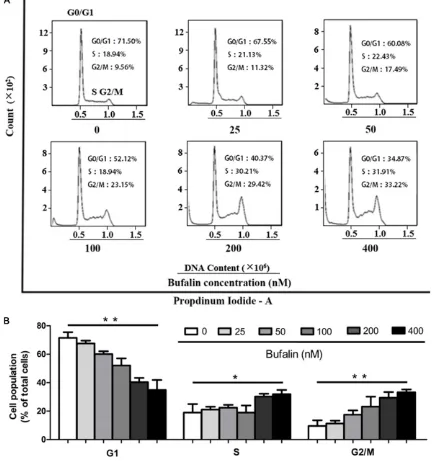
Bufalin blocks cell cycle in the G2/M phase in Lovo cells
Normal cell cycle progression is necessary for tumor cell proliferation. Thus, the cell cycle sta-tus of Lovo cells was measured after bufalin treatment at 25-400 nmol/L for 48 h. As shown in Figure 2, after treatment with different con-centrations of bufalin, the G0/G1
sub-popula-tion of Lovo cells was significantly increased
from 71.50% ± 4.2% (control) to 34.87% ± 7.3% (400 nmol/L, P < 0.01). G2/M phase cells were
significantly increased from 9.56 ± 4.70% (con -trol) to 33.22 ± 2.23% (400 nmol/L, P < 0.01), in a concentration dependent manner. S phase cells were increased at high bufalin
concentra-tions. These findings revealed that bufalin
could regulate the cell cycle of Lovo cells, block-ing the cancer cells in the G2/M phase, to inhibit proliferation (Figure 2).
Bufalin inhibits the ability of cell migration and invasion
Cell migration represents a critical event for tumor growth and metastasis. The effects of bufalin on the motility of Lovo cells were
[image:6.612.91.519.71.442.2]sured by scratch assay. As shown in Figure 3A, treatment with bufalin at 25-400 nmol/L for 48 h resulted in decreased migration rates in Lovo cells in a dose-dependent manner, i.e. from 35.2% ± 2.1% (25 nmol/L) to 3.5% ± 1.6% (400 nmol/L); non-treated control cells showed no
significant difference in migration ability. To
assess the effects of bufalin on the invasion ability of Lovo cells, the transwell invasion assay was adopted. The amounts of Lovo cells after 48 h of treatment with bufalin at 0 nmol/L, 12.5 nmol/L and 25 nmol/L concentration were 225.3 ± 10.54, 167.7 ± 9.61, and 115.4
± 10.52, respectively (200×, 5 fields). The dif -ferences between each treatment group and
controls were statistically significant (P < 0.01). Lovo cells were also treated with bufalin at 50 nmol/L or more (data not shown), but no adher-ent cells were found at the surface of the tran-swell chamber. These results suggested that low bufalin concentrations could inhibit the invasive ability of cells (Figure 3B).
Bufalin regulates miR-497 and the IGF1R-PI3K-Akt signaling pathway in Lovo cells
Recently, multiple studies demonstrated that MiR-497 plays a vital role in cancer develop-ment and relapse, with its downregulation closely associated with poor prognosis. Mean-
while, miR-497 causes a significant inhibition
of tumor cell viability, proliferation, and metas-tasis [22]. To assess the role of miR-497 in human colorectal adenocarcinoma cells, a len-tiviral system was used for the transfection of miR-497 in Lovo cells. Stable miR-497 express-ing Lovo cells (miR-497-Lovo cells) or no-load Lovo cells (Lovo NC cells) were established via
GFP-sorting by flow cytometry. Real-time PCR
(RT-PCR) demonstrated that the mRNA levels of miR-497 were close to 30,000 times higher in miR-497-Lovo cells compared with those of Lovo NC cells (P < 0.01, Figure 4A). These find -ings indicated that lentivirus over-expression vector increased miR-497 mRNA levels in Lovo cells. In addition, to assess whether bufalin affects miR-497 expression, Lovo cells were treated with different concentrations of bufalin for 24 h, after which miR-497 expression levels were determined by RT-PCR. In this assay, a lentiviral system was used for the transfection of miR-497 in Lovo cells as positive control. Interestingly, miR-497 expression was increa- sed by bufalin, in a concentration-dependent
manner (Figure 4A). Since one of the predicted targets of miR-497 is IGF1R, (http://www.tar-getscan.org; http://www.ebi.ac.uk/enright-srv/ microcosm/htdocs/targets/v5), which plays a critical role in colon cancer proliferation and survival [34, 37, 38], we also assessed the ex- pression levels of IGF1R and the downstream PI3K-Akt signaling effectors in Lovo cells. As shown in Figure 4B, Lovo cells treated with bufalin showed a decreasing trend of IGF1R expression. Meanwhile, the protein expres-sions levels of p-Akt Ser473 and p-Akt Thr308 were markedly reduced after treatment of Lovo cells with bufalin or LY294002 (PI3K/Akt sig-naling pathway inhibitor), while no change was observed in pan-Akt and IGF1R amounts (Fig- ure 4B), denoting impaired PI3K/Akt signaling.
These findings indicated that bufalin played a
role in the regulation of IGF1R-PI3K-Akt signal-ing mediated by miR-497.
Discussion
Bufalin is a compound extracted from Venenum Bufonis, and widely used due to its broad-spec-trum anti-tumor activities and the advantages of natural drugs. It is considered a potential anticancer agent in a variety of cancer models [39-42]. MiRNAs are highly conserved small non-coding regulatory RNAs with sizes of 17-25 nucleotides. As posttranscriptional regulators, miRNAs can negatively regulate gene expres-sion by binding directly to 3’ untranslated region (3’UTR) of corresponding target
messen-ger RNAs (mRNAs) in a sequence-specific man -ner; this induces mRNA degradation or protein translation repression [19, 43]. MiRNAs are involved in a number of important processes, including tumor occurrence, development and metastasis [43-46]. Among miRNAs, miR-497 has gained a lot of attention in recent years, due to its decreased level in tumors. One of the predicted targets of miR-497 is IGF1R (http:// www.targetscan.org; http://www.ebi. ac.uk/en- right-srv/microcosm/htdocs/targets/v5), whi- ch is an epidermal growth factor receptor that regulates the downstream PI3K-Akt signaling pathway and the malignant transformation. Several reports demonstrated the role of IGF1R in colon cancer cell survival, proliferation, and resistance to treatment [35, 37, 38, 47]. Pre-
the detailed mechanism remains elusive. In addition, it remains unclear whether IGF1R and its downstream PI3K-Akt pathway are involved in this regulation process. In this study, the human colorectal cancer Lovo cell line was assessed.
Our results showed that upon bufalin
adminis-tration, cell proliferation was significantly sup -pressed in a time- and dose-dependent man-ner. Flow cytometry and Annexin V-FITC/PI staining showed that rates of apoptosis were increased in a dose-dependent manner after bufalin treatment. In addition, treatment with bufalin increased the rates of Lovo cells in the G0/G1 phase. Besides, through transwell and scratch assays, we demonstrated that migra-tion and invasion in Lovo cells were impaired after treatment with bufalin. For mechanistic assessment, we focused on IGF1R since it is a miR-497 target, and has been reported to regu-late colon cancer. After treatment with the PI3K/Akt signaling pathway inhibitor LY294002 or bufalin, the protein expression levels of p- Akt Ser473 and p-Akt Thr308 were markedly reduced, indicating impaired PI3K/Akt signal-ing. Therefore, we speculate that IGF1R-PI3K-Akt signaling, which is mediated by miR-497, is responsible for the ef fects of bufalin in regulat-ing invasion and metastasis in colorectal can-cer cells.
Acknowledgements
This study was supported by the To Further Accelerate the Development of Chinese Tradi- tional Medicine Three Years Action Plan, Shang- hai (Grant No. ZY3-CCCX-3-3054) and the Chi- nese Clinical Medicine Diagnosis and Treatment Technology Innovation and Development Spe- cial Fund Projects of Wu Jieping Medical Foundation (Grant No. 320.6750.14202).
Disclosure of conflict of interest
None.
Address correspondence to: Qiang Hu, Department of General Surgery, Dahua Hospital, Xuhui District, 901 Laohumin Rd, Shanghai 200237, People’s Re- public of China.Tel: +86 21-34080686; Fax: +86 21-34080686;E-mail: hqdahua123@163.com;You- Rong Duan, State Key Laboratory of Oncogenes and Related Genes, Shanghai Cancer Institute, Renji Hospital, School of Medicine, Shanghai Jiaotong Uni-
versity, Luwan District, Shanghai 200025, People’s Republic of China.Tel: +86 21-64046550; Fax: +86 21-64046550;E-mail: yrduan@shsci.org
References
[1] Siegel R, Desantis C and Jemal A. Colorectal cancer statistics, 2014. CA Cancer J Clin 2014; 64: 104-117.
[2] Hawk ET, Limburg PJ and Viner JL. Epidemiolo-gy and prevention of colorectal cancer. Surg Clin North Am 2002; 82: 905-941.
[3] Bezerra-de-Souza DL, Bernal MM, Gomez FJ and Gomez GJ. Predictions and estimations of colorectal cancer mortality, prevalence and in-cidence in Aragon, Spain, for the period 1998-2022. Rev Esp Enferm Dig 2012; 104: 518-523.
[4] Figueredo A, Coombes ME and Mukherjee S. Adjuvant therapy for completely resected stage II colon cancer. Cochrane Database Syst Rev 2008; 16: CD005390.
[5] Hunter KW, Crawford NP and Alsarraj J. Mecha-nisms of metastasis. Breast Cancer Res 2008; 10 Suppl 1: S2.
[6] Dykxhoorn DM. MicroRNAs and metastasis: little RNAs go a long way. Cancer Res 2010; 70: 6401-6406.
[7] Shimada H, Tanaka K, Endou I and Ichikawa Y. Treatment for colorectal liver metastases: a re-view. Langenbecks Arch Surg 2009; 394: 973-983.
[8] Hong Z, Chan K and Yeung HW. Simultaneous determination of bufadienolides in the tradi-tional Chinese medicine preparation, liu-shen-wan, by liquid chromatography. J Pharm Phar-macol 1992; 44: 1023-1026.
[9] Panesar NS. Bufalin radioimmunoassays: in search of the endogenous digitalis-like sub-stance. J Immunoassay 1994; 15: 371-391. [10] Bagrov AY, Roukoyatkina NI, Fedorova OV,
Pinaev AG and Ukhanova MV. Digitalis-like and vasoconstrictor effects of endogenous digoxin-like factor(s) from the venom of Bufo marinus toad. Eur J Pharmacol 1993; 234: 165-172. [11] Eliades D, Swindall B, Johnston J, Pamnani M
and Haddy F. Hemodynamic effects of bufalin in the anesthetized dog. Hypertension 1989; 13: 690-695.
[12] Pamnani MB, Chen S, Bryant HJ, Schooley JF Jr, Eliades DC, Yuan CM and Haddy FJ. Effects of three sodium-potassium adenosine triphos-phatase inhibitors. Hypertension 1991; 18: 316-324.
[14] Qi F, Inagaki Y, Gao B, Cui X, Xu H, Kokudo N, Li A and Tang W. Bufalin and cinobufagin induce apoptosis of human hepatocellular carcinoma cells via Fas- and mitochondria-mediated path-ways. Cancer Sci 2011; 102: 951-958. [15] Sola S, Morgado AL and Rodrigues CM. Death
receptors and mitochondria: two prime trig-gers of neural apoptosis and differentiation. Biochim Biophys Acta 2013; 1830: 2160-2166.
[16] Kurosawa M, Numazawa S, Tani Y and Yoshida T. ERK signaling mediates the induction of in-flammatory cytokines by bufalin in human monocytic cells. Am J Physiol Cell Physiol 2000; 278: C500-508.
[17] Zhu Z, Li E, Liu Y, Gao Y, Sun H, Ma G, Wang Z, Liu X, Wang Q, Qu X and Yu Y. Inhibition of Jak-STAT3 pathway enhances bufalin-induced apoptosis in colon cancer SW620 cells. World J Surg Oncol 2012; 10: 228.
[18] Xie CM, Chan WY, Yu S, Zhao J and Cheng CH. Bufalin induces autophagy-mediated cell death in human colon cancer cells through re-active oxygen species generation and JNK acti-vation. Free Radic Biol Med 2011; 51: 1365-1375.
[19] Bartel DP. MicroRNAs: genomics, biogenesis, mechanism, and function. Cell 2004; 116: 281-297.
[20] He L and Hannon GJ. MicroRNAs: small RNAs with a big role in gene regulation. Nat Rev Gen-et 2004; 5: 522-531.
[21] Vasudevan S, Tong Y and Steitz JA. Switching from repression to activation: microRNAs can up-regulate translation. Science 2007; 318: 1931-1934.
[22] Guo ST, Jiang CC, Wang GP, Li YP, Wang CY, Guo XY, Yang RH, Feng Y, Wang FH, Tseng HY, Thorne RF, Jin L and Zhang XD. MicroRNA-497 targets insulin-like growth factor 1 receptor and has a tumour suppressive role in human colorectal cancer. Oncogene 2013; 32: 1910-1920.
[23] Luo Q, Li X, Gao Y, Long Y, Chen L, Huang Y and Fang L. MiRNA-497 regulates cell growth and invasion by targeting cyclin E1 in breast can-cer. Cancer Cell Int 2013; 13: 95.
[24] Zhu W, Zhu D, Lu S, Wang T, Wang J, Jiang B, Shu Y and Liu P. miR-497 modulates multidrug resistance of human cancer cell lines by tar-geting BCL2. Med Oncol 2012; 29: 384-391. [25] Shen L, Li J, Xu L, Ma J, Li H, Xiao X, Zhao J and
Fang L. miR-497 induces apoptosis of breast cancer cells by targeting Bcl-w. Exp Ther Med 2012; 3: 475-480.
[26] Luo M, Shen D, Zhou X, Chen X and Wang W. MicroRNA-497 is a potential prognostic marker in human cervical cancer and functions as a tumor suppressor by targeting the insulin-like
growth factor 1 receptor. Surgery 2013; 153: 836-847.
[27] Qiu YY, Hu Q, Tang QF, Feng W, Hu SJ, Liang B, Peng W and Yin PH. MicroRNA-497 and bufalin act synergistically to inhibit colorectal cancer metastasis. Tumour Biol 2014; 35: 2599-2606.
[28] Pollak M. Insulin and insulin-like growth factor signalling in neoplasia. Nat Rev Cancer 2008; 8: 915-928.
[29] Warshamana-Greene GS, Litz J, Buchdunger E, Garcia-Echeverria C, Hofmann F and Krystal GW. The insulin-like growth factor-I receptor ki-nase inhibitor, NVP-ADW742, sensitizes small cell lung cancer cell lines to the effects of che-motherapy. Clin Cancer Res 2005; 11: 1563-1571.
[30] Morrione A, DeAngelis T and Baserga R. Fail-ure of the bovine papillomavirus to transform mouse embryo fibroblasts with a targeted dis -ruption of the insulin-like growth factor I recep-tor genes. J Virol 1995; 69: 5300-5303. [31] Valentinis B and Baserga R. IGF-I receptor
sig-nalling in transformation and differentiation. Mol Pathol 2001; 54: 133-137.
[32] Hellawell GO, Turner GD, Davies DR, Poulsom R, Brewster SF and Macaulay VM. Expression of the type 1 insulin-like growth factor receptor is up-regulated in primary prostate cancer and commonly persists in metastatic disease. Can-cer Res 2002; 62: 2942-2950.
[33] Scharf JG, Schmidt-Sandte W, Pahernik SA, Ramadori G, Braulke T and Hartmann H. Char-acterization of the insulin-like growth factor axis in a human hepatoma cell line (PLC). Car-cinogenesis 1998; 19: 2121-2128.
[34] Jones HE, Goddard L, Gee JM, Hiscox S, Rubini M, Barrow D, Knowlden JM, Williams S, Wakel-ing AE and Nicholson RI. Insulin-like growth factor-I receptor signalling and acquired resis-tance to gefitinib (ZD1839; Iressa) in human breast and prostate cancer cells. Endocr Relat Cancer 2004; 11: 793-814.
[35] Sekharam M, Zhao H, Sun M, Fang Q, Zhang Q, Yuan Z, Dan HC, Boulware D, Cheng JQ and Coppola D. Insulin-like growth factor 1 receptor enhances invasion and induces resistance to apoptosis of colon cancer cells through the Akt/Bcl-x(L) pathway. Cancer Res 2003; 63: 7708-7716.
[36] Markman B, Atzori F, Perez-Garcia J, Tabernero J and Baselga J. Status of PI3K inhibition and biomarker development in cancer therapeu-tics. Ann Oncol 2010; 21: 683-691.
effect of IGF-1 on human cancer cell death. J Biol Chem 2010; 285: 6563-6572.
[38] LeRoith D and Helman L. The new kid on the block(ade) of the IGF-1 receptor. Cancer Cell 2004; 5: 201-202.
[39] Jiang Y, Zhang Y, Luan J, Duan H, Zhang F, Ya-gasaki K and Zhang G. Effects of bufalin on the proliferation of human lung cancer cells and its molecular mechanisms of action. Cytotechnol-ogy 2010; 62: 573-583.
[40] Jiang L, Zhao MN, Liu TY, Wu XS, Weng H, Ding Q, Shu YJ, Bao RF, Li ML, Mu JS, Wu WG, Ding QC, Cao Y, Hu YP, Shen BY, Tan ZJ and Liu YB. Bufalin induces cell cycle arrest and apoptosis in gallbladder carcinoma cells. Tumour Biol 2014; 35: 10931-10941.
[41] Wu SH, Hsiao YT, Chen JC, Lin JH, Hsu SC, Hsia TC, Yang ST, Hsu WH and Chung JG. Bufalin al-ters gene expressions associated DNA dam-age, cell cycle, and apoptosis in human lung cancer NCI-H460 cells in vitro. Molecules 2014; 19: 6047-6057.
[42] Chueh FS, Chen YY, Huang AC, Ho HC, Liao CL, Yang JS, Kuo CL and Chung JG. Bufalin-inhibit-ed migration and invasion in human osteosar-coma U-2 OS cells is carried out by suppres-sion of the matrix metalloproteinase-2, ERK, and JNK signaling pathways. Environ Toxicol 2014; 29: 21-29.
[43] Winter J, Jung S, Keller S, Gregory RI and Died-erichs S. Many roads to maturity: microRNA biogenesis pathways and their regulation. Nat Cell Biol 2009; 11: 228-234.
[44] Wu WK, Law PT, Lee CW, Cho CH, Fan D, Wu K, Yu J and Sung JJ. MicroRNA in colorectal can-cer: from benchtop to bedside. Carcinogenesis 2011; 32: 247-253.
[45] Wilmott JS, Zhang XD, Hersey P and Scolyer RA. The emerging important role of microRNAs in the pathogenesis, diagnosis and treatment of human cancers. Pathology 2011; 43: 657-671.
[46] Liu L, Chen L, Xu Y, Li R and Du X. microR-NA-195 promotes apoptosis and suppresses tumorigenicity of human colorectal cancer cells. Biochem Biophys Res Commun 2010; 400: 236-240.