much larger extent than at present, on the pathologic characteristics of the tumor and its properties [7-9]. Research on mechanisms of angiogenesis that support tumor development constitutes one of the directions in this area. Many reports point to the existence of mutual relation between the level of expression of angiogenesis factors, tumor pathologic propri-eties, and the disease recurrence after radical surgery [10-15].
The report analyses the levels of the vascular endothelial growth factor and its receptor-2 (VEGF/VEGFR-2) in the peripheral blood and in the tumor tissue, as well as the number of
Original Article
The value of tumor angiogenesis activity for
stratification of HCC patients
Włodzimierz Otto1, Janusz Sierdziński2, Maria Król3, Ewa Wolińska4, Magdalena Feliksbrot-Bratosiewicz3, Urszula Wilkowojska3
1Departments of General, Transplant & Liver Surgery, Central Teaching Hospital, Medical University of Warsaw,
Poland; 2Departments of Medical, Informatics & Telemedicine, Central Teaching Hospital, Medical University of
Warsaw, Poland; 3Departments of Oncology, Hematology & Internal Medicine, Central Teaching Hospital, Medical
University of Warsaw, Poland; 4Departments of Pathology, Central Teaching Hospital, Medical University of
Warsaw, Poland
Received May 18, 2016; Accepted December 13, 2016; Epub February 15, 2017; Published February 28, 2017
Abstract: The study aimed to evaluate whether the identification of angiogenesis factors improves the system of clinical classification and allows for choice of more appropriate candidates for surgery. The cohort study covered 180 patients. Ninety two of them underwent radical treatment: 48 liver resection, 44 liver transplantation. Eighty eight patients were selected for palliative treatment. Endothelial progenitor cells in the blood were assessed with a flow cytometer, serum VEGF concentration was measured by ELISA. HCC samples were examined for morphologic characteristics and stained for VEGF and VEGFR concentration within tumor tissue. Patients were followed up to 3 years after the surgery. With the use of the multivariate analysis model we identified the angiogenesis factors that are significant for the appropriate stratification of candidate for the radical treatment (P-value < 0.05). Patients considered as eligible for the radical surgical treatment had lower levels of angiogenesis factors than those chosen for the palliative care (P < 0.001). The optimal cutoff identified for the rate of circulating EPCs, serum VEGF level, and tumor VEGF concentration was 47.08%, 327.2 pg/mL and 83.16 morphometric units, respectively. In 30% of patients undergoing surgery HCC recurred within 3 years. The preoperative level of angiogenesis factors exceeded the value of the cutoff in all of them (P < 0.001). The multivariate analysis showed an increased EPCs level as the strongest independent factor indicating tumor unfavorable biology and the possibility of relapse after surgery (Odds Ratio = 15.5, P < 0.001). We conclude that the assessment of angiogenesis factors improves the clinical evaluation and allows for more accurate stratification of patients for surgery.
Keywords: Hepatocellular carcinoma, tumor angiogenesis, vascular endothelial growth factor, endothelial progeni-tor cells, HCC patients stratification
Introduction
endothelial progenitor cells (EPCs) circulating in the peripheral blood in patients with HCC undergoing surgical treatment. The assumption was that the eradication of the tumor is condi-tional upon the intensity of angiogenesis. If so, the assessment of angiogenesis factors should allow to distinguish patients with high probabil-ity of tumor recurrence already at the stage of qualification.
Material and methods
Study design & setting
The cohort study covered 180 patients with hepatocellular carcinoma treated at the Department in the years 2009-2012 in the research project supported by the National Science Center (Grant No. NN 403 177940) and approved by the Bioethics Committee of Medical University of Warsaw (Approval No. 235/209).
Patients
[image:2.612.91.525.86.189.2]The HCC patients were classified for either radi-cal or palliative therapy group on the basis of the clinical evaluation of their health status,
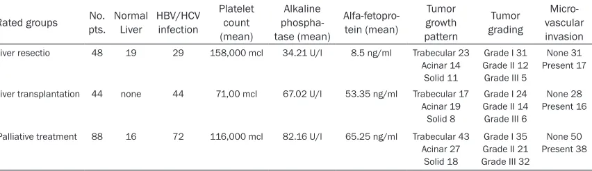
Table 1. Data indicating results of clinical evaluation of 180 HCC patients attending the study
Rated groups No. pts. Sex (mean)Age BCLC stage 0-A1 BCLC stage A2-A4 BCLC stage C-D Total bilirubin (mean) Serum Albumin (mean) INR
(mean) Ascites Encephalo-pathy
Okuda Score (mean points)
Liver resection 48 M 29 F 19
51 years 48 none none 26.75 mg/dl 49.04 g/l 1.06 None None 5.55
Liver transplantation 44 M 23 F 21
47 years none 47 none 27.29 mg/dl 50.65 g/l 1.13 Mild None/Mild 5.72
Palliative treatment 88 M 48
F 40 63 years none none 88 53.73 mg/dl 34.06 g/l 1.12 Severe Mild/Severe 11.30
Table 2. Data indicating liver status and tumor pathologic proprieties in 180 HCC patients attending the study
Rated groups No. pts. Normal Liver HBV/HCV infection Platelet count (mean) Alkaline phospha-tase (mean) Alfa-fetopro-tein (mean) Tumor growth pattern Tumor grading Micro-vascular invasion
Liver resectio 48 19 29 158,000 mcl 34.21 U/l 8.5 ng/ml Trabecular 23 Acinar 14
Solid 11
Grade I 31 Grade II 12 Grade III 5
None 31 Present 17
Liver transplantation 44 none 44 71,00 mcl 67.02 U/l 53.35 ng/ml Trabecular 17 Acinar 19
Solid 8
Grade I 24 Grade II 14 Grade III 6
None 28 Present 16
Palliative treatment 88 16 72 116,000 mcl 82.16 U/l 65.25 ng/ml Trabecular 43 Acinar 27
Solid 18
Grade I 35 Grade II 21 Grade III 32
None 50 Present 38
liver function and tumor stage in accordance with Child-Pugh classification and TNM as well as BCLC and the Milan Criteria [1-3, 16]. The radical therapy group (RT Group) consisted of 92 patients and included:
48 patients (29 M, 19 F; Mean age: 51 years) with Stage I tumor (0-A1 Stage acc. to BCLC) in whom a liver resection was performed. In 19 of them the tumor developed in a healthy liver, in 29 in a cirrhotic liver (but with normal synthetic function and without features of portal hyper-tension) after viral hepatitis (HBV/HCV). The blood serum alpha-fetoprotein level averaged 28.5 ng/ml (SD +/- 1457), the number of blood platelets 158,000 mcl (SD +/- 74,000), INR 1.06 (SD +/- 0.15).
[image:2.612.96.522.236.359.2]71,000 mcl (SD +/- 46,000), IRN 1.13 (SD +/- 0.21).
The post-operative approach was based on the overall condition, liver functioning, and a nutri-tional status in all patients after extended sur-gical intervention for HCC. In the immediate postoperative period after hepatic resection or liver transplantation all patients were held in the intensive care unit for 2-14 days (mean 9 days) depending on their cardiovascular and respiratory conditions as well as on other me- tabolic and functional disorders. Maintenance of an adequate fluid balance and a normal renal function were of primary importance. All patients underwent broad-spectrum antibiotic therapy. The patients transplanted for HCC received immunosuppressive therapy with cal-cineurin inhibitors, antimetabolites and cortico-steroids. Patients were followed up continu-ously for 3 years undergoing routine clinical examination and USG examination every 6 months. Their alpha-fetoprotein level was also determined every 6 months. Its presence, sug-gestive in any case of cancer relapse, was veri-fied with a CT scan.
The palliative therapy group (PT Group) includ-ed 88 patients (48 M, 40 F; Mean age 63 years) with an advanced neoplastic process (III/IV Clinical Stage, B-C acc. to BCLC). In 16 of the patients the tumor developed in a healthy liver, in 72 in a cirrhotic liver in the course of HBV/
HCV infection. Their alpha-pro-tein level averaged 65.25 ng/ ml (SD +/- 1,798), the number of blood platelets 116,000 mcl (SD +/- 82,000), INR 1.12 (SD +/- 0.11).
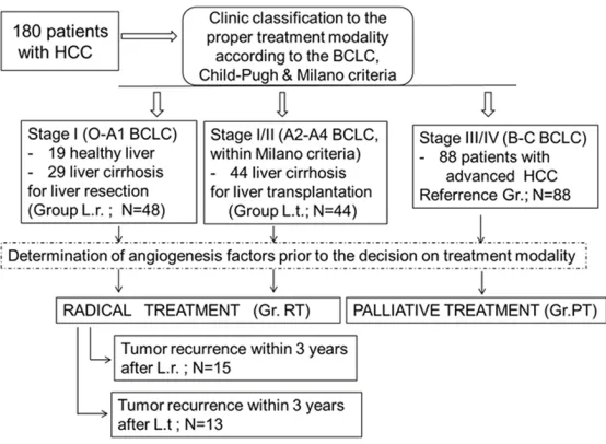
Demographic data are pre-sented in Tables 1 and 2. The flow of the patients is present-ed in Figure 1.
Determination of circulating EPCs
Prior to the treatment (1-3 days before the operation or upon a palliative therapy deci-sion) blood samples were col-lected from the patients. Two mL of venous blood were col-lected for K3 EDTA and ana-Figure 1. The flow of 180 patients with HCC evaluated for the level of
angio-genesis factors.
[image:3.612.94.371.73.276.2]endothe-Figure 2. The rating of CD309+ cells in the population of CD34+Cd45- and CD34+CD45+ cells of WBC in HCC pa-tients. The cells of CD309+ were identified in both populations in relation to the isothypic control. Pictures indicate the small number of the CD309+ cells.
lial progenitor cells in patients who remained asymptomatic and in patients with tumor recur-rence are presented in Figures 2 and 3. Examination of the blood serum VEGF concen-tration
Eight ml of blood were collected for the se- rological examination of the blood serum VEGF concentration. After centrifugation at 3000 rotations/minute for 10 minutes, the serum sample was stored at -80°C. The VEGF concentration was determined with the help of the immunoenzymatic assay Quan- tikine Human VEGF Immunoassay, R&D Sy- stems, Minneapolis, MN, USA). The sensitivity of the method was 9 pg/ml, and the precision coefficient was 4.4-6.7% and between exami-nations 6.2-8.8%.
Pathologic analysis
Pathologic analysis was performed on material from 92 tumors excised during surgery and 88
specimens collected during a core needle biop-sy in patients under palliative therapy. The preparations were fixed in 10% formalin and immersed in paraffin. Tissue blocks were sliced into 4 µm-thick sections and stained with hematoxylin/eosin. The degree of tumor cells differentiation acc. to the Edmondson-Steiner scale, the structural variant and the presence of microvascular invasion by the tumor were evaluated.
Morphometric determination of VEGF/VEGFR tissue concentration
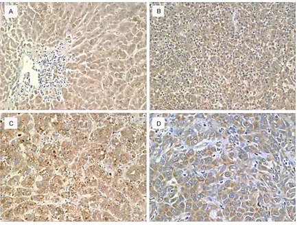
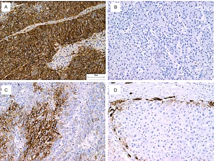
[image:4.612.93.524.73.402.2]VEGF proteins was determined by morphome-try with the use of an automatic image analy- zer in 5 representative fields of vision (Media Cybernetics). The analysis covered the immu-nological reaction surface field and the inten-sity of the reaction. The product of the mean surface field and the mean reaction intensity expressed in morphometric units (m.u.) was adopted as the mean level of VEGF protein immunoreactivity in the tissue. Pictures show-ing the results of the immunohistochemical staining for the expression of VEGF and VEGFR in the samples of HCC are presented in Figures 4 and 5, respectively.
Statistical analysis
The dataset was recorded on a disc and ana-lyzed in the SAS 9.4 statistical software (SAS Institute Inc. 100 SAS Campus Drive Cary, NC 27513-2414, USA). Continuous variables were expressed as a mean +/- SD, with a sample rep-resentativeness of 95% CI, discrete variables
[image:5.612.95.522.73.398.2]Figure 4. Results of immunohistochemical staining for expression of VEGF in the HCC samples. A: Immunoreactivity for VEGF in normal liver tissue; B: Weak immunoreactivity for VEGF in HCC cells; C: Mild intensity of VEGF expres-sion in HCC cells; D: Strong immunoreactivity for VEGF in HCC cells. Images A-C: Magnification of 10x, image D: Magnification of 20x.
Results
The differences in patients clinical-pathologic presentation
Although candidates for liver resection differed from candidates for liver transplantation in terms of HCC staging (0-A1 for L.r. and A2-A4 for L.t. group), underlying liver disease and the health status, they were analyzed as one radi-cal treatment group (RT Group).The reason was that the development of an early cancer (stage 0-A4 according to BCLC classification) was re- cognized in all of them in the assessment by the clinical classification systems (Child-Pugh, Okuda and BCLC). Candidates for liver resec-tion also had a higher number of blood plate-lets, a lower INR level and a lower blood serum AFP level. Post- HBV/HCV inflammation cirrho-sis was found only in 61% of them. The pathologic examination showed prevalence of tumors of trabecular and acinar structure
(85.8%),well or medium differentiated (90.2%). Relevant data are presented in Tables 1 and 2. There were statistically significant differences neither in the mean values of the VEGF/VEGFR protein expression in the blood serum and tumor tissue nor in the mean number of EPCs in circulation between patients treated with liver resection (L.r.) and with liver transplanta-tion (L.t.), as determined in preoperative set-tings (see Table 3).
Figure 5. Results of immunohistochemical staining for expression of VEGFR in the HCC samples. A: Strong immu-noreactivity for VEGFR in HCC cells.; B: Absence of immuimmu-noreactivity for VEGF in HCC; C: Mild intensity of VEGFR expression in HCC cells; D: Marking of HCC cells on the wedge of cancer. Images A-D: Magnification of 10x.
treatment (RT Group) by higher levels of angio-genesis factors (P < 0.001), except for the mean level of the VEGF blood serum concentra-tion, as shown in Table 3.
ROC curve and cutoff values for angiogenesis factors
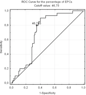
The mean value of each of the angiogenesis factors determined on the basis of measure-ments performed in all the 180 HCC patients presented as follows: 47.8% (SD +/- 17.5, 44.5-49.6 95% CI) for the percentage of EPCs, 327.2 pg/ml (SD +/- 284.0, 285.1-368.9 95% CI) for the VEGF concentration in blood serum and 83.1 m.u. (SD +/- 12.8, 81.2-85.4 95% CI) and 139.3 m.u. (SD +/- 58.11, 30.7-140.8 95% CI) for VEGF and VEGFR in tumor tissues. Details are given in Table 3. We adopted them as the cutoffs for the further analysis instead of val-ues established by the receiver operating char-acteristic (ROC curve) because they constituted
a better point of reference in the analysis of factors favoring cancer recurrence in the 92 patients after radical surgical treatment (RT Group). The ROC values were as follows: 46.75% for the percentage of EPCs, 221.9 pg/ml for the VEGF concentration in the blood serum, and 65.8 m.u. and 197.03 m.u. for VEGF and VEGFR in tumor tissues. The cutoff value for the percentage of EPCs was estab-lished at the same level in both cases (mean value 47.8 % versus ROC value 46.75%). The ROC analysis for EPCs is presented in Figure 6.
Clinical-pathologic predisposition for recur-rence
Table 3. Expression of angiogenesis factors in individual groups of 180 HCC patients
Rated groups No. pts. % EPCs VEGF (pg/ml)Mean +/- SD Tissue VEGF (units) Tissue VEGFR (units) Liver resection
(L.r. group) 48 36.4, +/- 12.9 356.3, +/- 208 77.9, +/- 14.2 110.1, +/- 58.5 Liver transplantation
(L.t. group) 44 40.5, +/- 13.1 309.7, +/- 245 80.9, +/- 13.1 126.4, +/- 50.1 Radical treatment
(L.r. + L.t. = R.T. group) 92 35.5, +/- 13.1 334.8, +/- 226 79.4, +/-13.7 117.9, +/- 55.1 Palliative treatment
(P.T. group) 88 56.1, +/- 17.6 319.6, +/- 336 87.8, +/-10.4 161.6, +/- 52.8 All HCC patients 180 47.8, +/- 17.5 327.2, +/- 284 83.1, +/- 12.8 139.3, +/- 58.1 Student t-Test
L.r. versus L.t. group NS NS NS NS
Student t-Test
R.T. versus P.T. group P < 0.001t = -6.88 NS P < 0.001t = -3.41 P < 0.001t = -5.51
the preoperative clinical settings (t = -1.7, P < 0.01). However, clinical evaluation systems revealed no significant differences between patients with and without cancer relapse in terms of the general health status and tumor staging (BCLC Staging = early cancer, Okuda score = NS). There were also no significant dif-ferences in tumor tissue differentiation, its structure variants and frequency of microvas-cular invasion found on the autopsy examina-tion of HCC preparaexamina-tions collected during sur-gery from patients with or without a tumor relapse.
High angiogenesis activity predisposes to tu-mor recurrence
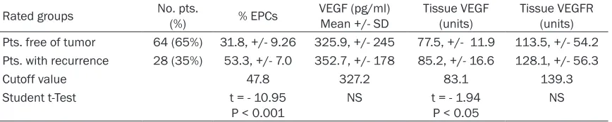
All patients with tumor relapse had an elevated level of VEGF/VEGFR-2 protein expression in peripheral blood and in HCC tissue as well as an increased number of EPCs in blood circula-tion, exceeding the cutoff value in each of them. The data are given in Table 4.
Factors indicating unfavorable tumor biology predisposing to early recurrence
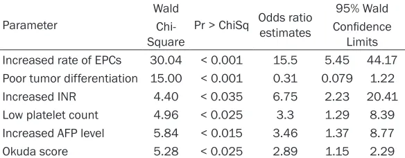
The multivariate logistic regression model (Wald test, Software SAS 9.4) was used to describe factors of unfavorable tumor biology predisposing to early tumor recurrence after radical surgery. When only the clinical and pathologic features of the tumor were used as dependent variables, the analysis indicated an increased INR (OR = 8.15), a low PLT count (OR = 3.12), tumor microvascular invasion (OR = 3.35) and an increased AFP level (OR = 2.74, P < 0.01) as significant independent predispos-ing factors (see Table 5). Unfortunately, patients with an abnormal INR and a low platelets count are not qualified for surgical treatment in prin-ciple and the knowledge about the tumor micro-vascular invasion is available only after opera-tion. However, when angiogenesis features of the tumor were used together with clinical/ pathological features in the model, the analysis indicated an increased level of endothelial pro-genitor cells in the blood circulation as the
Table 4. Expression of angiogenesis factors in patients with and without tumor recurrence at the time of 3 years of follow-up after radical surgical treatment
Rated groups No. pts.(%) % EPCs VEGF (pg/ml)Mean +/- SD Tissue VEGF (units) Tissue VEGFR (units) Pts. free of tumor 64 (65%) 31.8, +/- 9.26 325.9, +/- 245 77.5, +/- 11.9 113.5, +/- 54.2 Pts. with recurrence 28 (35%) 53.3, +/- 7.0 352.7, +/- 178 85.2, +/- 16.6 128.1, +/- 56.3
Cutoff value 47.8 327.2 83.1 139.3
Student t-Test t = - 10.95
[image:8.612.91.525.316.403.2]dominant factor predicting the unsuccessful outcome due to likely occurrence of the recur-rence of HCC (OR = 15.5, P < 0.001, Table 6). Notably, the angiogenesis activity of the tumor can be readily ascertainable at the time of pre-operative settings.
Discussion
[image:9.612.94.375.73.382.2]The number of HCC recurrences after surgical treatment indicates that patients qualified for surgery are these with a prognosis of an unlike-ly or impossible recovery due to the aggressive biological activity of the tumor. Unfortunately, none of the algorithms based on the system of the clinical assessment takes into account the tumor potential to stimulate processes that disturb the homeostasis of the host organism and determine the invasiveness of the neo-plasm [4, 5, 17]. The current study presents results of a multifactor analysis of clinical variables commonly used to determine the HCC stage as well as variables testifying to the biological activity of the neoplasm. The
Figure 6. The ROC curve analysis indicating cutoff value for the percentage of EPCs in the blood circulation in patients with HCC.
blood/tissue VEGF/VEGFR protein expression and the count of endothelial progenitor cells (EPCs) in the population of CD34+ stem cells in patients with the tumor recurrence significantly exceeded the mean values of angiogenesis fac-tors that were established in the preoperative settings for the group eligible for radical surgi-cal management (P < 0.001). Our findings indi-cate that the determination of angiogenesis factors can prove helpful in the proper discrimi-nation and stratification of patients in clinically ambiguous cases [9, 22-27]. And this is the main contribution of the present study.
Table 5. Analysis of maximum likelihood of unfavorable tumor biol-ogy predisposing to tumor recurrence
Parameter Chi-Square Pr > ChiSqWald Odds ratio estimates Confidence Limits95% Wald Increased INR 18.36 < 0.001 8.15 3.12 21.28 Low platelet count 7.74 < 0.005 3.12 1.40 6.98 Increased AFP level 6.50 < 0.01 2.74 1.26 5.93
Clinical features of BCLC classification were used as the dependent variables. Patients who stayed asymptomatic over 3 years after radical surgical treatment con-sisted of the reference group.
given therapy option. There is no doubt as to the presence of a tight link between the VEGF and VEGFR protein concentration in the tumor tissue and poor treatment outcome after surgi-cal treatment. Confirmation of this correlation can be found in numerous reports on the tumor angiogenesis from the 1996 findings of Mise [9, 31]. Numerous authors draw attention to the fact that the level of VEGF protein concen-tration in the blood serum of HCC patients dif-fers from its level in the blood serum of healthy people or patients with liver cirrhosis but with-out a neoplasm [9, 18, 20, 31, 32]. The prog-nostic importance of VEGF protein appears mostly in patients with advanced HCC and a level of blood serum VEGF concentration exceeding 400 pg/mL [27, 31]. In such cases, the level of the VEGF factor may prove a conve-nient indicator of the effectiveness of a bridg-ing or palliative treatment with TACE [27, 34]. In our study, the level of VEGF concentration in the blood serum of patients with HCC recur-rence exceeded the level of 400 pg/mL just in a few cases. Though the mean level of VEGF serum concentration was higher than the
deter-whole process of HCC growth and Sun et al. demonstrated the role of EPCs in HCC neovas-cularization [11, 13, 36]. EPCs constitute a rel-atively small, re-aching less than 3%, subpopu-lation of hematopoietic CD45+ stem cells and a larger,amounting to 35%, subpopulation of vascular endothelium CD45- cells [21]. The study showed that in patients with an early HCC the number of endothelial progenitor cells (EPCs) in the population of CD34+ stem cells is significantly lower than in patients with an advanced HCC (35.5% versus 56.1%; t = -6.88, P < 0.001). It also revealed that in all early HCC patients with a relapse within 3 years from radi-cal surgery, the number of EPCs determined at the time of the patients’ qualification for treat-ment was significantly higher (31.8%, SD +/- 9.26, 29.5-34.1, 95% CI versus 53.3%, SD +/- 7.0, 50.6-56.6, 95% CI). Also, it exceeded sig-nificantly the cutoff value set at 47.8% (t = -10.95, p < 0.001). These results indicate that the activity of angiogenesis is proportional to how advanced the tumor is. Consequently, the determination of the EPCs in circulation pro-vides an effective way of identifying patients
mined cutoff value, the dif-ference was not significant. The level of the VEGF serum concentration correlated wi- th increased levels of the VEGF and VEGFR protein immune reactivity in the tumor tissue of patients with HCC recurrence (t = -1.94, p < 0.05 for VEGFR). The findings correspond to the experience reported by others [9, 31, 32, 35]. What proved to be the best indi-cator was, however, the number of endothelial pro-genitor cells of CD34+, CD309+ phenotype circu-lating in the blood. The experience of many resear- ch centers indicates that the level of endothelial pro-genitor cells (EPCs) is essential for tumor growth [10, 12, 14, 15, 19, 31]. Recently, Zhu et al. revealed that EPCs are mobilized and incorporated into tumor vessels throughout the
Table 6. Analysis of maximum likelihood estimates of unfavorable tumor biology predisposing to tumor recurrence
Parameter
Wald
Pr > ChiSq Odds ratio estimates
95% Wald
Chi-Square Confidence Limits Increased rate of EPCs 30.04 < 0.001 15.5 5.45 44.17 Poor tumor differentiation 15.00 < 0.001 0.31 0.079 1.22 Increased INR 4.40 < 0.035 6.75 2.23 20.41 Low platelet count 4.96 < 0.025 3.3 1.29 8.39 Increased AFP level 5.84 < 0.015 3.46 1.37 8.77 Okuda score 5.28 < 0.025 2.89 1.15 2.29
[image:10.612.91.383.242.356.2]with an advanced tumor. Yet, most importantly, a high number of circulating EPCs indicates the presence, among early HCC patients deter-mined on the basis of the clinical classification system, of patients with a tumor of exception-ally high biological activity characteristic of more malignant, more advanced tumors with poor prospects for successful surgical treat-ment. Determination of the number of circulat-ing EPCs at the time of the qualification of can-didates for radical surgical treatment may thus constitute a valuable clue in the proper stratifi-cation of patients.
The study indicates that it is not enough to base the algorithm of clinical assessment on physical parameters, such as the size and num-ber of neoplastic foci, pathological data inform-ing on the microvascular invasion and clinical liver function parameters which constitute the foundation of BCLC, Child-Pugh, Milan Criteria and other, similar classification systems. The single end point of our study used to assess the performance of the different factors was tumor recurrence at the 3 year follow-up after a radi-cal surgery. The multifactorial analysis with the use of clinical factors, determined according to the standards of the qualification of patients for surgical treatment referred to above, show-ed that the risk of recurrence does not depend so much on the extent of the tumor impact as on disturbances resulting from a liver disfunc-tion. Significantly, the same analysis made with the use of standard clinical factors and angio-genesis factors revealed a major role of the tumor angiogenesis potential and the degree of tumor cell maturity in the appearance of HCC recurrences. It is therefore possible to ‘tighten’ the systems of clinical classification and impro-ve the stratification of patients for radical surgi-cal treatment. Determination of the EPCs in circulation is a non-invasive, simple and inex-pensive method and the obtained results seem to be much easier to interpret than the ‘caprici-ous’ VEGFR factor.
Expectations and limits of the study
There are at least several patient and tumor-related factors responsible for the ultimate out-come of the patient after surgical treatment. Coexisting HCV infection, tumor number and size, as well as tumor histologic grade and microvascular invasion are the most frequently
aware that the identification of the EPCs by flow cytometry is a “rare event”, depending exclu-sively on the number of positive events (n), we increased the total number of acquired events to at least 2,000,000. Despite the above men-tioned limitations we consider flow cytometry a method sensitive enough when the count of peripheral blood EPCs is conceived as a bio-marker for the stratification of early HCCs. We conclude that an increased number of EPCs in the blood is a reliable prognostic indicator. Radical surgery with an intention to cure the disease in patients with an over 47% higher rate of circulating EPCs should be proposed with full awareness of failure without an adjunc-tive therapy.
Acknowledgements
We acknowledge the work of all colleagues from the Central Teaching Hospital, Medical University of Warsaw for their cooperation. The project was supported by the National Science Center (Grant No. NN 403 177940) and approved by the Bioethics Committee of Medical University of Warsaw (Approval No. 235/209).
Disclosure of conflict of interest
None.
Abbreviations
HCC, hepatocellular carcinoma; VEGF, vascular endothelial growth factor; VEGFR-2, vascular endothelial growth factor receptor-2; EPCs, endothelial progenitor cells; BCLC, Barcelona Clinic Liver Cancer classification; TNM, tumor nodules metastases classification; AFP, alpha fetoprotein; ALP, alkaline phosphatase; PLT, platelets count; CD (34, 133, 45, 3019), clu- ster of differentiation antigens; K3 EDTA, ethyl-enediaminetetraacetic acid; INR, International Normalized Ratio of prothrombin time of blood coagulation; ISHAGE, International Society of Hematology and Graft Engineering; HBV/HCV, hepatitis B virus/hepatitis C virus.
Address correspondence to: Dr. Janusz Sierdziński, Department of Medical, Informatics & Telemedicine, Central Teaching Hospital, Medical University of Warsaw, Banacha 1a, Warsaw 02097, Poland. Tel: (+48) 22 658 29 97; E-mail: [email protected]. pl
References
[1] Pons F, Varela M and Llovet JM. Staging sys-tems in hepatocellular carcinoma. HPB (Ox-ford) 2005; 7: 35-41.
[2] Subramaniam S, Kelley RK and Venook AP. A review of hepatocellular carcinoma (HCC) staging systems. Chin Clin Oncol 2013; 2: 33. [3] Llovet JM, Fuster J, Bruix J; Barcelona-Clínic
Liver Cancer Group. The Barcelona approach: diagnosis, staging, and treatment of hepato-cellular carcinoma. Liver Transpl 2004; 10 Suppl 1: S115-20.
[4] Portolani N, Coniglio A, Ghidoni S, Giovanelli M, Benetti A, Tiberio GA and Giulini SM. Early and late recurrence after liver resection for hepato-cellular resection: patterns and therapeutic implications. Ann Surg 2006; 243: 229-235. [5] Welker MW, Bechstein WO, Zeuzem S and
Tro-jan J. Recurrent hepatocellular carcinoma af-ter liver transplantation - an emerging clinical challenge. Transpl Int 2013; 26: 109-118. [6] Tabrizian P, Jibara G, Shrager B, Schwartz M
and Roayaie S. Recurrence of hepatocellular cancer after resection: patterns, treatments, and prognosis. Ann Surg 2015; 261: 947-955. [7] Yamashita Y, Shirabe K, Toshima T, Tsuijita E,
Takeishi K, Harimoto N, Ikegami T, Yoshizumi T, Ikeda T, Soejima Y and Maehara Y. Risk factors for recurrence after curative resection of hetitis C-related hepatocellular carcinoma in pa-tients without postoperative interferon thera-py. Hepatol Res 2013; 43: 1313-1320. [8] Dituri F, Serio G, Filannino D, Mascolo A, Sacco
R, Villa E and Giannelli G. Circulating TGF-be-ta1-related biomarkers in patients with hepa-tocellular carcinoma and their association with HCC staging scores. Cancer Lett 2014; 353: 264-271.
[9] Tseng PL, Tai MH, Huang CC, Wang CC, Lin JW, Hung CH, Chen CH, Wang JH, Lu SN, Lee CM, Changchien CS and Hu TH. Overexpression of VEGF is associated with positive p53 immu-nostaining in hepatocellular carcinoma (HCC) and adverse outcome of HCC patients. J Surg Oncol 2008; 98: 349-357.
[10] Beerepoot LV, Mehra N, Vermaat JS, Zonnen-berg BA, Gebbink MF and Voest EE. Increased levels of viable circulating endothelial cells are an indicator of progressive disease in cancer patients. Ann Oncol 2004; 15: 139-145. [11] Zhu H, Shao Q, Sun X, Deng Z, Yuan X, Yu D,
[12] Yu DC, Chen J, Sun XT, Zhuang LY, Jiang CP and Ding YT. Mechanism of endothelial progenitor cell recruitment into neo-vessels in adjacent non-tumor tissues in hepatocellular carcino-ma. BMC Cancer 2010; 10: 435.
[13] Zhu K, Dai Z and Zhou J. Biomarkers for hepa-tocellular carcinoma: Progression in early diag-nosis, progdiag-nosis, and personalized therapy. Biomark Res 2013; 1: 10.
[14] Ma S. Biology and clinical implications of CD133(+) liver cancer stem cells. Exp Cell Res 2013; 319: 126-132.
[15] Yuan SX, Yang F, Yang Y, Tao QF, Zhang J, Huang G, Yang Y, Wang RY, Yang S, Huo XS, Zhang L, Wang F, Sun SH and Zhou WP. Long noncoding RNA associated with microvascular invasion in hepatocellular carcinoma promotes angiogenesis and serves as a predictor for hepatocellular carcinoma patients’ poor re- currence-free survival after hepatectomy. Hep-atology 2012; 56: 2231-2241.
[16] Mazzaferro V, Bhoori S, Sposito C, Bongini M, Langer M, Miceli R and Mariani L. Milan crite-ria in liver transplantation for hepatocellular carcinoma: an evidence-based analysis of 15 years of experience. Liver Transpl 2011; 17 Suppl 2: S44-57.
[17] Shah SA, Cleary SP, Wei AC, Yang I, Taylor BR, Hemming AW, Langer B, Grant DR, Greig PD and Gallinger S. Recurrence after liver resec-tion for hepatocellular carcinoma: risk factors, treatment, and outcomes. Surgery 2007; 141: 330-339.
[18] Prager GW, Poettler M, Unseld M and Zielinski CC. Angiogenesis in cancer: Anti-VEGF escape mechanisms. Transl Lung Cancer Res 2012; 1: 14-25.
[19] Nowak K, Rafat N, Belle S, Weiss C, Hanusch C, Hohenberger P and Beck G. Circulating en-dothelial progenitor cells are increased in hu-man lung cancer and correlate with stage of disease. Eur J Cardiothorac Surg 2010; 37: 758-763.
[20] Boige V, Malka D, Bourredjem A, Dromain C, Baey C, Jacques N, Pignon JP, Vimond N, Bou-vet-Forteau N, De Baere T, Ducreux M and Farace F. Efficacy, safety, and biomarkers of single-agent bevacizumab therapy in patients with advanced hepatocellular carcinoma. On-cologist 2012; 17: 1063-1072.
[21] Otto W, Krol M, Maciaszczyk M, Najnigier B, Si-erdzinski J, Krawczyk M. Levels and values of circulating hematopoietic and endothelial pro-genitor cells in patients with hepatocellular carcinoma. J Liver 2014; 3: 4-14.
[22] Kaseb AO, Hassan MM, Lin E, Xiao L, Kumar V, Pathak P, Lozano R, Rashid A, Abbruzzese JL, Morris JS. V-CLIP: integrating plasma VEGF into a new scoring system to stratify patients with
advanced hepatocellular carcinoma for clinical trials. Cancer 2011; 117: 2478-2488.
[23] Kaseb AO, Hanbali A, Cotant M, Hassan MM, Wollner I and Philip PA. Vascular endothelial growth factor in the management of hepatocel-lular carcinoma: a review of literature. Cancer 2009; 115: 4895-4906.
[24] Srivastava S, Wong KF, Ong CW, Huak CY, Yeoh KG, Teh M, Luk JM and Salto-Tellez M. A mor-pho-molecular prognostic model for hepatocel-lular carcinoma. Br J Cancer 2012; 107: 334-339.
[25] Zhu AX, Duda DG, Sahani DV and Jain RK. HCC and angiogenesis: possible targets and future directions. Nat Rev Clin Oncol 2011; 8: 292-301.
[26] Chan EY, Larson AM, Fix OK, Yeh MM, Levy AE, Bakthavatsalam R, Halldorson JB, Reyes JD and Perkins JD. Identifying risk for recurrent hepatocellular carcinoma after liver transplan-tation: implications for surveillance studies and new adjuvant therapies. Liver Transpl 2008; 14: 956-965.
[27] Shim JH, Park JW, Kim JH, An M, Kong SY, Nam BH, Choi JI, Kim HB, Lee WJ, Kim CM. Associa-tion between increment of serum VEGF level and prognosis after transcatheter arterial che-moembolization in hepatocellular carcinoma patients. Cancer Sci 2008; 99: 2037-2044. [28] Sarasin FP, Majno PE, Llovet JM, Bruix J,
Men-tha G and Hadengue A. Living donor liver trans-plantation for early hepatocellular carcinoma: A life-expectancy and cost-effectiveness per-spective. Hepatology 2001; 33: 1073-1079. [29] Belghiti J and Kianmanesh R. Surgical
treat-ment of hepatocellular carcinoma. HPB (Ox-ford) 2005; 7: 42-49.
[30] El-Serag HB. Hepatocellular carcinoma. N Engl J Med 2011; 365: 1118-1127.
[31] Yang ZF and Poon RT. Vascular changes in he-patocellular carcinoma. Anat Rec (Hoboken) 2008; 291: 721-734.
[32] Zhan P, Qian Q, Yu LK. Serum VEGF level is as-sociated with the outcome of patients with he-patocellular carcinoma: a meta-analysis. Hep-atobiliary Surg Nutr 2013; 2: 209-215. [33] Zhang W, Kim R, Quintini C, Hashimoto K,
Fuji-ki M, Diago T, Eghtesad B, Miller C, Fung J, Tan A, Menon KV and Aucejo F. Prognostic role of plasma vascular endothelial growth factor in patients with hepatocellular carcinoma under-going liver transplantation. Liver Transpl 2015; 21: 101-111.
[35] Yamaguchi R, Yano H, Iemura A, Ogasawara S, Haramaki M and Kojiro M. Expression of vas-cular endothelial growth factor in human hepa-tocellular carcinoma. Hepatology 1998; 28: 68-77.
[36] Sun XT, Yuan XW, Zhu HT, Deng ZM, Yu DC, Zhou X and Ding YT. Endothelial precursor cells promote angiogenesis in hepatocellular carcinoma. World J Gastroenterol 2012; 18: 4925-4933.