Case Report
Multiple primary malignancies of the
thyroid gland and the brain: a case report
Xiao-Qiang Zheng1, Dong-Li Zhao1, Yan-Xia Sui2
Departments of 1Radiotherapy, 2Pathology, First Affiliated Hospital, Xi’an Jiaotong University, Xi’an 710061,
Shaanxi, P. R. China
Received February 18, 2017; Accepted March 30, 2017; Epub June 15, 2017; Published June 30, 2017
Abstract: A majority of studies have reported cases on patients with primary thyroid cancer who developed sub-sequent malignancies, following their initial thyroid cancer diagnosis. Notably glioma has been reported as a sec-ondary cancer following thyroid cancer diagnosis. A case of a 50-year-old woman with a history of thyroid nodules and symptom of throat discomfort is reported in the present study. A fine needle aspiration of the thyroid nodules confirmed the presence of papillary thyroid carcinoma. The patient received a total thyroidectomy followed by io -dine 131 (131I) treatment. The patient was admitted to the hospital following 5 years of the initial treatment, due to
headache and nausea. Magnetic resonance imaging (MRI) of the head region revealed an irregular lesion at the left temporal lobe. Following frontal tumor resection, the biopsy revealed brain astrocytoma. Postoperatively, the patient underwent intensity modulated radiation therapy (IMRT) at a total dose of 60 Gy in combination with oral temozolo -mide chemotherapy. The frequency of radiation treatment was 2 Gy/day ×30 fractions. The data are not conclusive of a correlation between the incidence of glioma and 131I treatment. But, the incidence of long-term survival of the
patients with thyroid cancer and second primary cancer warrants considerable attention by medical professionals. In addition, during radioactive iodine therapy, the indication and the total dose of the radioactive iodine require careful monitoring.
Keywords: Thyroid, glioma, cancer
Introduction
Multiple primary carcinomas, refers to the de- velopment of two or more independent primary malignant tumors, simultaneously or succes-sively, in a single or multiple organs of the same
individual. Thyroid cancer is one of the most
common solid tumors. It has been estimated that the incidence of thyroid cancer in China in 2012 was approximately 11.9 million new
cases [1]. Glioma is a common cancer of the
nervous system, accounting for 80% of primary central nervous system malignancies in the
United States [2]. Thyroid cancer can develop
concurrently with primary glioma, although very
few cases have been documented. To our
know-ledge, one case of an American young woman with the aforementioned malignancies has been reported to date. In the present study, a thyroid cancer case with glioma is reported.
Case report
The patient was a 50 years old woman, who
presented with symptoms of throat discomfort
and was diagnosed with thyroid nodules. The patient had no history of smoking or alcohol
abuse. During physical examination, two mass-es with diameters of approximately 1.5 cm and
1 cm in size were observed in the left and right lobes of the thyroid, respectively. The masses
were homogeneous, hard and exhibited smooth surfaces. In addition, they could move up and
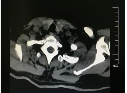
down during swallowing. The computed tomog
-raphy (CT) scan of the neck region indicated
that the left thyroid lobe exhibited a low density mass with dimensions of approximately 1.4
cm×1.3 cm, whereas the right lobe exhibited an
uneven low density mass with dimensions of
approximately 1.1 cm×0.8 cm (Figure 1). The
papillary thyroid carcinoma was diagnosed by
patient underwent a total thyroidectomy in
2011. The postoperative pathological examina -tions indicated papillary thyroid carcinoma, invading the local capsule (Figure 2). Following surgery, the patient was subjected to radioio-dine ablation therapy at a total dose of 100 mCi in one month duration. No local recurrence or distal metastasis was present during follow-up examinations.
The patient was admitted to hospital due to
continuous headaches for a total period of 3 months, following 5 years of the radioiodine ablation therapy. No nausea, vomiting, or blur-
red vision was reported at first. The headache
symptoms were progressively exacerbated and were associated with nausea and projectile
vomiting. The physical examination indicated
muscle strength of grade 5 of upper and lower extremities, normal muscle tone, pain and nor-mal sensation. Magnetic resonance imaging (MRI) of the head region revealed an irregular lesion at the left temporal lobe with dimensions
of 4.1 cm×3.5 cm×3.0 cm. The lesions were subjected to garland like enhancement (Fig- ure 3). The supratentorial cranial craniotomy for
tumor resection was conducted in December of
2015. The pathological examinations revealed
brain astrocytoma that developed from grade II
to grade III (WHO classification 2007) (Figure 4). Following a period of one month after
surgi-cal resection, the patient received IMRT at a total dose of 60 Gy. The frequency of radiation therapy was 2 Gy/day ×30 fractions at 4 MV. Concomitant oral temozolomide chemotherapy
(100 mg, 150 mg alternate on days 1-42) was
administered to the patient. The subject exhib
-ited no major toxicities. The patient showed no
signs of tumor, following 4 months of the initial diagnosis.
Discussion
The incidence of thyroid cancer has increased
recently, notably with regard to the papillary thyroid carcinoma. Following standard treat-ment, the majority of the thyroid cancer patients exhibit optimal survival rates and quality of life. However, certain cohort and population-based studies have shown that the incidence of thy-roid cancer with a second primary cancer has
increased significantly compared with the nor
-mal population. This increase has been notably
observed in young females [3, 4]. A previous study indicated that the 5 most common sites
of initial cancer localization included the breast, prostate, colon, skin and lung tissues in the patients with secondary thyroid cancer [5]. The
combination of clinical thyroid cancer with
glio-ma is relatively rare. Murali Krishna Gurram
et al. reported a young woman who was diag-nosed with two synchronous tumours namely,
glioblastoma multiforme (GBM) and papillary thyroid carcinoma [6]. The major symptoms of the patient were dizziness, headache and vom -iting. MRI of the brain demonstrated a non-enhancing and non-hemorrhagic component of the lesion along the lateral margin of the hem-orrhage that was indicative of a tumor. In
addi-tion, CT scan of the head and neck region dem -onstrated that the left lobe of the thyroid exhib-ited a nodule with a diameter of approximately
1.1 cm. The brain biopsy confirmed GBM, which
was associated with an extremely low survival
rate. The thyroid fine needle aspiration biopsy confirmed the diagnosis of the thyroid papillary
cancer. In contrast to the diagnosis, the pres-ent study does not report on the treatmpres-ent and prognosis of the patient. However, the present case is a middle-aged female who had been diagnosed with metachronous multiple primary
carcinomas. The secondary tumor was an
as-trocytoma, originating from a relatively low gra-
de glioma than GBM. The patient currently has
a high quality of life following the combined radiation therapy and chemotherapy.
[image:2.612.89.291.72.221.2]Several studies demonstrated that the expo-sure to radioactive iodine could affect the de- velopment of thyroid cancer and subsequent primary cancers, with the exception of the con-tributing factors namely, age, environment and Figure 1. Preoperative CT scan of the neck region.
genetic predisposition. A retrospective analysis
iodine treatment increased the probability of occurrence of a second primary cancer com-pared with non radioactive iodine treatment
[7]. Teng and co-workers demonstrated that 131I
treatment of thyroid cancer patients was
asso-ciated with an increased risk of lymphoma,
whereas it was not associated with the inci-dence of other solid tumors [8]. However, Ko et
al. reported an increased risk of urinary system cancer and head and neck cancer in patients
that received 131I treatment [9].
It is well established that the exposure to high
doses of ionizing radiation, and the inherited
mutations of highly penetrant genes are
associ-ated with rare syndromes. The aforementioned
[image:3.612.90.524.71.398.2]parameters are considered the two main patho-genetic factors of glioma [10]. Furthermore, certain studies have reported recently that thy-Figure 2. Pathological examination of the thyroid. A. Thyroid fine needle aspiration pathological examination indi -cated that the tumor cells were in papillary arrangement. Staining: hematoxylin and eosin (H&E); Magnification ×100. B. Histopathological sections from resection of thyroid cancer: tumor cells were in papillary arrangement. The central of the nipple indicated fibrovascular and psammoma body formation. Staining: H&E; Magnification ×100. C. Immunohistochemistry (IHC) of thyroglobulin (Thyroglobulin, Tg) stain. Tg expression in the cytoplasm of the tumor cells was positive. Magnification ×100. D. IHC for the thyroid transcription factor-1 (thyroid transcription factor-1, TTF-1) staining. TTF-1 expression in the nucleus of the tumor resection tissue was positive. Magnification ×100.
[image:3.612.91.289.497.646.2]influence the development of glioma cells
[11-13]. Cristiana Perrotta proposed that 3. 3’.5-
triiodothyronine (T3) indirectly affects glioma
growth via the modulation of microglia [12].
Nauman demonstrated that TH affects the pathogenesis of GBM via the activation of vari
-ous signalling pathways, such as the EGFR/ PTEN/Akt/mTOR and the TP53/MDM2/p14ARF
pathways [13]. Clinical evidence further sup-ports to some extent that the cancer remiss- ion and lifespan of patients with high grade gli-oma can be extended by reducing their
corre-sponding TH levels in the blood [14]. Although
further investigations are required to fully clari-fy the exact causes of cancer remission, the
aforementioned clinical and epidemiological evidence may provide insight in the develop-ment of novel treatdevelop-ment strategies to patients with glioma, such as chemical-induced hypo- thyroidism.
In addition, in the present study, the patient had undergone radioactive iodine therapy
fol-lowing thyroidectomy. The correlation between
the incidence of glioma and 131I treatment in
[image:4.612.91.524.72.446.2]py, the indication and the total dose of the radioactive iodine require careful monitoring. Future studies can address the development of new therapeutic methods for thyroid cancer patients.
Acknowledgements
We acknowledge this research did not receive any specific grant from funding agencies in the public, commercial, or not-for-profit sectors.
Disclosure of conflict of interest
None.
Address correspondence to: Dr. Yan-Xia Sui, De- partment of Pathology, The First Affiliated Hospital of Medical College, Xi’an Jiaotong University, No. 277, West Yanta Road, Xi’an 710061, Shaanxi, P. R. China. Tel: +86-13772009970; E-mail: sui [email protected]
References
[1] Ferlay J, Soerjomataram I, Dikshit R, Eser S, Mathers C, Rebelo M, Parkin DM, Forman D and Bray F. Cancer incidence and mortality worldwide: sources, methods and major pat-terns in GLOBOCAN 2012. Int J Cancer 2015; 136: E359-386.
[2] Ostrom QT, Gittleman H, Stetson L, Virk SM and Barnholtz-Sloan JS. Epidemiology of glio -mas. Cancer Treat Res 2015; 163: 1-14. [3] Hakala TT, Sand JA, Jukkola A, Huhtala HS,
Metso S and Kellokumpu-Lehtinen PL. In -creased risk of certain second primary malig -nancies in patients treated for well-differenti-ated thyroid cancer. Int J Clin Oncol 2016; 21: 231-239.
[4] Canchola AJ, Horn-Ross PL and Purdie DM. Risk of second primary malignancies in wom -en with papillary thyroid cancer. Am J Epidemiol 2006; 163: 521-527.
[5] Ronckers CM, McCarron P and Ron E. Thyroid cancer and multiple primary tumors in the SEER cancer registries. Int J Cancer 2005; 117: 281-288.
[6] Pulivarthi S, Haglind E, McGary CT and Gurram MK. Glioblastoma multiforme and papillary thyroid carcinoma-a rare combination of mul-tiple primary malignancies. J Neurosci Rural Pract 2015; 6: 241-244.
[7] Sawka AM, Thabane L, Parlea L, Ibrahim-Zada I, Tsang RW, Brierley JD, Straus S, Ezzat S and Goldstein DP. Second primary malignancy risk after radioactive Iodine treatment for thyroid cancer: a systematic review and meta-analy-sis. Thyroid 2009; 19: 451-457.
[8] Teng CJ, Hu YW, Chen SC, Yeh CM, Chiang HL, Chen TJ and Liu CJ. Use of radioactive iodine for thyroid cancer and risk of second primary malignancy: a nationwide population-based study. J Natl Cancer Inst 2016; 108.
[9] Ko KY, Kao CH, Lin CL, Huang WS and Yen RF. (131)I treatment for thyroid cancer and the risk of developing salivary and lacrimal gland dys-function and a second primary malignancy: a nationwide population-based cohort study. Eur J Nucl Med Mol Imaging 2015; 42: 1172-1178. [10] Schwartzbaum JA, Fisher JL, Aldape KD and
Wrensch M. Epidemiology and molecular pa-thology of glioma. Nat Clin Pract Neurol 2006; 2: 494-503; quiz 491 p following 516.
[11] Davis FB, Tang HY, Shih A, Keating T, Lansing L, Hercbergs A, Fenstermaker RA, Mousa A, Mou -sa SA, Davis PJ and Lin HY. Acting via a cell surface receptor, thyroid hormone is a growth factor for glioma cells. Cancer Res 2006; 66: 7270-7275.
[12] Perrotta C, De Palma C, Clementi E and Cervia D. Hormones and immunity in cancer: are thy-roid hormones endocrine players in the mi-croglia/glioma cross-talk? Front Cell Neurosci 2015; 9: 236.
[13] Nauman P. Thyroid hormones in the central nervous system (CNS) and their effect on neo-plasm formation, particularly on the develop-ment and course of glioblastoma multiforme-research hypothesis. Endokrynol Pol 2015; 66: 444-459.